Advances in Animal and Veterinary Sciences
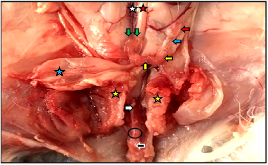
Reproductive system of the female kitten in situ showed vagina (white arrow) and vestibule (black arrow) inside the pelvic cavity, cut of pelvic symphysis (yellow stars), site of urethra cut (black circle) as well as ovary (red arrow), uterine tube (blue arrow), uterine horn (yellow arrows), cervices (green arrows), urinary bladder (blue star), abdominal aorta (red star), caudal vena cava (white star).
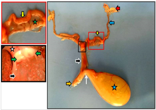
Dissected reproductive system of immature doe showed vagina (black arrows), vestibule (orange arrow), urethra (white arrow) as well as ovary (red arrow), uterine tube (blue arrow), uterine horn (yellow arrows), urinary bladder (blue star), cervices (white star), portio vaginalis uteri (green arrows).
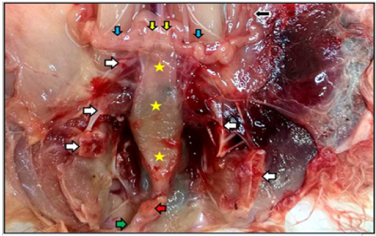
Reproductive system of the mature doe in situ showed vagina (yellow stars), vestibule (green arrow) inside the pelvic cavity, cut of pelvic symphysis (white arrows), site of urethra cut (red arrow) as well as uterine tube (black arrow), uterine horns (blue arrows), and cervices (yellow arrows).
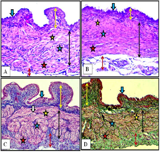
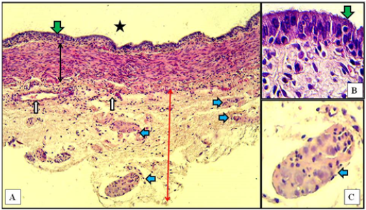
Cranial (A) and middle (B) regions of the vagina of the female kitten (upper panel) and cranial (C) and middle (D) regions of the vagina of the immature doe (lower panel) showed mucosa (double heads yellow arrows), simple columnar epithelium (blue arrow) and muscularis (double heads black arrows) of inner circular (yellow stars), middle longitudinal (blue stars) and outer circular (red stars), adventitia (double heads red arrow), nerve plexuses (white arrow). A & B: X200, H&E. C: X200, MTC. D: X200, Gomori Trichrome
Caudal region of vagina of immature doe showed mucosa (green arrows) of pseudostratified columnar epithelium, muscularis (double heads black arrow), adventitia (red arrow), sympathetic ganglia (blue arrows), blood vessels (white arrows), and vaginal lumen (black star). A: X10, H&E. B: X100, H&E. C: X40, H&E
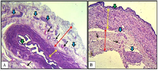
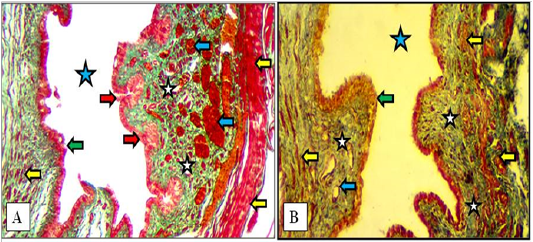
Cranial (A) and caudal (B) regions of the vagina of the mature doe showed mucosa (green arrows), muscularis (double heads yellow arrows), adventitia (double heads red arrows), sympathetic ganglia (blue arrows) and blood vessels (black arrows). A: X40, H&E. B: X100, H&E
Cranial (A) and caudal (B) parts of vestibule of the female kitten showed mucosa of stratified squamous epithelium (yellow arrows), mucosal invaginations to form vestibular glands (blue arrows), lamina propria of loose connective tissue filled with blood vessels (red stars), superficial blood capillaries (short black arrow) and large vessels deeply (long black arrow), thin circular muscularis (green arrows), adventitia (blue stars) and lumen of vestibule (green stars). A & B: X100, H&E.
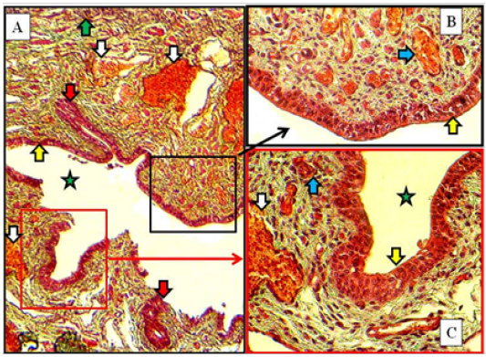
Caudal part of vestibule of immature doe showed stratified squamous epithelium (yellow arrows), lamina propria filled with blood capillaries superficially (blue arrows) and large vessels deeply (white arrows), vestibular glands (red arrow) and lumen (green star). A: X100. B & C: X400, MTC
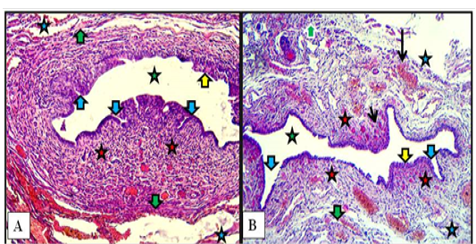
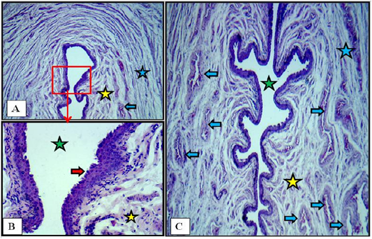
Cranial (A) and caudal (B) parts of the vestibule of the mature doe showed mucosa of stratified squamous epithelium (green arrows), lamina propria of loose connective tissue (white stars) filled with blood vessels (blue arrows), newly formed vestibular glands (red arrows), thin circular muscularis (yellow arrow) and lumen of vestibule (blue stars). A: X100, Gomori Trichrome. B: X200, MTC
Cross section at the labia minor of vulva of immature doe showed vulvar cleft (green stars), stratified squamous epithelium (red arrows), cavernous vessels (blue arrows), inner longitudinal (yellow stars) and outer circular smooth muscle fibers (blue stars). A: X40. B: X400. C: X40. H&E


