Advances in Animal and Veterinary Sciences
Research Article

Morpho-Histological Comparisons of Liver between the Broiler Chickens and Wild Boar in Algeria
Khenenou Tarek1,3*, Berghiche Amine1,3, Rahmoun Djalal Eddine1,2 , Miloudi Abdelhafidh1 , Athamna Hanane1
1Institute of Agronomic and Veterinarian Sciences, University of Mohamed Cherif Messaâdia, Souk Ahras, Algeria; 2Laboratory of Animal Production, Biotechnology and Health, University of Mohamed Cherif Messaâdia, Souk Ahras, Algeria; 3Laboratory science and technic of living, Institute of Agronomic and Veterinarian Sciences, University of Mohamed Cherif Messaâdia, Souk Ahras, Algeria.
Abstract | Aim: The objective of this study is to compare the normal macro and microscopic appearance of the liver in two very different species, one is an omnivorous mammal; the wild boar and the other belongs to the family of poultry; broiler chicken from the region of Bouhmama (Khenchela). Materials and methods: Eight broilers (58 days of age) and eight wild boars were included in the experiment to obtain information about the morpho-histological appearances of liver in two species.Results: There is a big difference in the liver appearance between the two species, in the wild boar it is of firm consistency with a tiger aspect and divided into four lobes, whereas in the broiler, the liver is brown and sometimes pale during the first 10-14 days, so it was divided into two lobes. Concerning the liver parenchyma, we used the Russian LOMBO MBS-10 stereo microscope, our results showed that the liver parenchyma was well developed in wild boar than in broiler chickens whereas, in broiler chickens; an excessive development of the sinus; the latter were less developed in the wild boar. Conclusion: The macroscopic observation showed a marked difference in liver between the two species. The microscopic examination of liver showed that the parenchyma is less pronounced in broilers while the sinuses were highly developed in the wild boar.
Keywords | Broiler chicken, Liver, Macro and microscopic appearances, Wild boar, Algeria.
Editor | Kuldeep Dhama, Indian Veterinary Research Institute, Uttar Pradesh, India.
Received | August 25, 2018; Accepted | October 14, 2018; Published | November 02, 2018
*Correspondence | Khenenou Tarek; Institute of Agronomic and Veterinarian Sciences, University of Mohamed Cherif Messaâdia, Souk Ahras, Algeria; Email: tarekkhenenou@yahoo.fr
Citation | Tarek K, Amine B, Eddine RD, AbdelHafidh M, Hanane A (2019). Morpho-histological comparisons of liver between the broiler chickens and wild boar in Algeria Adv. Anim. Vet. Sci. 7(1): 24-29.
DOI | http://dx.doi.org/10.17582/journal.aavs/2019/7.1.24.29
ISSN (Online) | 2307-8316; ISSN (Print) | 2309-3331
Copyright © 2019 Tarek et al. This is an open access article distributed under the Creative Commons Attribution License, which permits unrestricted use, distribution, and reproduction in any medium, provided the original work is properly cited.
Introduction
The liver, by its functions, is an indispensable organ for life, so it is considered the largest gland of the body and can be considered as the central organ for the maintenance of the energy supply. Moreover, it catalyzes the biosynthesis and process of biodegradation and excretion of final metabolic products. Blood drains to it from the intestines in the hepatic portal vein, and the products of digestion are metabolized, harmful material detoxified, senescent erythrocytes removed from the circulation and bile secreted (Dellman, 1979; Whitlow, 2000).
In the organoleptic properties of this organ, scientific research considers that the liver of wild boar as well as that of chicken is a by-product of the first category; that our ethnic and religious principles do not allow it for wild boar. The average boar liver weight is 2 kg. It has a light brown color, on the cut, it is porous and moist. The surface of a good quality fresh boar liver should be smooth and crumbly, with no dry, green stains, slightly sweet, pleasant (Calhoun, 1954).
There should be no strong, sour smell. It is not of high quality. It is considered that the chicken liver comes with a weight of a chicken liver depends on the weight of the whole chicken, compared to the other offal at the largest volume. Considered a delicate product, consisting of two unequal halves with an intense brown-red colour, glossy protective film, consistency - dense and elastic, with a pressure on the fresh liver, it immediately restores the shape.
A lot of research shows that the caloric content of the chicken liver is 136 kcal per 100 g of product. The composition and useful properties of the chicken liver contains a highly digestible protein, many nutrients such as vitamins A and B12, the first of which is essential for the process of skin cell renewal useful for improving vision (Belabbas, 2006). The mineral substances needed to allocate copper which is an essential element for the construction of all of the body’s cells and the iron participates in the formation of increased level of haemoglobin in the blood and prevents anemia. The chicken liver must be with an intact glossy film, a saturated burgundy colour, without bruising and signs of friability (Quentin et al., 2005).
In recent years, there has been a confusion of consumption of porcine liver taken as broiler chickens by this population .thus , the objective of present study was to compare the normal macro and microscopic appearance of the liver in two very different species, one is an omnivorous mammal; the wild boar and the other belongs to the family of poultry; broiler chicken from the region of Bouhmama (Khenchela).
Materials and Methods
Ethical Approval
The ethical approval is not necessary for such type of study. We have used the liver of chickens and Wild boar which were presented for post-mortem examination.
Sample Collection
8 wild boars obtained by hunting in Khenchla area, Algeria and 8 broiler chickens (Hubbard F15) were used in this experiment.
Macroscopic Study
The livers obtained, from the two species (chickens, wild boars) undergoing a morphometric study (weight gain using a “Tehniprot-WTW” scale with an error point of 0.002 mg) and the measurement of the length and width dimensions of the organs with a GOCT17435-72 ruler set to 1mm.
Histological Study
The study was performed in the histology laboratory at the agro-veterinary institute of Taoura (Souk Ahras University, Algeria). The collected livers were subjected to a macroscopic and histological study. The following technique was adopted to prepare histological slide; Tissues obtained from chickens were fixed in 10% formalin for 24 hours and then underwent successive passes through the various compartments; dehydrated in increasing concentration of ethanol, then cleared in xylene and finally soaked in paraffin (Berghiche et al., 2017; Khenenou et al., 2017).
The residence time of the fragments in the automate is 24 hours. The blocks were then cut to a thickness of 2 µm with a microtome. The sections were placed into a flotation bath at 37° C. Then, they were placed on the slides with adhesive (egg white) and dried on a hot plate. The sections were stained by hematoxylin and eosin method of staining as per method suggested by Khenenou (Khenenou et al., 2013).
Microscopic examination was performed with an ocular microscope, the morphometric measurement of the parenchyma and the sinus was performed on five ocular areas, using the Russian LOMBO MBS-10 stereo microscope.
Results and discussion
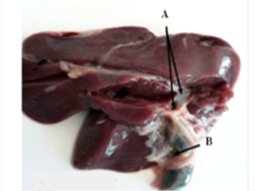
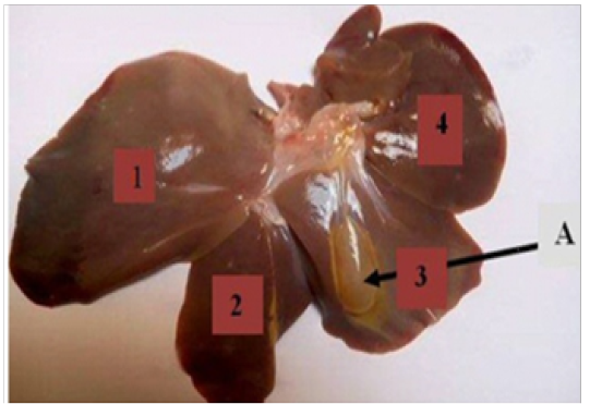
Normal macroscopic and histological aspect of broiler liver: Broiler chicken liver is a large gland, covered by a mesothelium beneath which is a layer of connective tissue (Glisson’s capsule). Lobes of the liver are subdivided into many lobules indistinctly separated from each other . The broiler liver is dark brown. It is closely associated with the pro-ventricle and the spleen. The right lobe is larger than the left lobe (Figure 1).
Microscopic examination revealed that the hepatic parenchyma of broiler chicken is composed of hepatocytes radially organized around a central vein. These hepatocytes are polyhedral and angular in shape and are larger than mammalian cells. In the intermediate layer of the connective tissue between the lobes, individually located sampling veins were determined (Figure 2, 3 and 4).
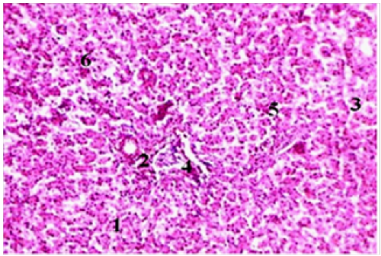
Figure 2: Normal histological aspect of broiler chickens liver (H& E x400). 1- Hepatocyte. 2- Central vein 3 - sinusoidal capillary. 4 - Central parenchyma nucleus. 5 - Triad. 6-inter lobular artery .
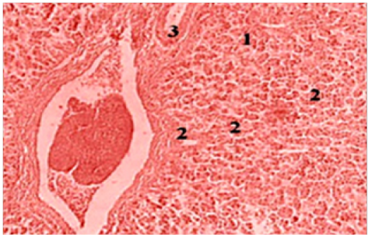
Figure 3: Normal histological aspect of broiler chickens liver (H& E x100). Sinusoidal capillary 2- hepatocyte. 3 - sinusoidal capillary.
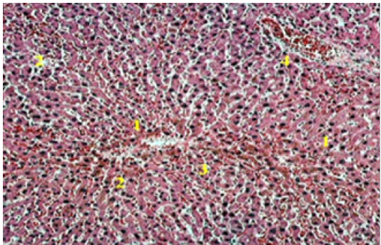
Figure 4: Liver parenchyma of broiler chickens at high magnification (H& E X100). 1- Hepatocyte. 2- Central vein 3 - sinusoidal capillary. 4 - Central parenchyma nucleus
Normal macroscopic and microscopic aspect of wild boar liver: Wild boar liver is located behind the diaphragm in the abdominal cavity, connected to the digestive system through the bile ducts, gall bladder and duodenum. We observed that the liver has deep interlobular fissures and a large amount of interlobular connective tissue (Glisson capsule) and its appearance is mottled (Figure 5)
The deep inter-lobular fissure divides the liver into four lobes: left, right, medial and lateral; the parenchyma is divided into lobules, which are partially separated by Glisson’s capsule. We have observed that the blood vessels supplying the liver (portal vein and hepatic artery) enter the hilum (Portahepatis), from which the common bile duct and the lymphatic vessels also flow.
We noted under optical microscope x400 that the parenchymal tissue of the liver is composed of hepatocytes, which are grouped into lobules. These lobules are limited by thin interlobular septa of appropriate collagen-supporting tissue that are particularly easy to identify in wild boar liver. The lobules of liver are almost hexagonal, polyhedral (Figure 6). Under the microscope, the hepatocytes have a roughly cubic form in situ, take a rounded shape. The dissociated cells have an average diameter on the scale of approximately thirty microns, but considerable variations in size are observed, with some cells reaching twenty microns while others measure more than fifty microns. The nuclei are rounded. Different granulations are present in the cytoplasm but their size is variable.
It is also noted that the lobules of the liver are composed of hepatocytes, forming plaques of liver. In the center of the lobule is located the central vein and around the lobule are inter-lobular arteries and veins, from which the inter-lobular capillaries derive. We observed that the hepatic parenchyma overlaid all central zones which narrowed towards the peripheries; the sinusoidal capillaries of central venous origin plungedthroughout the periphery. The inter-lobular capillaries penetrate the lobule and pass through the sinusoidal vessels, which are located between the liver’s plaques. The sinusoidal vessels flow into the central, the right, middle and left hepatic veins. Among the most developed functional areas are those near to the conjunctive tissue, bordering on the deep liver parenchyma, especially in the liver of chicken, wild boar liver contains small amount of sinus (Figure 7).
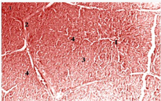
Figure 6: Normal histological aspect of wild boar liver (H& E x100). Hepatocyte. 2- trabecule . 3 - sinusoidal capillary. 4 - Central parenchyma nucleus.
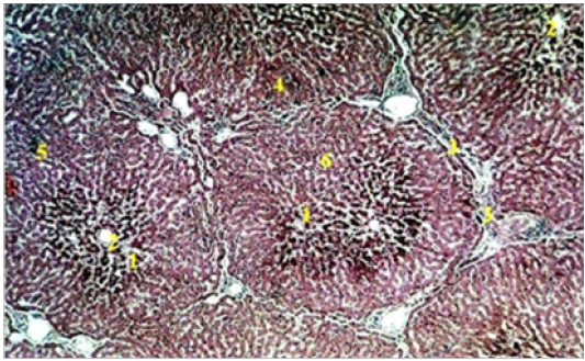
Figure 7: Histological section of hepatic parenchyma of wild boar (H& E X100). Hepatocyte. 2- Central vein. 3- Sinusoidal capillary. 4 - Inter lobular artery. 5 - Triad. 6-lobular parenchyma (H & E X100).
the examination of the liver at low magnification (ocular x 10) shows that; the parenchymal cells are arranged in rows, which come from the periphery of the lobule and converge towards its central vein, between these irregular rows of hepatocytes, there are bright spaces, also the hepatic lobules are generally separated from each other by inter-layers of loose connective tissue (inter-lobular septa) their shape is polyhedric and appear hexagonal in the cut.
The examination of the hepatic parenchyma of broiler chicken showed that the five parenchymal surfaces less pronounced than those of the liver parenchyma of the wild boar with a maximum area of 12.9 ± 0.87% and a minimum of 8.33 ± 0.37%.The number of hepatic chicken sinuses depends on the regional characteristics of the parenchyma with a gradual increase in their volume especially in zones 1, 4 and 5, it is intermediate in zones 2 and 3, we also note that the amount of sinuses are in reduction when approaching the deep peripheral zones. With regard to the sinus of the liver of the wild boar, are less developed than that of the chicken because of the excessive volume occupied by the parenchyma, it is of 4.2 ± 0.30% in zone 1 and maximum 8, 7 ± 0 , 31 in zone 3 (Table 1 and Figure 8).
Table 1: Relative surface of parenchyma and hepatic sinuses of broiler chickens and wild boar
|
Area |
Parenchyma %
Chicken Wild Boar |
Sinus %
Chicken Wild Boar |
| Area 1 |
11.23 ± 0.76 16.55 0.34 |
15.2 ± 0.016 4.2 ± 0.30 |
| Area 2 |
12.9 ± 0.87 19.56 ± 0.43 |
11.1 ± 0.021 8.5± 0.51 |
| Area 3 |
11.91 ± 0.78 21.13 ± 0.75 |
11.1 ± 0.041 8.7± 0.31 |
| Area 4 |
11.73 ± 0.94 18.83 ± 0.92 |
14.1 ± 0.051 5.5± 0.43 |
| Area 5 |
8.33 ± 0.37 19.03 ± 0.94 |
13.2 ± 0.011 6.5± 0.31 |
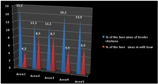
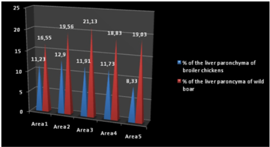
In broilers the parenchyma is less pronounced, the sinuses are highly developed, whereas in the wild boar is the opposite, the surface of the sinuses is less developed the parenchyma is very pronounced (Figure 9).
Sus scrofa is a wild and productive mammal living in the forests of the North Africa. Represents an exploration target for scientific, despite much research carried out on this species, these vital organs are still a new topic in the histo-morphological field, in order to better understand the anatomy and hepatic histology of the animal.. The liver of wild boar represented by its structure a metabolisms regulation plant, therefore in other researches, they mention that the liver is an organ promoting a high adaptation for the fight against metabolic attacks, also against bacterial and parasitic infections, at the same time research showed that the liver is considered as an immune defence organ for its synthesis of immunological elements (Van Soest, 2013).
The nutritional study for chicken (Hubbard F15) at the industrialized breeding phase is an asset, because it is an important source of animal protein for the human population. (Villate, 2001).
The progress made in the study of the digestive system of the chicken include the oral cavity, tongue, salivary glands, esophagus, stomach, intestines and auxiliary glands, which are the organs that grap food, transport it, digests and excreta. (Beghoul, 2006).
Our results are confirmed with those of other authors (Whitlow, 2002; Taşçi et al., 2018; Iqbal, 2013) who noted that the right lobe of the poultry liver is larger than the left lobe, which are composed of two lobes whose left lobes are small and subdivided into dorsal and ventral parts, there was no other lobular subdivision in the chicken liver. (Sturkie, 2012; Saran, 2017), also they are declared that chicken liver is a large lobed gland surrounded by a serous coating composed of a thin capsule of continuous connective tissue, subdividing the liver into lobes, to a lesser extent into lobes that provide physical support; they also indicate that the chicken liver is covered by the Glisson capsule. Quentin (2005), has proved that the fibers are reticular type, supporting the livre cords and elastic fibers in capsule and vessels. However, According to (Das et al., 2018) the hepatic lobules are indistinct (except which are close to the hilum) because of a lack of periobular connective tissue. The bile of the gallbladder helps to emulsify fat through its plenty in amylase and lipase.
(Shiojiri et al., 2018) Mentioned that wild boars liver is located behind the diaphragm in the abdominal cavity, connected to the digestive system through the bile ducts, gallbladder and duodenum. According to Zhiru et al. (2013) the liver has deep inter-lobular fissures and a large amount of inter-lobular connective tissue (Glisson’s capsule), they also noted that his appearance is mottled.
Das et al. (2018) reported that deep interlobular fissure is divided in the liver into 4 lobes - left, right, medial and lateral; The liver parenchyma is divided into lobules, which are partially separated by the capsule of Glisson.
According to (Shiojiri et al., 2018) blood vessels that supply the liver (portal vein and hepatic artery) penetrated through the hilum (or porta hepatis), from which the common bile canal (the secretion of bile through the liver) from where the outflow of lymphatic vessels as well.
(Eurell and Frappier, 2013; Hristov et al., 2017; Hanan, 2013) demonstrated that the parenchyma of chicken liver is composed of hepatocytes, overlaid in radial form limiting around the central vein, so they noted that hepatocytes are polyhedral and angular shape, larger than mammalian cells. The cells have a large spherical nucleus and the base of the cell formed the the sinusoidal wall. The top of the cells communicate with the bile ducts its cytoplasmis grainy.
In wild boar, the length of the lobules slightly exceeds their width. They are located at any section of the liver; the lobes have a wide variety of planes. The first landmark is the central vein, which has a circular cross-section.
Conclusion
The comparative morpho-histological study of species is less carried out by previous research, especially between mammals and birds. This type of study provides a lot of data for researchers in the different fields, and calls for other studies of the same type. Comparison between histological structures between different species, genera and families can help researchers in the field of animal origin and evolution. The hepatic morpho-histological differential study in both species (broiler chickens and wild boar) shows that; the liver is located in the epigastrium of the abdominal cavity, filling the right hypochondrium. The left part of the liver in the broiler is composed of 2 lobes, whereas in the wild boar it has 4 lobes on which the gallbladder is well fixed for both animals. In the broiler, the weight arrives on average to 205, 88 g, while in the wild boar it is 750 g.
The microscopic examination of liver showed that In broilers the parenchyma is less pronounced, the sinuses are highly developed, whereas in the wild boar is the opposite, the surface of the sinuses is less developed the parenchyma is very pronounced.
Acknowledgements
Authors are thankful to the Veterinary department of Souk Ahras and research laboratory for providing all the facilities and funds to carry out the present study and Dr. Nacir, M. in manuscript preparation.
Conflict of interest
There is no conflict of interest.
Authors Contribution
TK, BA, RD: Research was designed and executed by these authors as a part of diagnostic services to the poultry farmers. Postmortem, histological examination, sample collection, photography, and interpretation of related data were done by these authors. BA: Supervised the work, analysis data and helped in manuscript preparation. MA, AH: Supervised the work, analysis data and helped in manuscript preparation. All authors read and approved the final manuscript.
References.


