Advances in Animal and Veterinary Sciences
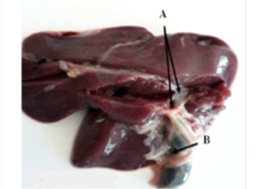
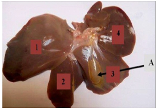
Dissection of liver lobes of broiler chicken.
A. Bile ducts . B. Gallbladder
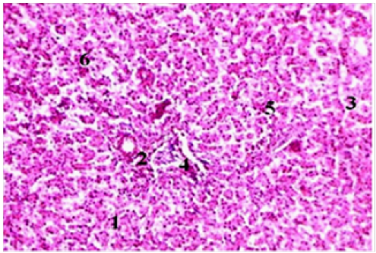
Normal histological aspect of broiler chickens liver (H& E x400).
1- Hepatocyte. 2- Central vein 3 - sinusoidal capillary. 4 - Central parenchyma nucleus. 5 - Triad. 6-inter lobular artery.
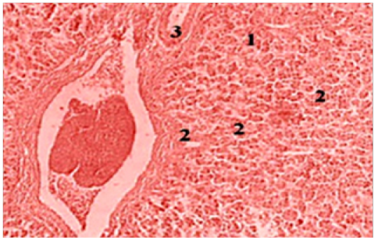
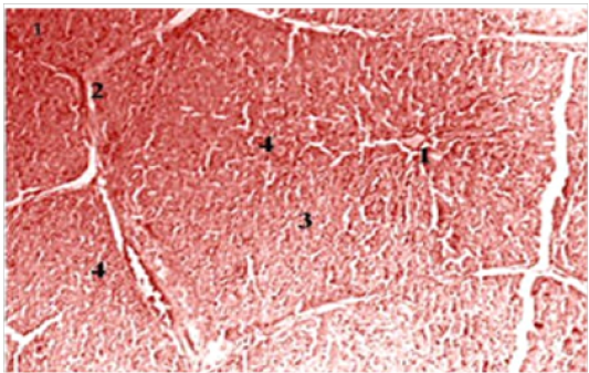
Normal histological aspect of broiler chickens liver (H& E x100).
Sinusoidal capillary 2- hepatocyte. 3 - sinusoidal capillary.
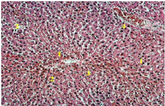
Liver parenchyma of broiler chickens at high magnification (H& E X100).
1- Hepatocyte. 2- Central vein 3 - sinusoidal capillary. 4 - Central parenchyma nucleus
The liver lobes of wild boar (1, 2, 3 and 4)
Normal histological aspect of wild boar liver (H& E x100).
Hepatocyte. 2- trabecule . 3 - sinusoidal capillary. 4 - Central parenchyma nucleus.
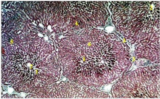
Histological section of hepatic parenchyma of wild boar (H& E X100).
Hepatocyte. 2- Central vein. 3- Sinusoidal capillary. 4 - Inter lobular artery. 5 - Triad. 6-lobular parenchyma (H & E X100).
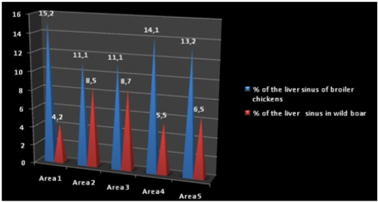
Morphometric indices of liver sinus of broiler chicken and wild boar.
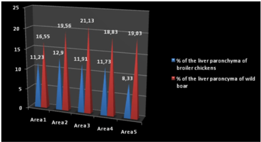
Morphometric indices of liver parenchyma of broiler chickens and wild boar.


