Journal of Infection and Molecular Biology
Research Article
Journal of Infection and Molecular Biology. 1 (4): 58 – 63Antimicrobial Resistance Profile of Sorbitol Non–fermenting Shiga Toxin Producing Escherichia coli Isolated from Small holdings Cattle
Muhammad Zohorul Islam*, Abdul Ahad, Paritosh Kumar Biswas
-
Department of Microbiology, Chittagong Veterinary and Animal Sciences University, Chittagong–4225, Bangladesh
*Corresponding author:zahir_vet@yahoo.com
ARTICLE CITATION:
Islam MZ, Ahad A and Biswas PK (2013). Antimicrobial resistance profile of sorbitol non–fermenting Shiga toxin producing Escherichia coli isolated from smallholdings cattle. J. Inf. Mol. Biol. 1 (4): 58 – 63.
Received: 2013–09–15, Revised: 2013–09–24, Accepted: 2013–09–25
The electronic version of this article is the complete one and can be found online at
(
http://nexusacademicpublishers.com/table_contents_detail/2/146/html
)
which permits unrestricted use, distribution, and reproduction in any medium, provided the original work is properly cited
ABSTRACT
Cattle and other ruminants are natural reservoir of sorbitol non–fermenting shiga toxin producing Escherichia coli (SNF–STEC), and human beings can be infected through food chain. Likewise, anti-microbial resistant strain of animal origin may be transmitted to human. The objectives of this study were to determine the antimicrobial resistance profile of SNF–STEC isolated from smallholdings cattle. A total of 57 SNF E. coli isolates were tested, of which 88%, 84% and 82% were sensitive to chloramphenicol, gentamicin and ciprofloxacin, respectively. All the isolates (100%) were resistant to penicillin, whereas, 53% were resistant to trimethoprim–sulfamethoxazole. Among the 57 SNF E. coli, 28 were shiga toxin producing (carrying stx1 or stx2 gene) which exhibited the highest resistance (57%) against trimethoprim–sulfamethoxazole and tetracycline, and the lowest (4%) against chloramphenicol. Of the Stx1 genotypic isolates, 60% and 40% were resistant to trimethoprim–sulfamethoxazole and ampicillin, respectively. On the other hand, 58% Stx2 genotypic isolates were resistant to tetracycline, whereas 44% both of hly and eae genotypic isolates were resistant to trimethoprim–sulfamethoxazole. About 60% isolates carrying all the three STEC virulent genes were resistant to ≥ 2 antimicrobials; among them one isolate was resistant to six antimicrobials – ceftriaxone, nalidixic acid, ciprofloxacin, tetracycline, doxycycline and trimethoprim–sulfamethoxazole. Isolates having no virulent gene, but harboring 54.2 kb sized plasmid were resistant to tetracycline. sulfamethoxazole, ampicillin and amoxicillin.
INTRODUCTION
The gut of endotherms is home to very diverse type of Escherichia coli. Among them shiga toxin producing E. coli (STEC) is major concern for public health. Shiga like toxin is the principal factors in the virulence of STEC (Acheson, 2000). There are two major types of shiga toxin and are identified namely stx1 and stx2 (Paton and paton, 1998). Some members of this group are unable to ferment sorbitol which are known as sorbitol non–fermenting STEC. Of the sorbitol non–fermenting STEC, E. coli O157 is the notorious serogroup causing hemolytic–uremic syndrome (HUS) in humans (Karch et al. 1999; Mead and Griffin, 1998). Cattle and other ruminants are the primary reservoir of STEC (Bettelheim, 2000) and these animals are the principal sources for human infection. The emergence of STEC as a global public health threat has been established since its first outbreak in 1982 (Riley et al. 1983). Treatment of an infectious disease like STEC by antimicrobials is questionable because the lytic action of antimicrobials upon bacterial cell may liberate shiga toxins from organism (Karch et al. 1986; Walterspiel et al. 1992; Wong et al. 2000). But some studies reported that early administration of antimicrobials in STEC infection may inhibit the disease progression (Fukushima et al. 1999; Ikeda et al. 1999; Shiomi et al. 1999). But, the fear is in the development of resistance of the organism against antimicrobial agents. The phenomenon of antimicrobial resistance of a bacterium can spread from one to another via transferable plasmid (Winokur, 2001) if the resistance is due to plasmid encoded resistance gene. Drug resistant strain can transmit from animal to human through food channel.
In Bangladesh, use of antimicrobials as therapeutics and growth promoter in food animals is very common and extensive. The abuse of antimicrobials leads to inevitable selection and spreading of resistance among gut commensals of food animal that is threatening for human health (Witte, 1998). In developed countries attention is paid to commercial dairy farms but in developing countries maximum effort is given to smallholdings cattle farm. The objective of this study was to determine the antimicrobial resistance profile of sorbitol non–fermenting shiga toxin producing E. coli isolated from smallholdings cattle.
MATERIALS AND METHODS
A total of 57 sorbitol non–fermenting E. coli were collected from a previous prevalence study (Islam, 2012). The organisms were isolated from smallholdings cattle population in Bangladesh from three randomLy selected districts. The samples were collected from different age groups of smallholdings cattle.
All the sorbitol non–fermenting isolates were investigated for their antimicrobial susceptibility profiles. Bauer–Kirby disk–diffusion procedure (Bauer et al. 1966) was used on Mueller–Hinton (MH) agar, prepared according to the manufacturer’s instructions (Oxoid). A 0.5 McFarland standard was prepared by adding 0.5 mL of 1% (11.75g/L) BaCl2.2H2O to 99.5mL of 1% (0.36N) H2SO4 (Carter and Cole, 1990). The panel of antibiotics used for the assays along with the sizes of zone of inhibition of them to be considered as “resistant (R)”, “intermediately resistant (I)” and “sensitive (S)” against the tested isolates are shown in Table 1. These characterizations were based on the recommendations from Clinical and Laboratory Standards Institute (CLSI, 2007).

Table 1: Panel of antibiotics used, their concentrations and zone diameter interpretative standards for E. coli (CLSI, 2007)
The isolates (Islam, 2012) having no virulent gene (stx1/stx2/hly) but sorbitol non–fermenter were tested for their plasmid content. Plasmid profiling was conducted by alkaline–lysis method according to the protocol described by Kado and Liu (1981) with minor modifications. One mL overnight shaking culture grown in LB broth at 37°C was used for plasmid isolation. The extracted plasmid DNA was subjected to electrophoresis using 0.8% agarose (Sea Kem LE® agarose; Lonza, Rockland, ME USA) gel in Tris–acetate–EDTA (TAE) buffer at 120V for 3 hours at room temperature and subsequently stained with ethidium bromide (10 µg/mL; E1510; Sigma–Aldrich, USA). The gel picture was taken under UV–transillumination using GelDoc EQ system with Quantity One® (Version 4.2.1) software (Bio–Rad Laboratories, Hercules, California, USA). Plasmids in E. coli 39R861 (Threlfall et al. 1986) and E. coli V517 (Macrina et al. 1978) were used as references for standard plasmid sizes. The sizes of plasmids were estimated by calculating the migration of plasmid mobility relative to that of the reference plasmids (Rochelle et al. 1985).
All data were entered into a spreadsheet programme (Excel 2003, Microsoft Corporation) and analyzed by using Stata 9.2 (Intercooled Stata 9.2, Stata Corp., College Station, Texas, USA).
RESULTS
All the 57 sorbitol non–fermenting E. coli isolates were tested for susceptibility to 11 different antimicrobial agents. The frequencies of isolates showing sensitive, intermediately resistant and resistant to the antimicrobials tested are shown in Figure 1. Of the tested isolates 88%, 84% and 82% were sensitive to chloramphenicol, gentamicin and ciprofloxacin, respectively; 100% isolates were resistant to penicillin, and 53% to trimethoprim–sulfamethoxazole.
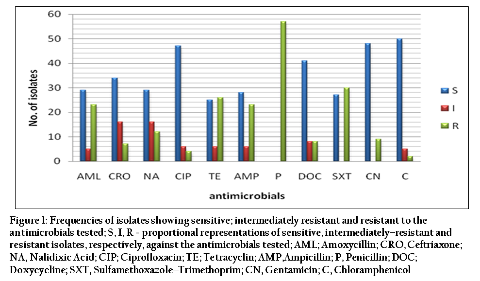
Figure 1: Frequencies of isolates showing sensitive; intermediately resistant and resistant to the antimicrobials tested
The frequencies at which different zones of inhibition to 11 different antimicrobials tested are displayed in Figure 2. The Stx1, Stx2 and hly genotypic isolates’ susceptibility profiles are demonstrated in Table 2. Among 28 shiga toxin producing E. coli isolates (carrying stx1 and/or stx2 gene), the highest resistance (57%) was found against trimethoprim–sulfamethoxazole and tetracycline, and the lowest resistance (4%) was against chloramphenicol. Of the Stx1 genotypic isolates 60% and 40% were resistant to trimethoprim–sulfamethoxazole and ampicillin, respectively; 58% Stx2 genotypic isolates were resistant to tetracycline, whereas 44% both of hly and eae genotypic isolates were resistant to trimethoprim–sulfamethoxazole

Figure 2: Frequency distributions of the isolates showing different zones of inhibition to 11 antimicrobials tested (zone diameter in millimeter)

About 60% isolates carrying all the three EHEC O157 virulent genes were resistant to > 2 antimicrobials; among them one isolate was resistant to six antimicrobials – ceftriaxone, nalidixic acid, ciprofloxacin, tetracycline, doxycycline and trimethoprim–sulfamethoxazole. Isolates having no virulent gene, but harbouring 54.2 kb sized plasmid were resistant to tetracycline, sulfamethoxazole, ampicillin and amoxicillin.
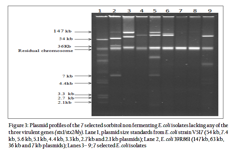
Figure 3: Plasmid profiles of the 7 selected sorbitol non fermenting E. coli isolates lacking any of the three virulent genes (stx1/stx2/hly)
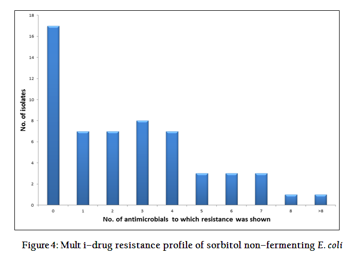
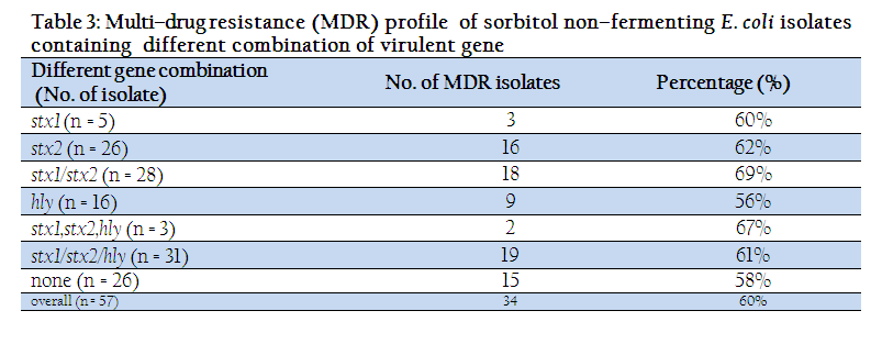
All the seven isolates had a >54.2 kb size plasmid (Figure 3). Six isolates contained > 1– <3 plasmids. Four of the isolates contained very large plasmid of 147 kb size in addition to other smaller plasmid(s). Other isolates contained a plasmid of 95 kb size. Only 17 sorbitol non–fermenting isolates was showing no resistance to any of the 11 antimicrobials tested and 14 isolates were resistant to one or two antimicrobials. On the other hand 23 isolates (40%) were resistant to more than two antimicrobial agents (Figure 4). A variable level of multidrug resistant profile was found in the isolates containing different combination of virulent gene (Table 3).
DISCUSSION
There exist a paucity of information on the antimicrobial resistance profile of sorbitol non–fermenting E. coli isolated from smallholdings cattle. Identification of drug resistant sorbitol non–fermenting shiga toxin producing E. coli in smallholder’s cattle might have potential public health impacts owing to smallholders’ closer contacts with their cattle. It is thinkable that antimicrobial resistant microorganisms may be transmitted from food animal to human beings through several channels including food chain, direct exchange through professional exposure, or from animal production surroundings (Van den Bogaard and Stobberingh, 1999; Witte, 1998).
In this study, majority of isolates showing resistance to antimicrobials tested were diverse based on the variable numbers of antimicrobials against which they showed resistance. However, the present study observed a high prevalence of resistant isolates to sulfamethoxazole, tetracycline and ampicillin, an agreement with some previous reports (Galland et al., 2001; Meng et al., 1998; Zhao et al., 2001). Obviously penicillin was resistant to all isolates tested which is a normal phenomenon of Gram negative bacteria. Because antimicrobial–resistant bacteria from food animals may colonize in humans and smallholders’ contacts with their cattle heads are close and more frequent therefore such antimicrobial resistant strains might have more zoonotic consequences. Interestingly, a small percentage of the tested isolates were resistant to ceftriaxone, chloramphenicol, and nalidixic acid. These antibiotics are not commonly used to treat any bacterial diseases of smallholders’ cattle in the study areas; consequently, why a few isolates were resistant to them is hard to explain from this study. But one possibility is that ceftriaxone and nalidixic acid resistant bacteria from human have colonized smallholdings cattle via environmental cross–contamination. However, coselection via genetic linkage of resistance determinants may have a significant contribution in the development of resistance (Zhanel et al. 1995).
Isolates lacking Stx1/Stx2/hly gene, but harbouring 54.2 kb sized plasmid were resistant to tetracycline, sulfamethoxazole, ampicillin and amoxicillin, suggesting that such resistance might be plasmid–borne (Winokur, 2001). There is common perception; antibiotic is generally used in commercial dairy farm where infectious disease is more prevailed compared to smallholder’s cattle. However, in this study it was shown that antibiotic resistant bacteria were isolated from smallholder farm indicating random uses of antibiotic or it might be cross infection from the environment shed by commercial dairy farm. Therefore, awareness against random uses of antibiotic in food animals should be strengthened in commercial as well as smallholder farm.
CONCLUSION
The antimicrobial resistance profiles of the isolated organisms to the 11 antimicrobials tested are varied. There was also evidence to multidrug resistance of E. coli Isolated from smallholding’s cattle. This result is more significant and alarming for public health if the organism colonizes to human body from animal.
ACKNOWLEDGEMENTS
The study was financed by Regional Fisheries and Livestock Development Component (RFLDC), Noakhali though the Poultry Research and Training Centre, CVASU, Bangladesh. A special thanks to Dr. Himel Barua for providing some valuable technical supports.
REFERENCES
Acheson DW (2000). How does Escherichia coli O157:H7 testing in meat compare with what we are seeing clinically? J. Food Prot. 63: 819 – 821.
PMid:10852579
Bauer AW, Kirby WMM, Sherris JC and Turck M (1966). Antibiotic susceptibility testing by a standardized single disk method. American J. Clin. Pathol. 36: 493 – 496.
Bettelheim KA (2000). Role of none–O157 VTEC. J. App. Microbiol. 88: 385 –405.
http://dx.doi.org/10.1111/j.1365-2672.2000.tb05331.x
Carter GR and Cole JRJ (1990). Diagnostic Procedure in Veterinary Bacteriology and Mycology, Fifth eition, Academic Press, Inc., Harcourt Brace Jovanovich, Publishers, New York, P. 484.
Clinical Laboratory Standards Institute (CLSI) (2007). Performance standards for antimicrobial susceptibility testing; Seventeenth information Supplement. CLSI document M100–S17. 27: 3
Fukushima H, Hashizume T, Morita Y, Tanaka J, Azuma K, Mizumoto Y, Kaneno M, MatsuuraM, Konma K and Kitani T (1999). Clinical experiences in Sakai city hospital during the massive outbreak of enterohemorrhagic Escherichia coli O157 infections in Sakai City, 1996. Ped. Int. 41: 213 – 217.
http://dx.doi.org/10.1046/j.1442-200X.1999.4121041.x
Galland JC, Hyatt DR, Crupper SS and Acheson DW (2001). Prevalence, antibiotic susceptibility, and diversity of Escherichia coli O157:H7 isolates from a longitudinal study of beef cattle feedlots. App. Envir. Microbiol. 67: 1619 – 1627.
http://dx.doi.org/10.1128/AEM.67.4.1619-1627.2001
PMid:11282614 PMCid:PMC92778
Ikeda K, Ida O, Kimoto K, Takatorige T, Nakanishi N and Tatara K (1999). Effect of early fosfomycin treatment on prevention of hemolytic uremic syndrome accompanying Escherichia coli O157:H7 infection. Clin. Nephrol. 52: 357 – 362.
PMid:10604643
Islam MZ (2012). An investigation on clonal diversity of Escherichia coli from cattle in smallholdings while using selective media for O157 strains. MS thesis, PP 23 – 24.
Kado CI and Liu ST (1981). Rapid procedure for detection and isolation of large and small plasmids. J. Bacteriol. 145: 1365 – 1373.
PMid:7009583 PMCid:PMC217141
Karch H, Bielaszewska M, Bitzan M and Schmidt H (1999). Epidemiology and diagnosis of Shiga toxin producing Escherichia coli infections. Diag. Microbiol. Infect. Dis. 34: 229 – 243.
http://dx.doi.org/10.1016/S0732-8893(99)00031-0
Karch H, Stockbine N and O'Brien A (1986). Growth of Escherichia coli in the presence of trimethoprim–sulfamethoxazole facilitates detection of Shiga– like toxin producing strains by colony blot assay. FEMS Microbiol. Lett. 35: 141 – 145.
http://dx.doi.org/10.1111/j.1574-6968.1986.tb01516.x
Macrina FL, Kopecko DJ, Jones KR, Ayers DJ and McCowen SM (1978). A multiple plasmid–containing Escherichia coli strain: convenient source of size reference plasmids molecules. Plasmid. 1: 417 – 420.
http://dx.doi.org/10.1016/0147-619X(78)90056-2
Mead PS, Griffin PM (1998). Escherichia coli O157:H7. Lancet. 352: 1207 – 1212.
http://dx.doi.org/10.1016/S0140-6736(98)01267-7
Meng J, Zhao S, Doyle MP and Joseph SW (1998). Antibiotic resistance of Escherichia coli O157:H7 and O157: NM from animals, food, and humans. J. Food Prot., 61: 1511 – 1514.
PMid:9829195
Paton JC and Paton AW (1998). Pathogenesis and diagnosis of Shiga toxin–producing Escherichia coli infections. Clin. Microbiol. Rev. 11: 450 – 479.
PMid:9665978 PMCid:PMC88891
Riley LW, Remis RS, Helgerson SD, McGee HB, Wells JG, Davis BR, Hebert RJ, Olcott ES, Johnson LM, Hargrett NT, Blake PA and Cohen ML (1983). Hemorrhagic colitis associated with a rare Escherichia coli serotype. New Eng. J. Med. 24: 681 – 685.
http://dx.doi.org/10.1056/NEJM198303243081203
PMid:6338386
Rochelle PA, Fry JC, Day MJ and Bale MJ (1985). An accurate method for estimating sizes of small and large plasmids and DNA fragments by gel electrophoresis. J. Gen.l Microbiol. 132: 53 – 59.
Shiomi M, Togawa M, Fujita K and Murata R (1999). Effect of early oral fluoroquinolones in hemorrhagic colitis due to Escherichia coli O157:H7. Ped. Int. 41: 228 – 232.
http://dx.doi.org/10.1046/j.1442-200X.1999.4121038.x
Threlfall EJ, Rowe B, Ferguson JL and Ward LR (1986). Characterization of plasmids conferring resistance to gentamicin and apramycin in strains of Salmonella typhimurium phage type 204c isolated in Britain. J. Hygiene. 97: 419 – 426.
http://dx.doi.org/10.1017/S0022172400063609
Van den Bogaard AE and Stobberingh EE (1999). Antibiotic usage in animals: impact on bacterial resistance and public health. Drugs. 58:589 – 607.
http://dx.doi.org/10.2165/00003495-199958040-00002
PMid:10551432
Walterspiel J, Ashkenazi S, Morrow A and Cleary TG (1992). Effect of subinhibitory concentrations of antibiotics on extracellular Shiga–like toxin 1. Inf. 20: 25 – 29.
http://dx.doi.org/10.1007/BF01704889
Winokur PL, Vonstein DL, Hoffman JJ et al. (2001). Evidence for transfer of CMY–2 ampC B–lactamase plasmids between Escherichia coli and Salmonella isolates from food animals and humans. Antimicrobial Agents Chemotherapy. 45: 2716 – 2722.
http://dx.doi.org/10.1128/AAC.45.10.2716-2722.2001
PMid:11557460 PMCid:PMC90722
Witte W (1998). Medical consequences of antibiotic use in agriculture. Sci. 279: 996 – 997.
http://dx.doi.org/10.1126/science.279.5353.996
Wong CS, Jelacic S, Habeeb RL, Watkins SL and Tarr PI (2000). The risk of hemolytic–uremic syndrome after antibiotic treatment of Escherichia coli O157:H7 infections. New Eng. J. Med. 342: 1930 – 1936.
http://dx.doi.org/10.1056/NEJM200006293422601
PMid:10874060 PMCid:PMC3659814
Zhanel GG, Karlowsky JA, Saunders MH, Davidson RJ, Hoban DJ, Hancock RE, McLean I and Nicolle LE (1995). Development of multipleantibiotic– resistant (Mar) mutants of Pseudomonas aeruginosa after serial exposure to fluoroquinolones. Antimicrobial Agents Chemotherapy. 39: 489 – 495.
http://dx.doi.org/10.1128/AAC.39.2.489
PMid:7726519 PMCid:PMC162565
Zhao S, White DG, Ge D, Ayers S, Friedman S, English L, Wagner D, Gaines S and Meng J (2001). Identification and characterization of integron– mediated antibiotic resistance among Shiga toxin–producing Escherichia coli isolates. Appl. Envir. Microbiol. 67: 1558 – 1564.
http://dx.doi.org/10.1128/AEM.67.4.1558-1564.2001
PMid:11282605 PMCid:PMC92769


