Research Journal for Veterinary Practitioners
Case Report

Surgical Management of Canine Vaginal Leiomyoma of a Bitch in SAQTVH, Bangladesh
Tuli Dey1*, Bhajan Chandra Das1, Syed Imran1, Mohammed Bayazid Bostami1, Bibek Chandra Sutradhar1, Sonnet poddar2
1Department of Medicine and Surgery, Chittagong Veterinary and Animal Sciences University, Khulshi, Chittagong-4225, Bangladesh; 2Department of Anatomy and Histology, Chittagong Veterinary and Animal Sciences University, Khulshi, Chittagong-4225, Bangladesh.
Abstract | Vaginal tumors are more common benign, solid tumors in bitch as compared to tumors of upper reproductive organs. A 20 kg, seven years old street bitch was admitted to SAQTVH of CVASU, Chittagong, Bangladesh with a gross lesion of large mass around vagina from two years ago and it was increasing day by day. Clinical examination revealed round, solid mass of 20 cm width and 16 cm diameter with 110 gm in weight. After proper diagnosis, the bitch was anesthetized with general anesthesia using diazepam and ketamine combination and local anesthetics also used for maintenance. The bitch was stabilized with 0.9% NaCl isotonic solution. Surgical correction was performed by total resection of tumor mass and the resected part was transferred to pathology lab for histo-pathological diagnosis resulting leiomyoma of vagina. Post-operative treatment was maintained with antibiotic and the owner was advised to spay her dog to reduce recurrence of tumor. After 10 days the bitch was completely recovered from surgical wound and follow up history for 5 months revealed no recurrence of tumor.
Keywords | Vaginal leiomyoma, Surgical management, Bitch
Editor | Muhammad Abubakar, National Veterinary Laboratories, Islamabad, Pakistan.
Received | October 20. 2017; Accepted | November 18, 2017; Published | December 25, 2017
*Correspondence | Tuli Dey, Department of Medicine and Surgery, Chittagong Veterinary and Animal Sciences University, Khulshi, Chittagong-4225, Bangladesh; Email: shantacvasu@gmail.com.
Citation | Dey T, Das BC, Imran S, Bostami MB, Sutradhar BC, Poddar S (2017). Surgical management of canine vaginal leiomyoma of a bitch in saqtvh, Bangladesh. Res. J. Vet. Pract. 5(4): 40-43.
DOI | http://dx.doi.org/10.7582/journal.rjvp/2017/5.4.40.43
ISSN (Online) | 2308-2798
Copyright © 2017 Dey et al. This is an open access article distributed under the Creative Commons Attribution License, which permits unrestricted use, distribution, and reproduction in any medium, provided the original work is properly cited.
Introduction
Dog is one of the best companion animals for men from the time being. In Bangladesh day by day keeping a dog as companion animal is going to be a tradition. There are many dog breeds available in Bangladesh, but local breed or street dog is most common. These local breed or street dogs are suffering from different reproductive diseases and leiomyoma one of them (Ahuja et al., 2017). Before going to treat the animals, it is very important to distinguish whether these neoplasms are benign or malignant and to differentiate them from other conditions such as hyperplasia, granulation tissue or abscessation.
Neoplasms of the female tubular genitalia account for 3% of all canine tumors and among these, 85-90% occur in the vagina and vulva (Noakes et al., 2009). Tumors of mesenchymal origin: leiomyomas, fibro- leiomyomas and fibromas occur most commonly and leiomyosarcomas, lipomas, mastocytomas, adenocarcinomas, squamous cell carcinomas and transmissible venereal tumors occur much less frequently (Rollon et al., 2008).
Leiomyoma is a tumor of smooth muscle cells that may arise in any organ with a connective tissue or mesenchymal component and have been found in many organs including female reproductive tract (James et al., 2012, Singh et al., 2014). Leiomyomas of the reproductive tract in bitch are frequently associated with estrogen secreting tumors or ovarian follicular cysts and cystic endometrial hyperplasia, mammary hyperplasia and/or neoplasia may also be concurrently found (Yuefei et al., 2012).
Previous reports have shown 73-94% vaginal tumours as benign and pedunculated often with a narrow stalk (Salomon et al., 2004). Vaginal leiomyomas may be single or multiple, intraluminal or extraluminal. The tumor is usually round or oval, well defined and encapsulated. The size and consistency may vary depending upon duration of growth, becoming firmer due to an increase in connective tissue. Large intraluminal tumors may protrude through the vulva, while extraluminal tumors tend to cause perineal swelling (Umamageswari et al., 2016).
Leiomyomas must be differentiated from leiomyosarcomas by histological examination and as vaginal leiomyoma is benign, the prognosis is good. Here the study is planned to execute the properly diagnosis of vaginal leiomyoma and manage by surgical procedure to find the follow up result.
Materials and Methods
Study Area and Duration
The presented case study was conducted at Shahedul Alam Quadary teaching veterinary hospital, (SAQTVH), Chittagong Veterinary and Animal Sciences University (CVASU) Khulshi, Chittagong, Bangladesh on 29 January, 2017.
Case History
In SAQTVH a seven years old bitch of 20 kg body weight was presented with a gross lesion of large mass around vagina. From history of that bitch, it was informed that this bitch was suffered from this condition 2 years ago and it was increasing day by day. The bitch was facing problem with its locomotion and parturition. Owner noticed that its appetite was normal but body condition was not good or improved.
Clinical Findings
After clinical examination it was found that a big soft fat like mass was hanging from the outside of vulvar lips and palpation revealed that it was from vagina.The mass was measured as 20 cm in length and 16 cm in diameter with no hernial sign was palpated (Figure 1, a,b, c) The temperature of the mass was normal. No discharge was coming from it. The bitch was facing trouble in locomotion. Body condition was normal and body temperature was 38.8°C. No abnormalities were noted regarding her appetite, respiration and pulsation values. Heart rate was 45 per minute and pulse rate was 60 per minute.
Radiological and Histo-pathological Findings
Radiological image revealed white solid mass near vulva, involving vagina. (Figure 1, d). In presented case, connective tissue coating covered by a thin layer of stratified epithelium was found histo-pathologically and cells were of smooth muscle origin, lying in parallel bundles with collagen fibers interposed.
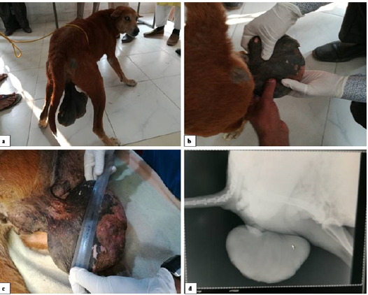
Figure 1: Clinical findings a. Dog before surgical correction, b. Palpation of tumor mass c. measurement of tumor mass and d. Radiological findings
Pre-Anesthesia and Patient Preparation
As premedication, atropine sulphate @ 0.04 mg/kg bodyweight and xylazine @ 1 mg/kg body weight were administered intramuscularly. The surroundings of the tumor site was perfectly shaved and aseptically prepared.
Anesthesia
The patient was anesthetized with ketamine and diazepam combination @ 1: 4 ratios and stabilized with 0.9% sodium chloride saline as fluid therapy.In the presented case, local anesthesia was also used in the mid time of the operation as maintenance drug. The animal was placed as dorsal recumbence.
Surgical Procedure
After aseptic preparation of members of surgery team and surgical site, incision was performed at the base of the tumor. Small blood vessel was sealed by artery forceps and ligation was done in case of larger one. The mass was easily visualized and it was noted that the vagina was extremely dilated. It was firm to the touch and had a well-defined stalk attached to the left ventral vaginal wall. The growth was excised and portions were sent immediately to pathology laboratory for hsito-pathological slide preparation and other portions were fixed in 10% formalin. Closure of the excision site was achieved by approximating the mucosa of the vaginal floor as well as occluding the submucosal dead space using 3-0 chronic catgut. Skin was appositioned by using silk. (Figure 2).
Post-operative Care
The wound was dressed with povidone iodine for 7 days. The bitch was received antibiotic-Ceftriaxone @ 3 ml intramuscularly and 1.5 ml of anti-histaminic- Pheneaminmaliet for five days; and 2 ml of meloxicum intramuscularly for five days.
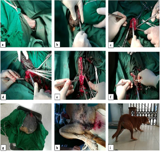
Figure 2: Surgical procedure, a. : Aseptic preparation of surgical site, b. Incision at base of tumor, c. Blunt dissection to separate subcutaneous tissue, d. Hemostasis by clamping and ligation, e. Subcutaneous closure by catgut, f. Skin closure by silk, g. Resected tumor mass, h. After surgery tumor area, i. dog after surgery and recovery from anesthesia
Patient care Instruction
It was advised that animal should be kept in close observation after taking in house. The postoperative treatment should also be continued for 7 days. The patient was advised as to do spaying after complete recovery from this condition not to recur the condition
Results and Discussion
The dog recovered uneventfully from anesthesia and did not develop postoperative complications such as urine retention or incontinence. She was sent home after recovery from anesthesia on treatment with systemic antibiotics for the next five days.
Histo-pathologically the mass had a connective tissue coating covered by a thin layer of stratified epithelium (Singh et al., 2014). The tumor cells were of smooth muscle origin, lying in parallel bundles with collagen fibers interposed. Final diagnosis of a leiomyoma was made and a good prognosis was given to owner and suggests spaying the bitch to prevent regrowth of the tumor. Regrowth or problems associated with the tumor have not occurred after three months of surgery.
Koestner and Higgins, (2008) describe that the incidence of leiomyomas is highest between five and 16 years of age. And in the presented case it was at seven years of age. So this study is supported by previous study. The incidence of vaginal leiomyomas in multiparous bitches is considered to be increased by some authors yet other statistics reveal no significant difference in the rate of occurrence between multiparous and nulliparous animals. In canine generally with the exception exception of canine transmissible venereal tumor, the exact causes of canine vaginal tumors are unknown (Umamageswari et al., 2016).
The role of estrogens in the etiology of leiomyomas is unclear. Estrogens are known to be carcinogenic, while the progestational phase of estrus inhibits tumor formation Hormonal influence on the growth of vulvar or vaginal tumour has been reported (MacLachlan and Kennedy, 2002). In a previous reported case (Sontaş et al., 2010, Singh et al., 2014) the tumour was accounted in a hysterectomized poodle dog which had an ovarian remnant, in current case the bitch was intact and hormonal background might have been acted on the formation of the mass detected in the vagina as reported by Sontaş et al. 2010.
Local treatment of vaginal leiomyomas primarily involves surgical excision of the mass (Klein, 2001). If few discrete metastatic foci are present, these may also be removed. Because most tumors arise from the vestibule or the smooth muscle wall of the vagina, they are usually removed per vulva (Withrow et al., 2013). An episiotomy may be necessary for larger tumors (Rollon et al., 2008). Radiation therapy may be considered if surgical removal of the tumor and/or its metastatic foci is not possible.
Iatrogenic damage to the urethra or accidental injury to other perineal structures is possible surgical complications. Urethral catheterization will greatly assist in avoiding damage to this structure. Prevention and control of the disease is best achieved by ovariohysterectomy (MacLachlan and Kennedy 2002).
Conclusion
Vaginal leiomyoma of a bitch are common and in possible cases it requires special and careful consideration during surgery. Surgical management of vaginal leiomyoma of bitch reveled that no recurrence of tumor again.
Acknowledgments
The author sincerely desires to express her deepest sense of gratitude to Prof. Dr. Bhajan Chandra Das and Prof. Dr. Bibek Chandra Sutradhar, Department of Medicine and Surgery, Chittagong Veterinary and Animal Sciences University, Chittagong, Bangladesh for their guidance at the time of surgery.
Author Contributions
Prof. Dr. Bhajan Chandra Das and DR. Tuli Dey did that surgery; Syed Imran planned and formatted the manuscript. Md. Bayazid Bostami helped during surgery, Prof.Dr. Bibek Chandra Sutradhar helped by instructing about manuscript and Sonnet Poddar helped to write manuscript.
Conflicts of Interest
The authors declare no conflict of interest
References


