Research Journal for Veterinary Practitioners
Case Report

Therapeutic Management of Canine Ehrlichiosis in a German Shepherd Bitch: A Case Report
Subhash Chand Gahalot1*, Dharm Singh Meena2, Rashmi Singh3, Yogendra Pal Singh4, Madan Mohan Mali5, Jitendra Bargurjar6, Nirmal Jeph7, Sarjana Meena8
1*,4Department of Veterinary Clinical Complex, PGIVER, Jaipur; 2,3,5,6,7Department of Veterinary Medicine, PGIVER, Jaipur; 8Department of Veterinary Pathology, PGIVER, Jaipur, India.
Abstract | Current case report describes the successful treatment of canine ehrlichiosis in a German shepherd bitch presented to department of Veterinary Clinical Complex (VCC), PGIVER, Jaipur. Clinical observation showed the signs of high fever (105oF), lethargy, anorexia, fits and slight nasal bleeding. On the basis of blood report and clinical signs, case was diagnosed as canine ehrlichiosis and successfully treated with antibiotic, antipyretics, haemostatics and supportive therapy with multivitamin, haematinics and liver tonics.
Keywords | Canine Ehrlichiosis, Doxycycline .
Editor | Muhammad Abubakar, National Veterinary Laboratories, Islamabad, Pakistan.
Received | October 03, 2017; Accepted | November 23, 2017; Published | December 24, 2017
*Correspondence | Subash Chand Gahalot, Department of Veterinary Clinical Complex, PGIVER, Jaipur, India; Email: subhashgvet@gmail.com
Citation | Gahalot SC, Meena DS, Singh R, Singh YP, Mali MM, Bargurjar J, Jeph N, Meena S (2017). Therapeutic management of canine ehrlichiosis in a german shepherd bitch: a case report. Res. J. Vet. Pract. 5(4): 37-39.
DOI | http://dx.doi.org/10.17582/journal.rjvp/2017/5.4.37.39
ISSN (Online) | 2308-2798
Copyright © 2017 Gahalot et al. This is an open access article distributed under the Creative Commons Attribution License, which permits unrestricted use, distribution, and reproduction in any medium, provided the original work is properly cited.
Ehrlichiosis in dogs is a rickettsial infection caused by the various species of genus Ehrlichia but most commonly by E. canis, E. chaffeensis, E. ewingii, and possibly E. ruminantium (Vieira et al., 2011). Ehrlichia are transmitted by two ticks species mainly, Brown Dog Tick, (Rhipicephalus sanguineus) (Groves et al., 1975) and the Lone Star Tick (Amblyomma americanum). Ehrlichia is a type of bacteria that have tropism for hematopoietic cells and causes mainly two types of canine ehrlichiosis namely monocytic ehrlichiosis by E. canis (Harrus and Waner, 2011) and granulocytic ehrlichiosis by E. ewingii (Anderson et al., 1992). Co-infections of Ehrlichia with Anaplasma, Rickettsia, Babesia or Bartonella spp. occur frequently as dogs are naturally exposed to multiple tick-borne pathogens (Straube, 2010). Generally all breeds of dogs are equally predisposed to ehrlichiosis but German shepherd and Siberian Huskies develops more severe form of ehrlichiosis because of reduced cell-mediated immune response to E. Canis in these breeds therefore; these breeds have a worse prognosis (Nyindo et al., 1980). Ehrlichiosis can manifest in three phases (Skotarczak, 2003). Signs of the acute phase of the disease usually include anemia, fever, depression, lethargy, loss of appetite, shortness of breath, joint pain and stiffness. In the subclinical phase the animal may appear normal or show only slight anemia and can last for months or years. The chronic phase can be either mild or severe and characterized by weight loss, anemia, neurological signs, bleeding, inflammation of the eye, edema in the hind legs, and fever. Blood tests show that one or all of the different blood cell types are decreased. One cell type, the lymphocyte may increase and be abnormal in appearance.
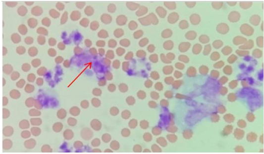
A one year old German shepherd bitch was presented to VCC with the signs of anorexia, fever (105 oF) lethargy, nasal bleeding (Figure 1), ecchymotic haemorrhages on various spots (Figure 2) and occasional fits. History revealed tick infestation previously. On the basis of clinical signs the animal was tentatively diagnosed with canine ehrlichiosis infection and treatment was started with Inj oxytetracyclin HCL @20mg/kg body wt IV with normal saline, Inj. Analgin @ 2.0g IV, Inj. Pheneramine maleate @0.5mg/kg body wt IM, Inj. Tranexamic acid @ 8mg/Kg body weight IM and Inj. Multivet @ 1ml /10kg body wt IM. For confirmatory diagnosis a whole blood sample with EDTA was send to the Central Testing Facility for complete blood count and blood protozoan before starting of the treatment. Consecutively owner was advised to cold fomentation with ice above nasal area at home regularly. On the next day the blood report shows bitch having infection of Ehrlichia in monocytes & Neutrophils (Figure 3). A differential leucocyte count shown Lymphocytes 23%, Neutrophils 70%, Eosinophils 0% , Basophils 0 %, Monocytes 7% and total leucocyte count was 8.45 thousand per ml. The concentration of haemoglobin was 7g/dl. So owner was advised to give Tab. doxycyclin @ 5mg/ kg body wt twice in a day, liver tonics Syrup All liv it @ 2tsf and haemetenics Syrup a-Rbc 1tsf twice in a day for 20 days. After 20 days owner was contacted telephonically and the animal was doing all well.
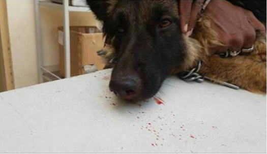
Figure 1: Showing nasal bleeding
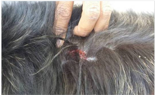
Figure 2: Showing haemorrhage on the back
Generally ehrlichiosis should be suspected in dogs with pale or whitish mucous membrane, pancytopenia, thrombocytopenia, nasal bleeding, ehymotic haemorrhages, neurological signs and have been exposed to ticks previously (Frank and Breitschwerdt, 1999). Similar clinical signs were also observed in this case also. Tetracyclines are the treatment of choice for many hemoprotozoan in canines. For canine ehrlichiosis, tetracycline (22 mg/kg q 12H) or doxycycline (10 mg/kg every q 24H) (Iqbal and Rikihisa, 1994) administered for four weeks is the well established treatment that also responded well in current case. As tetracycline are known to be hepatotoxic effect (Liénart et al., 1992), liver tonic are added to protect from adverse effect. Most dogs recover from the acute and subclinical phases when treated with doxycycline or other tetracyclines at appropriate dosages for an adequate period of time. Persistent infections with E. canis often remain as complete bacterial clearance is not guaranteed following antibiotic therapy. Supportive therapy such as blood or fluid transfusion and anabolic steroids may be required in severe cases. The prognosis becomes poor once dogs enter the chronic phase of disease. Co-infections with other pathogens like Babesia or Bartonella may contribute to the fatal outcome of chronic infections.
Acknowledgements
The authors are highly thankful to Dean, PGIVER, Jaipur for providing facilities to carry out diagnosis and treatment of the case.
Conflict of Interests
There is no conflict of interest
Author’s Contribution
SC Gahlot, DS Meena, R Singh, YP Singh, J Bargujar, MM Mali, Nirmal Jeph were involved in case diagnosis and treatment. S meena was involved in complete blood Count and smear preparation. SC Gahlot, DS meena R singh were involved in manuscript preparation.
References


