Research Journal for Veterinary Practitioners
Case Report

Schistosomus Reflexus Monster Fetus in Bovine and its Successful Management
Anand Kumar Pandey*, Sandeep Kumar, Pankaj Gunwant, Ajit Verma, Jagat Bir Phogat
Veterinary Clinical Complex, College of Veterinary Sciences, LUVAS, Hisar-125004, Haryana, India.
Abstract | Two Murrah buffaloes and one Hariana cow were brought to the VCC with the history of severe abdominal straining along with hanging of intestines out of the vulva. On per-vaginum examination the cases were diagnosed as dystocia due to (i) conjoined twin Schistosomus reflexus (ii) second and third cases as simple schistosomus reflexus. The caesarian operations were performed to remove the fetus in case I and III, however, vaginal delivery became possible to remove the fetus in IInd case.
Keywords | Bovine, Conjoined twin, Caesarian, Monster, Schistosomus reflexus
Editor | Muhammad Abubakar, National Veterinary Laboratories, Islamabad, Pakistan.
Received | May 13, 2017; Accepted | June 21, 2017; Published | June 28, 2017
*Correspondence | Anand Kumar Pandey, Veterinary Clinical Complex, College of Veterinary Sciences, LUVAS, Hisar-125004, Haryana, India; Email: dranandpandey@gmail.com
Citation | Pandey AK, Kumar S, Gunwant P, Verma A, Phogat JB (2017). Schistosomus reflexus monster fetus in bovine and its successful management. Res. J. Vet. Pract. 5(2): 25-27.
DOI | http://dx.doi.org/10.17582/journal.rjvp/2017/5.2.25.27
ISSN (Online) | 2308-2798
Copyright © 2017 Pandey. This is an open access article distributed under the Creative Commons Attribution License, which permits unrestricted use, distribution, and reproduction in any medium, provided the original work is properly cited.
Schistosomus reflexus is a developmental defect characterized by a marked ventral curvature of the spine, deformed pelvis and the body and chest walls bent laterally with exposed thoracic and abdominal viscera (Roberts, 2004). Schistosomus reflexus monster is an often reported cause of dystocia in cattle (Pandey et al., 2015). Schistosomus reflexus along with perosomus elumbis defect has been delivered through caesarian section in buffalo (Pandey et al., 2012). The present paper describes three cases of dystocia due to: i) Conjoined twin Schistosomus reflexus monster fetus and its delivery through caesarian section, (ii) Schistosomus reflexus monster fetus and its delivery through vagina and caesarian section in Murrah buffalo and Hariana cow, respectively.
Case I:
Eight years aged Murrah buffalo on its 3rd parity was brought to the Veterinary Clinical Complex, LUVAS, Hisar with history of abdominal straining since last 14 hours. Local vets made their effort to relieve dystocia but could not get success. Per-vaginum examination revealed four hind limbs and one fore leg in the birth canal. Head was felt at the level of sacrum along with two tails. The abdomen of the fetus was exposed. All the four hind legs were broken from tarsal-metatarsal joints; one fore leg from carpal-metacarpal joint. Even though the birth canal was severely lacerated and edematous, the animal was quite active and clinical parameters ranged within the normal limits. The condition was diagnosed as dystocia due to conjoined twin Schistosomus reflexus monster fetus. Due to inadequate space in the birth canal, it was decided to perform the caesarian operation to relieve dystocia.
Case II:
A Twelve years old Murrah buffalo of its 8th parity following completion of gestation period with hanging intestines out of vulva was reported at Veterinary Clinical Complex, LUVAS, Hisar. Owner reported that the buffalo is suffering with the labour pains since last 10 hours and water bags had also ruptured. Owner itself tried to remove the fetus, but he was unable to remove it. Clinical examination of buffalo revealed clinical parameters within normal physiologic range. Vaginal examination revealed fully dilated cervix along with all the fetal limbs and exposed abdominal cavity in the birth canal. The fetal head was felt close to its sacrum. However, the size of fetus seemed to be smaller as compared to the normal. No any fetal reflexes were present. The condition was confirmed as dystocia due to Schistosomus reflexus.
Case III:
A Seven years aged Hariana cow of 8.5 months pregnant in its 3rd parity with hanging intestines out of vulva was reported at Veterinary Clinical Complex, LUVAS, Hisar. Owner reported that the cow was suffering from the labour pains since last 18 hours. Local veterinarian failed to remove the fetus. All the clinical parameters of cow were within normal range. Vaginal examination showed one hand dilated cervix and uterus was completely contracted over the fetus. The abdominal organs were felt due to exposed abdominal cavity. The condition was confirmed as dystocia due to Schistosomus reflexus. Since, there was no adequate space in the birth canal and fetus was deep in the abdomen so, it was decided to perform caesarian section to relieve the dystocia.
Case I and III:
Caesarian operation was performed in left lateral recumbency under local anaesthesia (2% Lignocaine hydrochloride solution) with linear infiltration along the site of incision following all aseptic precautions. The fetus was removed and surgical wound was sutured as per standard procedure. The buffalo and cow had uneventful recovery following operation.
Case II:
Ample space was present in birth passage with fully dilated cervical os, and small sized deformed fetus was present in the uterus, therefore; it was determined that the fetus could be removed out with slight judicious manual manipulation. Before starting the manipulations, epidural anaesthesia (5 ml, Lignocaine hydrochloride, 2%) was done thereafter; birth passage was lubricated with infusion of Carboxymethyl cellulose gel. The successful delivery was made by application of judicious slight traction in the direction of right and left side moving the fetus. Fortunately, the fetus removed out within 5 minutes. The fetal membranes were also come out along with fetus. The buffalo had uneventful recovery.
Description of the fetus: In case I, fetus was having one head, two fore limbs, single thorax, and abdomen. The fetus had two separate pelvis containing four hind limbs (two hind limbs of each fetus) and two tails. The thoracic and abdominal viscera were exposed. The fetus was having scrotum. The liver and rumen was of enlarged size (Figure 1). The internal organs were present as in a single fetus. The hind quarters were twisted towards the head, thoracic and
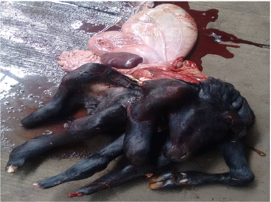
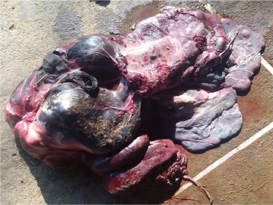
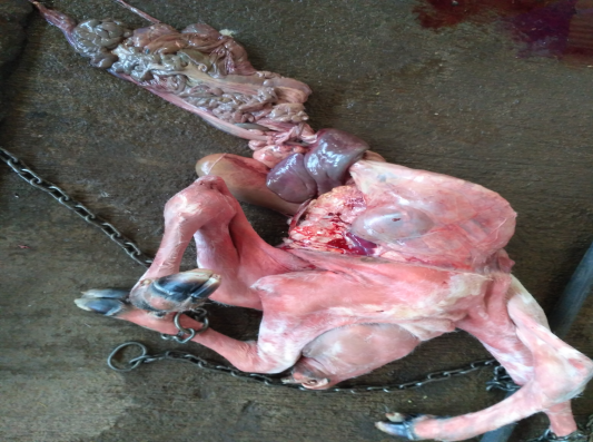
Figure 3: Schistosomus reflexus cow fetus
abdominal viscera were exposed therefore the monster fetus was termed as “monocephalus dibrachius tetrapus conjoined twin Schistosomus reflexus fetus” (Roberts, 2004). In case II and III, gross examination of the fetus (Figure 2 and 3, respectively) revealed that every joint of fetal legs were rigid along with entirely exposed visceral organs. The fetus had characteristic ventral curvature of the vertebral column and head was resting over the sacrum. The thoracic and abdominal organs were normal in size and shape. It was diagnosed as a case of Schistosomus reflexus with teratological defects (Roberts, 2004). The possibility of genetic predisposition cannot be ignored. The interplay of multiple genes is a frequent and most important genetic mechanism for the causation of such extensive anomalies (Jana and Ghosh, 2001). In monozygotic twins, incomplete division of embryo into two components at the primitive streak state leads to conjoined monstrosities (Noden and Delahunta, 1985). Schistosomus reflexus causes dystocia due to defective formation of spinal chord and exposure of viscera in cattle and buffaloes and either caesarian section or fetotomy is warranted to relieve this kind of dystocia (Roberts, 2004). Nevertheless, Schistosomus reflexus monster fetus delivered without caesarian section or fetotomy presently in case II might be due to relatively small sized of fetus and/or presence of large size of pelvic girdle of pluriparous buffalo.
Summary
Partial fetotomy of the fetal parts is suggested if the spinal curvature is acute and thus preventing passage of the fetus through the birth canal. If fetotomy is not possible, a caesarean operation is the only choice to deliver this kind of monster fetus. Per-vaginal expulsion without any obstetrical assistance is noticed in small sized monster fetus. Present communication reports three rare cases of dystocia due to Schistosomus reflexus and its delivery through caesarian section is possible.
ACKNOWLEDGEMENTS
Authors are highly thankful to Dr. Nitin Soni, Dr. Sonu Ghadwal for their help during caesarian section and relieving of dystocia.
CONFLICT OF INTEREST
The authors have no conflict of interest to declare.
AUTHORS CONTRIBUTION
AKP, SK, PG and AV were involved in caesarian section, relieving of dystocia and follow up the cases. AKP and JBP were involved in manuscript preparation.
References


