Research Journal for Veterinary Practitioners
Case Report

Spaying as a Tool for Birth Control: A Case Report
Azizunnesa1, Delower Hossain1, Preety Chaudhary2, Md. Anowar Parvez1, Pranab Paul1, Saroj Kumar Yadav1, Tanjila Hasan1*
1Department of Medicine and Surgery,Faculty of Veterinary Medicine,Chittagong Veterinary and Animal Sciences University, Khulshi, Chittagong-4225; 2Patuakhali Science and Technology University, Patuakhali, Bangladesh.
Abstract | A 3 years old local breed stray bitch with a history of parity 5 was reported to Teaching Veterinary Hospital (TVH) of Chittagong Veterinary and Animal Sciences University (CVASU), Chittagong, Bangladesh for birth control. As there was no birth control pill available for bitch, we decided to do spaying as a birth control measure. Before surgery the bitch was kept in TVH to evaluate its all physical parameters e.g. anemia, dehydration etc. Ultrasonographic screening was done to know whether the bitch was pregnant or not. Vaginal cytological test was performed using Fleishmann and Giemsa stain to observe the state of estrus cycle of the bitch. Presence of intermediate and parabasal cells and decline in superficial cells indicated bitch was in diestrus period, thus suitable for spaying. The surgery was aseptically controlled under general anaesthesia. Laparotomic mid line incision 2-3 cm behind the umbilicus was performed. The bitch was kept in TVH for 7 days for following up. After 14 days the suture was removed. The bitch recovered uneventfully without any noticeable complication. So spaying can be an effective method to control dog population in Bangladesh.
Keywords | Bitch, Spaying, Birth control
Editor | Muhammad Abubakar, National Veterinary Laboratories, Islamabad, Pakistan.
Received | March 23, 2017; Accepted | May 22, 2017; Published | May 28, 2017
*Correspondence | Tanjila Hasan, Lecturer, Dept. of Medicine and Surgery, Faculty of Veterinary Medicine, Chittagong Veterinary & Animal Sciences University Khulshi, Chittagong-4225, Bangladesh; Email: tanjila.cvasu@gmail.com
Citation | Azizunnesa, Hossain D, Chaudhary P, Parvez MA, Paul P, Yadav SK, Hasan T (2017). Spaying as a tool for birth control: A case report. Res. J. Vet. Pract. 5(2): 19-24.
DOI | http://dx.doi.org/10.17582/journal.rjvp/2017/5.2.19.24
ISSN | 2308-2798
Copyright © 2017 Azizunnesa et al. This is an open access article distributed under the Creative Commons Attribution License, which permits unrestricted use, distribution, and reproduction in any medium, provided the original work is properly cited.
Ovariohysterectomy (OVH) is a medical term used to indicate spaying or neutering a female dog. Ovariohysterectomy is one of the most routinely performed major abdominal surgeries in the veterinary practice (Pearson, 1973; Jason, 2009).
Ovariohysterectomy (OVH) is an irreversible technique which is used for the sterilization of the female animals (David, 2010; Kirsan et al., 2013) where surgery was done under proper general anesthesia and sterile operating technique (Virginia et al., 2012). The traditional OVH usually is accessed through caudal midline incision, which frequently encompasses the half or the middle third of the umbilicopubic distance (Machado et al., 2012) and involves surgical removal of the ovaries and uterus. Surgery is usually performed at 4 ½ to 9 months of age.
OVH in the bitch is a common procedure for birth control, to prevent oestrus and pseudopregnancy (Janssens and Janssens, 1991). Due to for these reasons it is the most common among elective surgeries (Pollari and Bonnett, 1996).
It has some immediate or short–term surgical complications that include hemorrhage from uterine and ovarian vessels, anesthesia accidents, tissue reaction to suture material, wound infection, or delayed healing (Pearson, 1973; Muir et al., 1991; Burrow and Batchelor, 2005).
It is also used to prevent or decrease the risk of the development of mammary cancer and pyometra and the inconvenience of vaginal discharge and male attraction during estrus (Al-Badrany et al., 2012).
There is no such strong case report about spaying in Bangladesh and this is one of the reasons for our study. The aim of this study was to evaluate the traditional surgical technique of spraying and make it as a strong tool of birth control in bitch.
Case History and Description
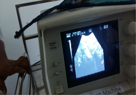
On 12th November 2016, a 3 years-old, 15kg weighing local bred bitch with history of parity 5 was brought to the Teaching Hospital (TVH) of Chittagong Veterinary and Animal Sciences University (CVASU), Chittagong. At first general physical examination was done. The bitch had fair body condition. Then blood sample was collected to do routine examination of blood (TLC, TEC, DLC, calcium,phosphorus etc). Almost all the parameter was within normal limit (Table 1 and Table 2) except low Hb% and high ESR, TLC level. Then ultrasonographic screening was accomplished to know physiological status of bitch. Ultrasonographic examination revealed that bitch was non-pregnant. There found no abnormality on ultrasound. After that vaginal cytology was performed with Fleishmann and Giemsa stain. By vaginal cytology the oestrous cycle of bitch was demonstrated. Presence of intermediate and parabasal cells, also decreased number of superficial cells confirmed that the bitch in diestrous. After 7 days of observation it was decided to spay the bitch.
Restraining and Anesthesia
Both physical and chemical methods were used to control the bitch. The bitch’s mouth was tied with a leash to prevent her from biting during restraining. The surgical site was located in the caudal midline 2 cm behind the umbilicus. After cleaning and shaving, the surgical site was soaked in tincture iodine. The bitch was kept under fasting condition for 24 hours. As pre anesthetic xylazine hydrochloride solution @1.5 mg/Kg body weight (Inj. Xylazine®, Indi-
Table 1: Blood routine examination
|
Name of the test |
Results |
Normal range |
|
Haemoglobin |
11.4 |
12-17gm % |
|
ESR ( Wintrobe tube) |
25 |
6-9 (mm in 1st hour) |
|
Total Count of TEC |
4.4 |
5-9 million/cumm |
|
Total Count of TLC |
17.2 |
9-15(thousand/cumm) |
|
PCV |
45.0 |
37-55% |
|
Differential count for WBC |
||
|
Lymphocytes |
30.0 |
20-25% |
|
Neutrophils |
60.0 |
60-75% |
|
Eosinophils |
6.0 |
2-5% |
|
Monocytes |
4.0 |
0-4% |
|
Basophils |
0 |
0-1% |
Table 2: Biochemical serum examination of blood
|
Name of the test |
Results |
Normal range |
|
Creatinine |
1.0 |
0.5-15 mg/dl |
|
Calcium |
9.2 |
9-11.3 mg/dl |
|
SGOT |
36.8 |
23-66 U/L |
|
SGPT |
39.9 |
21-102 U/L |
|
S. Total Protein |
114.1 |
54-71 U/L |
|
S. Glucose |
155.4 |
65-118mg/dl |
|
S. Phosphorous |
3.2 |
2.6-6.2mg/dl |
an Immunologicals Ltd., India) intramuscularly followed by general anesthetic injection ketamine hydrochloride @ 15 mg/Kg body weight (G-ketamine®, Gonoshasthaya Pharmaceuticals Ltd., Bangladesh) intravenously was administered. The maintenance anaesthetic dose was given @half of the initial dose during the surgery. The normal saline (Inj. Normasol®, Libra Pharmaceuticals Ltd, Bangladesh) was infused intravenously @500 ml during the surgery.
Surgical Procedure
The patient was kept on the operation table and covered with sterilized draper keeping the operative site open. The surgery was aseptically controlled under general anesthesia. Laparotomic mid line incision 2-3 cm behind the umbilicus was performed. At first 4 cm long incision was made on skin. The bleeding was checked by applying gauge pressure and artery forceps. The subcutaneous tissues and fats were removed. Then muscles and peritoneum were incised layer by layer. Large veins were ligated to check hemorrhage. The uterine horn was identified by fingers and ovaries were found following the horn to their ends. The broad ligament
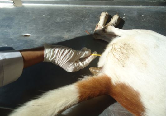
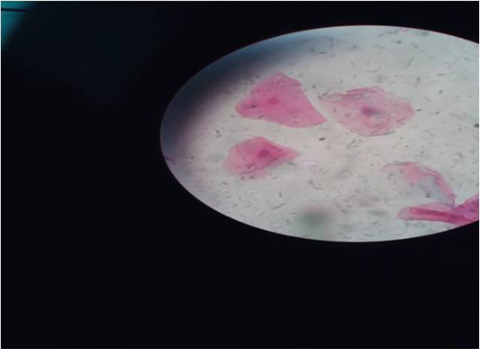
Figure 2: Intermediate cells under microscope
(mesovarium) attached to the ovaries were torn so the ovaries could be identified. The right ovarian artery was ligated and transfixed with absorbable suture material (catgut 1-0). Then right ovarian artery was cut. The same procedure was followed for left ovary. The uterine body and related arteries were ligated just in front of the cervix leaving the cervix as the natural barrier. The entire uterus and ovaries were then removed. The abdomen was checked for bleeding. The peritoneum and muscle layers were sutured with simple continuous pattern with catgut (1-0). The subcutaneous layer was sutured with subcuticular pattern using catgut (1-0). The skin was then closed with cross-mattress suture pattern using silk. The sutured wound was covered with the benzoin seal.
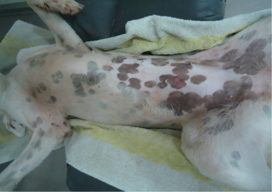
Figure 4: Clipping and shaving the incised area

Figure 5: Ligating uteus
Postoperstive Treatment and Care
After surgery, antibiotic ceftriaxone @20 mg /Kg body weight (Inj. Trijectvet 1gm®, SK+F Pharmaceuticals, Bangladesh) was administered intramuscularly daily for 7 days. Antihistaminic chlorpheneramine maleate @1mg/Kg body weight (Inj. Astavet®, Acme Laboratories Ltd., Bangladesh) was administered intramuscularly daily for 7 days. Analgesic meloxicam @40 mg/Kg body weight (Inj. Melvet®, Acme Laboratories Ltd., Bangladesh) was administered subcutaneously daily for 5 days for pain management. The patient was kept in clean squeeze cage and observed for 7 days. No complication was noted and the bitch recovered uneventfully. On the 14th day, the suture was removed and it was noticed that the surgical site was healed completely.
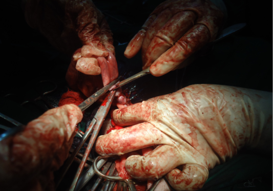
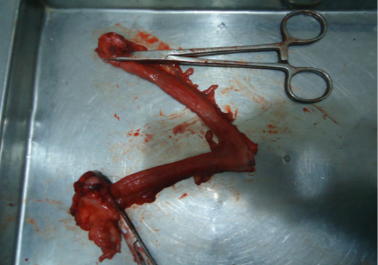
Figure 7: Uterus along with ovary after surgery

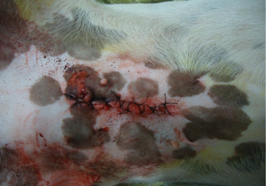
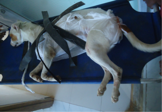
Figure 10: Bitch after surgery.
DISCUSSION
Every year millions of unwanted puppies are born. About 75% of the worldwide dogs, often referred to as stray, are free to roam and reproduce (ICAMC, 2007). The Humane Society of the USA describes that every year between 8 and 10 million dogs enter shelters and 4 to 5 million of these animals are euthanized due to only lack of homes (Kutzler et al., 2006). Many animals die agonizing death from starvation, disease, accidents and abuse. By spaying, we can prevent these horrible deaths.
(Macpherson et al., 2013) identified free roaming dog as a source of transmitting diseases to livestock and humans. Free-roaming dogs are mostly responsible for bites (Jackman et al., 2007). For example in Samoa, according to (Farnworth et al., 2012), 56% of bites occurred in a public place. In India, 64% of dog bites were associated with stray dogs (Sudarshan et al., 2001). Among the zoonoses, rabies is of particular concern for humans and livestock where dogs are responsible for more than 90% of the estimated 55,000 human deaths and for the millions of people that every year takes post exposure vaccine following a bite (Knobel et al., 2005). So we can follow the contraception rule by surgical method as easy method that describe by (Jackson, 1984) to know control overpopulation of stray dog. The article in US Today reported that spayed female dogs live 23% longer than unspayed female dogs and have less chance to develop pyometra and mammary tumor. Burrow and Batchelor (2005) described the Complications associated with the suture material or ligatures which included haemorrhage, abdominal wall dehiscence, surgical wound infection, stump granuloma, fistulous draining tracts, inadvertent ureteral ligation, and chewed-out sutures. We did not face any complications like that. However Kutzler and Wood (2006) said intratesticular injection of calcium chloride as a non-surgical methods which can effectively affect spermatogenesis, androgenesis and libido, while lacking toxicity and serious side effects for contraception of male dogs.
The post operative management was performed for 7 days and animal recovered without any complications. But still there is no available paper regarding spaying as birth control measure. So the aim of this study is to know the public about spaying procedure so that they can easily control the street dog. It will also support the animal welfare.
CONCLUSION
Dogs and cats are born five to ten at a time and are adopted out one at a time. Those that are not adopted, and that’s most of them, either suffer in homelessness or are destroyed in shelters. By spaying the suffering of these street or non-adopted dogs can be minimized. This paper will be helpful for field veterinarian and owner to know how they can control their pet population. In conclusion we can say that spaying can be considered as 100% safe birth control measure and also humane rather than euthanizing the homeless dogs.
ACKNOWLEDGMENTS
The authors were greatly acknowledged to Teaching Veterinary Hospital, Chittagong Veterinary and Animal Sciences University, Chittagong, Bangladesh.
CONFLICT OF INTERESTS
No conflict of interest.
AUTHORS’ CONTRIBUTION
All authors contributed equally.
REFERENCEs


