Research Journal for Veterinary Practitioners
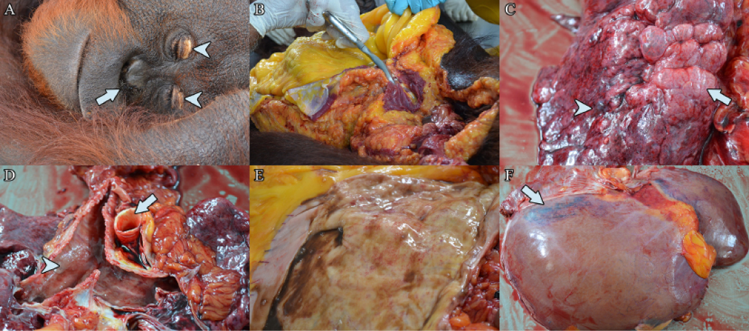
(a) Bilateral dried haemorrhagic nasal discharge (arrow), with bilateral yellowing of the eyelid (arrow heads). (b) Severe jaundice of the subcutaneous and visceral fat. (c) Pulmonary emphysema (arrow), with areas of pulmonary petechiation (arrow head). (d) Yellowing of the aorta (arrow) with presence of frothy serosanguineous exudate along the congested trachea (arrow head). (e) Mild haemorrhage on the stomach mucosa. (f) Yellow discoloration and generalised hepatomegaly, with hepatic haemorrhage (arrow)
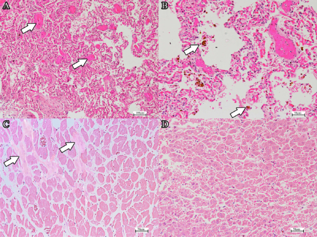
(a) DIC in the pulmonary capillaries (arrows) (H and E, bar=100µm). Note the extensive atelectasis. (b) Haemosiderin-laden alveolar macrophages (H and E, bar=50µm). (c) Mild fibrosis of the myocardium (arrow) (H and E, bar=50µm). (d) Generalised mild to moderate hepatic lipidosis (H and E, bar=50µm).


