Research Journal for Veterinary Practitioners
Research Article
Res. J. Vet. Pract. 5(1): 5-11
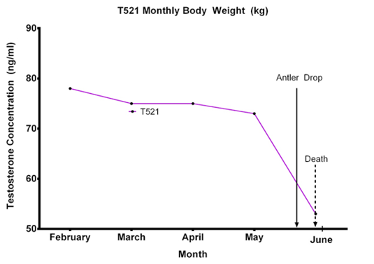
Figure 1
T521 body weight (kg) from February to May 2016. Note the severe reduction of body weight from 4th May 2016 with 30th May 2016
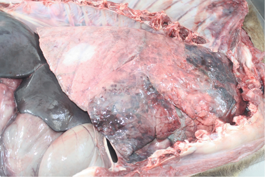
Figure 2
Severe pulmonary hepatisation at the cranioventral aspect of the lungs
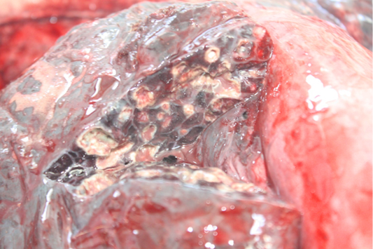
Figure 3
Multiple pulmonary abscesses with thickening of interlobular septa
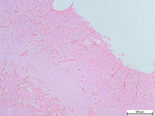
Figure 4
Fibrous tissue capsule in the lungs, with congested remnant of alveoli. (H and E, bar = 500µm)
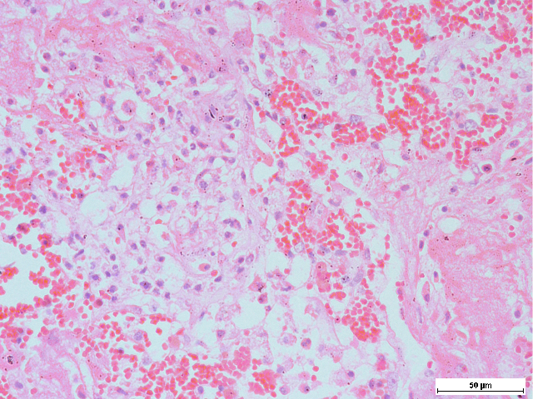
Figure 5
Presence of alveolar macrophages and neutrophils, intermixed with fibrin along with haemorrhage in the lungs

Figure 6
Swollen and shrinking of hepatocytes, with occasional cytoplasmic eosinophilia, Note that the sinusoids were widened


