Research Journal for Veterinary Practitioners
Short Communcation

Surgical Correction of Ventral Abdominal Hysterocele in Gravid Uterus of a Doe
Md. Anowar Parvez1*, Bibek Chandra Sutradhar1, Monoar Sayeed Pallab1, Tanjila Hasan1, Rokeya Khatun2
1Department of Medicine and Surgery, Faculty of Veterinary Medicine, Chittagong Veterinary and Animal Sciences University, Khulshi, Chittagong-4225; 2Additional Veterinary Surgeon (AVS), ECTAD-FAO (HPAI), Department of Livestock Services, Farmgate, Dhake-1215, Bangladesh.
Abstract | A four years old high yielding variety goat was admitted at Teaching Veterinary Hospital (TVH) in CVASU with the history of prolong gestation period, distended ventral abdominal wall and excessive enlargement of the udder. The doe exhibited anorexia, discomfort with dull and depression. Physical examination and abdominal ballottement were ensured that the extremities and head of the fetus were easily palpable in swollen udder but movement of the fetus was not found. Both the hind limbs of doe were seen to be extended laterally due to distended abdomen. As the gestation period has prolonged and there was no sign of parturition, so it was decided to perform caesarean section for delivering the fetuses and subsequently herniorrhaphy. Post operative care was given to the affected goat and it was recovered after fourteen days. So final recommendation is that after fulfillment of gestation period, any types abnormalities in ventral abdominal region or excessive enlargement of udder contact with the veterinarian for its diagnosis, correction and quick recovery.
Keywords | Correction, Hyserocele, Hernia, Multiple fetus, Gravid uterus
Editor | Muhammad Abubakar, National Veterinary Laboratories, Islamabad, Pakistan.
Received | November 31, 2016; Accepted | December 27, 2016; Published | December 30, 2016
*Correspondence | Md. Anowar Parvez, Assistant Professor, Dept. of Medicine and Surgery, Faculty of Veterinary Medicine, Chittagong Veterinary & Animal Sciences University, Khulshi, Chittagong-4225, Bangladesh; Email: aparvez0445@gmail.com
Citation | Parvez MA, Sutradhar BC, Pallab MS, Hasan T, Khatun R (2016). Surgical correction of ventral abdominal hysterocele in gravid uterus of a doe. Res. J. Vet. Pract. 4(4): 76-79.
DOI | http://dx.doi.org/10.14737/journal.rjvp/2016/4.4.76.79
ISSN (Online) | 2307-8316; ISSN (Print) | 2309-3331
Copyright © 2016 Parvez et al. This is an open access article distributed under the Creative Commons Attribution License, which permits unrestricted use, distribution, and reproduction in any medium, provided the original work is properly cited.
INTRODUCTION
Hernias are a passage of an organ or tissue through an opening which may be natural or acquired. Almost all animals are affected by it. Hernias can be divided on the basis of etiology e.g. congenital and acquired hernia, on the basis of location e.g. abdominal, umbilical, scrotal, inguinal, femoral, perinial and diaphragmatic hernia or clinically into reducible or irreducible hernia (Fahd and Ahmed, 2007). Typically a hernia is consist of hernia sac, hernia ring, hernia contents. Any viscera that migrate through any part of the abdominal wall ventral to the stifle skin fold other than natural orifice can be defined as ventral hernia (Yasin, 2004). Severe trauma on abdominal wall, violent force or injury originating by blunt object, horn thrust in cattle, kicks in camel, and abscess in abdominal wall, overstretching or straining of abdominal muscles due to pregnancy and parturition causing weak abdominal muscle may lead to ventral abdominal hernias. Some atraumatic causes e.g. weakness or rupture of prepubic tendon so that the gravid uterus cannot be supported also resposible for ventral abdominal hernias. (St Jean and Anderson, 2004; McILwraith, 1984). The sites of the Ventral abdominal hernias may vary from the Lateral site of the thoracic cavity to the iliac crest. Both abdominal and umbilical hernias occur mostly in females than in the males. Congenital, inguinal hernias are often seen in swine (St Jean and Anderson, 2004) but unusual in bulls. The prevalence of ventral hernia in sheep is 58.34%. ventral abdominal hernia are acquired in nature and more common in the older ages than younger ages (Mahdi, 2015). The incidence abdominal hernia is higher in females and of inguinal hernia is and abdominal hernias have excellent healing history with uneventfull recovery (Al-Sobayil and Ahmed, 2007). Ventral abdominal hernias are generally observed in ruminants. It is mostly occurs in pluriparous animal in advanced pregnancy incase of multiple fetus which leads to fragility of abdominal muscles or prepubic tendon (Vijayanand et al., 2009). A hernia of gravid uterus is very rare in goat. A case of ventral abdominal hernia in a goat is reported.
Case Presentation and History
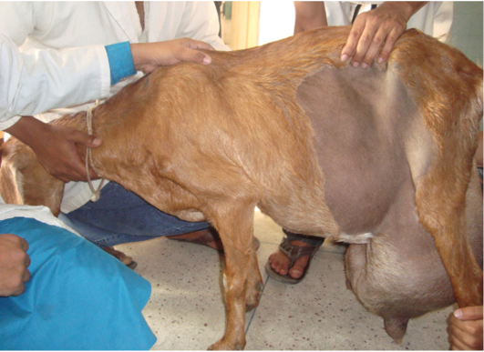
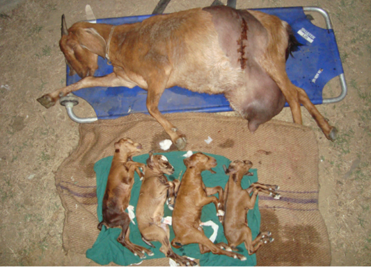
In present study, a referral case of four years old Jamunapari goat weighing 70 kg was registered at Teaching Veterinary Hospital in Chittagong Veterinary and animal Sciences University with the history of excessive enlargement of ventral abdominal wall and udder during pregnancy (Figure 1). The owner claimed that its gestation period has already completed fifteen day before. The physical examination was done by taking temperature, heart take, respiration rate, dehydration. The abdominal ballottement was done to ensure multiple fetuses existing in udder. The extremities and head were easily palpable on swollen udder. As the gestation period has prolonged and there was no sign of parturition decision was taken by the veterinary surgeon to do caesarean section for delivery of the fetuses. After cesearean section there are four fetuses within the gravid uterus of the doe, due to multiple fetuses in the gravid uterus the abdominal muscle could not bear the weight thus create a opening through the gravid uterus goes within the udder.
Surgical Management And Post Operative Care
The caesarean section was done in left lateral recumbent condition. Before surgery the skin was clipped, shaved, soaked with alcohol and painted with tincture iodine. As preoperative care 5% dextrose saline was infused and the doe was sedated by sedil (diazepam) @ 0.5 mg/kg body weight local anaesthesia was done using 2% lidocaine hydrochloride @ 1ml/ cm area for inverted seven blocks in front of incision site. Chronologically the skin, fascia, abdominal muscle layer (obliqqus abdominis externus, obliqqus abdominis internus, transverse abdominis, retus abdominis), peritonium, was incised. Then the gravid uter
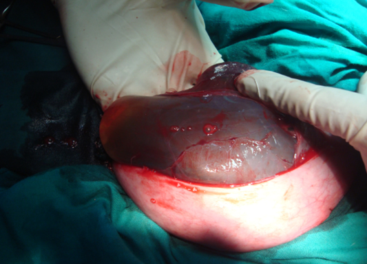

Figure 3: Suturing of the uterus)
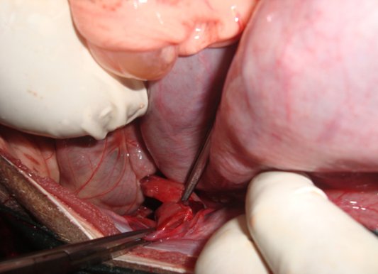
us was exteriorized and made an incision at greater curvature of uterus (Figure 2). Bleeding was checked using artery forceps and pressure pad. We expelled out four kids, but all the kids were dead. After that the uterus was sutured with czerny-lembert using catgut no. size 2 (Figure 3). The abdominal rupture was exposed (Figure 4) and closed with simple interrupted pattern using catgut no. size 1 (Figure 5). The uterus was replaced into the abdominal cavity. Then
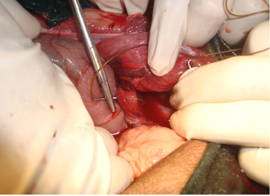
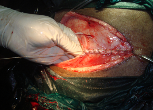
Figure 6: Suturing of abdominal muscle layer
the peritonium and the entire muscle layer were sutured layer by layer separately in a simple continuous pattern using catgut size no.2 (Figure 6). Horizontal mattress using nylon size no.2 was applied on skin for a positioning the two cutting ends. Tincture benzoin seal was applied on closed cutting edges as antiseptic which gives the protection from bacterial contamination (Figure 7). As Post operative care antibiotic, Streptopenicillin (Streptopen®, 2.5 gm vial, The Renata Limited, Dhaka, Bangladesh) 6ml and antihistaminic Pheneramine Meleate (Histavet® The ACI Limited, Dhaka, Bangladesh) 3ml daily intramuscularly for 7 days and pain killer Melvet (®, Acme Pharmaceuticals, Dhaka, Bangladesh) 2ml for 3 days and dressing was done as alternative day. Surgical wound was healed at 10th day of postoperative care and sutures were removed at the same day.
DISCUSSION
Hernias cause economic problem as well as welfare problem. They hampers of production and reproduction profitability (Keown, 1974) similar with the findings of this study. These are very frustrating for commercial producers e.g. scrotal hernia, ventral abdominal hernia (hysterocele). The incidence of ventral abdominal hernia in animals 32.3% but exact causes of hernia were not traced by (Jettennavar et al., 2010). In my present study as it was a advance pregnant doe with multiple (four) kids so there were weakling of abdominal muscle, ultimately the gravid uteus came to abdominal cavity near mammary gland. The fetuses we delivered were dead which is almost similar to the observation of (Venkatesan, 2007). A case of ventral hysterocele in a goat with a automobile accident followed by a tear in the abdominal muscles leading to herniation of gravid uterus with live full grown fetuses was reported earlier by (Vijayanand et al., 2009) dissimilar with this study it may be due to different condition. the rupture of the prepeubic tendon due to hydroallantois which increase weight of gravid uterus leads to ventral abdominal hernia ( Selvaraju et al., 2010) similar with the findings of this present study four fetus with fetal fluids which increase weight of uterus resulting ventral abdominal hernia. Rupture of the prepubic tendon is more complicated in small ruminants as it usually inures the udder due to the closeness of the udder to the ground and the animal heights, if not treated earlier it becomes complicated and more frequently observed in Shami breed does reported by (Mavrogenis, 2000; Mavrogenis et al., 2006) similar with this study. Therefore, Does with multiple (Three of four) fetus within the uterus should be diagnosed by expert veterinarian with the help of ultrasonography and safe the life of fetus and does after complete gestation period either normal delivery or caesarean section.
CONCLUSION
The doe survived successfully after operation, but the fetuses were not survived due to unknown causes; prolong gestation is one of them Early diagnosis of multiple fetuses by ultrasound, proper management and support during pregnancy and record of complete gestation period would be helpful for save the life of newborn and prevention of ventral abdominal hernia.
ACKNOWLEDGEMENTS
Authors are thankful to the Director of Teaching Veterinary Hospital, Chittagong Veterinary and Animal Science University for providing necessary facilities to this case study.
CONFLICT OF INTERESTS
No conflict of interest.
AUTHORS’ CONTRIBUTION
All authors contributed equally.
REFERENCES


