Research Journal for Veterinary Practitioners
Case Report

Clinical Management of Babesiosis in Cattle: A Case Report
Saritha Gungi*, Gurram Sreeramamurthy Haritha, Karumuri Nalini Kumari
Department of Veterinary Medicine, College of Veterinary Science, SVVU, Tirupati, Andhra Pradesh, India.
Abstract | Babesiosis is a tick-transmitted disease caused by protozoans of the genus Babesia and it is characterized by haemolytic anemia and fever, with occasional hemoglobinuria and death. In the present study 4 cross bred cows aged between 4-6 years were presented to the college hospital with the history of fever, anorexia, passing coffee coloured urine, reduced milk yield, depression and reluctance to move. Examination of the blood smears confirmed the Babesia spp. in all the cows. Microscopy detection methods are still the cheapest and fastest methods used to identify Babesia parasites although their sensitivity and specificity are limited. Haematological studies revealed reduced Hb, PCV and TEC. Serum chemistry revealed hyperglycemia, hyperbilirubinemia, BUN, AST and hypoprotienemia. Urine was positive for haemoglobin, glucose and bile pigments. Three cows were treated successfully with diminazene aceturate (Berenil) at 3 mg/kg body weight, together with supportive therapy. Whereas, one cow died due to severe anemia.
Keywords | haemoglibinuria, cattle, anemia, jaundice, BUN
Editor | Muhammad Abubakar, National Veterinary Laboratories, Islamabad, Pakistan.
Received | April 25, 2016; Accepted | May 24, 2016; Published | May 28, 2016
*Correspondence | Saritha Gungi, Department of Veterinary Medicine, College of Veterinary Science, SVVU, Tirupati, Andhra Pradesh, India; Email: drsaritha.vet@gmail.com
Citation | Gungi S, Haritha GS, Kumari KN (2016). Clinical management of Babesiosis in cattle: A case report. Res. J. Vet. Pract. 4(2): 30-33.
DOI | http://dx.doi.org/10.14737/journal.rjvp/2016/4.2.30.33
ISSN | 2308-2798
Copyright © 2016 Gungi et al. This is an open access article distributed under the Creative Commons Attribution License, which permits unrestricted use, distribution, and reproduction in any medium, provided the original work is properly cited.
Introduction
It is a disease with a world-wide distribution affecting many species of mammals with a major impact on cattle and man (Bock et al., 2004, Zintl et al., 2003). The disease is particularly severe in naive animals introduced into endemic areas and is a considerable constraint on livestock development in many part of the world (Urquhart et al., 2003). It occurs most commonly in exotic crossbred cattle under stress conditions (Radostits et al., 2000) particularly in tropical and subtropical countries including India (Tufani et al., 2015) when tick population is very high.
Case presentation and history
In the present study, 4 cross bred cows aged between 4-6 years were presented to the college hospital with the history of fever, anorexia, passing coffee coloured urine, reduced milk yield, depression and reluctance to move. On clinical examination elevated temperature ranging from 103oF to 104.2oF, accelerated heart rate and respiration, dyspnoea, suspended rumination, presence of icteric mucus membranes (Figure 3 and 4) with mild to moderate tick infestation and enlarged lymphnodes with haemoglobinuria (Figure 2) were observed.
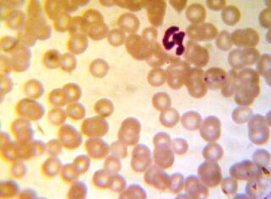
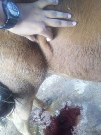
Figure 2: Enlarged lymph node and haemoglobinuria
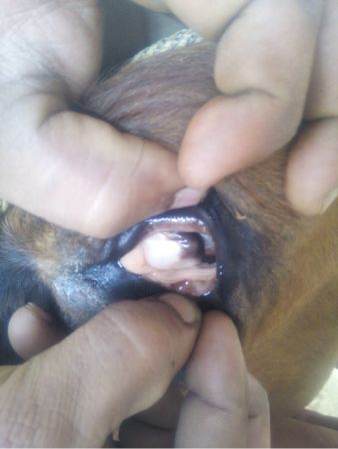
Figure 3: Icteric conjunctival mucous membranes
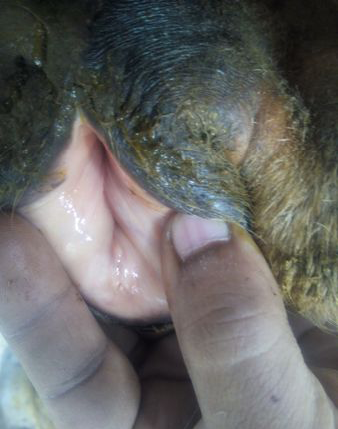
Figure 4: Icteric vaginal mucous membranes
Blood and serum were collected for laboratory investigation. Blood smears revealed presence of Babesia spp. in 40% of RBCs of all the smears (Figure 1). Haemogram revealed on average extremely low levels of Hb, PCV, TEC and platelets counts. Serum chemistry revealed hyperglycemia, hyperbilirubinemia, BUN, AST and hypoprotienemia. The values are presented in Table 1. Urine was coffee coloured and positive for haemoglobin, glucose and bile pigments in all the four cows.
Treatment
The animals were treated with a single dose of Diminazine accurate (Inj. Berenil RTU, Hoechst®) 3 mg/kg Bwt i/m at two different sites in neck muscles, long acting oxytetracycline (Inj. Intamycin-LA, Intas Pharmaceuticals) @ 20 mg / kg body wt i/m at 48 hours intervals on two occasions, haematinic (Inj. Feritas, Intas Pharmaceuticals®) 10 ml i/m thrice weekly for one week, rumenotoric (liquid Brotone, Virbac® Animal Health) 40 ml daily orally for 10 days and Injection Rintose (Wockhardt®) 500 ml i/v daily for 3 days. After 3 days temperature reduced drastically to 1020F in three animals. Hb and PCV levels improved after 3 wks. Treatment was successful with diminazene aceturate (Berenil) at 3 mg/kg body weight, together with supportive therapy in three cows. Whereas, one cow that was presented at delayed stage died due to severe anemia and delay in initiation of therapy.
Table 1: Average Hemato-biochemical values and urinalysis of affected cows
|
Parameters |
Apparently healthy cow values |
Avg Pre- treatment values |
Avg Post- treatment values |
|
Hemoglobin (g/dL) |
8 - 15 |
4.5 |
6.3 |
|
PCV (%) |
24 - 46 |
15 |
22 |
|
TEC X 106/μL |
5 - 10 |
1.3 |
3.2 |
|
TLC X 103/μL |
4 - 12 |
14.25 |
10.3 |
|
MCV (fL) |
40 - 60 |
37.23 |
42.7 |
|
MCH (pg) |
11 - 17 |
11.54 |
15.2 |
|
MCHC (g/dL) |
30 - 36 |
23.21 |
31.1 |
|
Platelets / μL |
100000 - 800000 |
57000 |
92000 |
|
Neutrophils (%) |
20 - 45 |
40 |
46 |
|
Lymphocytes (%) |
45 - 75 |
51 |
48 |
|
Monocytes (%) |
2 - 7 |
2 |
3 |
|
Eosinophils (%) |
2 - 8 |
7 |
3 |
|
Basophils (%) |
0 - 1 |
- |
- |
|
Serum Biochemical Values |
|||
|
AST (U/L) |
78 - 132 |
167 |
124 |
|
TP (g/dl) |
5.7 – 8.1 |
5.8 |
6.1 |
|
BUN (mg/dl) |
6 - 27 |
32 |
24 |
|
Tot. Bilirubin (mg/dl) |
0.01 - 0.5 |
0.9 |
0.56 |
|
Glucose (mg/dl) |
45 - 75 |
110 |
65 |
|
Creatinine (mg/dl) |
1 - 2 |
0.9 |
0.83 |
|
Urinanalysis |
|||
|
Blood (Hb) |
- |
+++ |
- |
|
Glucose |
- |
± |
- |
|
Bile pigments |
- |
++ |
± |
Discussion
Babesiosis is a tick-transmitted disease caused by protozoans of the genus Babesia and it is characterized by haemolytic anemia and fever, with occasional hemoglobinuria and death (Ristic et al., 1981). The minimum infective dose required to produce overt disease is thought to be 103 parasites inoculated intravenously. Variations in the number of parasites injected result in highly significant changes to the prepatent period, peak parasitemia, and the hematological response. In addition to the number of infected ticks that feed on an animal, the immune status of the host and the virulence of the infecting strain. Subclinical infections are quite common and are usually missed by the farmer and clinician. Affected animals have low parasitemia, may suffer mild fever and anorexia, and make an uneventful recovery (Zintl et al., 2003). Hemoglobinuria, frequently the clinical sign first detected by the owner, occurs at the peak of the hemolytic crisis is in accordance with the present cases. Immediately after the hemolytic crisis, a brief lymphocytosis and monocytosis combine to cause a leukocytosis (Gray and Murphy, 1985).
Detection and treatment of babesiosis are important tools to control babesiosis. Microscopy detection methods are still the cheapest and fastest methods used to identify Babesia parasites although their sensitivity and specificity are limited (Mosqueda et al., 2012). When tick population is very high, the disease may be so acute as to cause death within a few days, during which the PCV falls below 20% and the parasitaemia, which is usually detectable once the clinical signs appear and may involve 0.2% to 45% of the red cells, depending on the species of Babesia (Urquhart et al., 1996). Most of the clinico-haemotological findings observed in our cases were similar to those reported earlier by Tufani et al. (2015). For years, babesiosis treatment has been based on the use of very few drugs like imidocarb or diminazene aceturate. Recently, several pharmacological compounds were developed and evaluated, offering new options to control the disease (Mosqueda et al., 2012). Diminazene aceturate consists of an organic base and organic acid but once dissolved in water, it dissociates. It is usually given by intra-muscular injection at doses of 3-5 mg/kg (Kuttler, 1981). Long acting oxytetracyclinehas been shown to have a prophylactic effec against Babesia divergens infection (Urquhart et al., 1996). In humans treatment with quinine and clindamycin, successfully eradicated the organisms. Subsequent studies in animals have supported the usefulness of this combination of antimicrobial agents (Mary et al., 2000). Prolonged convalescent period results in considerable loss of production for a long period in babesiosis (Urquhart et al., 1996). B-complex and oral haematenics were continued for 3 weeks till the animals were completely recovered from anemia.
Conflict of interest
There exists no conflict of interest.
Authors’ contribution
All authors contributed equally.
References


