Research Journal for Veterinary Practitioners
Case Report

Surgical Management of Olecranon Bursitis in Belgium Shepherd Dog
Arvind Kumar Sharma1*, Pankaj Kumar1, Laxmi Kumari1, Lalita Kumari1, Chandrakala1, Madhurendra Kumar Gupta2, Sanjit Kumar2, Praveen Kumar2,3
1Department of Surgery and Radiology; 2Department of Veterinary Pathology; 2Department of Veterinary Medicine, College of Veterinary Science and Animal Husbandry, Birsa Agricultural University, Kanke, Ranchi-834006, Jharkhand, India.
Abstract | An 8 months-old female Belgium Shephered dog was presented at Department of Surgery & Radiology,Clinical complex, Ranchi Veterinary College with a history of golf-ball sized growth over left elbow and damaged skin due to long duration. The case was treated earlier medically without successful outcome. Clinical approach included history, clinical examination, radiography, surgical management along with supportive medication. Growth over left elbow was removed surgically. Skin sutures were removed 12th day post-operatively with observable healing. Furthermore, the dog was observed for 1month period. Dog recovered uneventfully. The aim of this presentation is to report the surgical treatment of long standing olecranon bursitis with ulceration not responded with intralesional medication of dexamethasone and hyluronidase.
Keywords | Capped elbow, Dog, Hygroma, Olecranon bursitis, Surgical management
Editor | Muhammad Abubakar, National Veterinary Laboratories, Islamabad, Pakistan.
Received | August 15, 2015; Revised | September 22, 2015; Accepted | September 23, 2015; Published | October 12, 2015
*Correspondence | Arvind Kumar Sharma, Birsa Agricultural University, Kanke, Jharkhand, India; Email: arsham10@rediffmail.com
Citation | Sharma AK, Kumar P, Kumari L, Kumari L, Chandrakala, Gupta MK, Kumar S, Kumar P (2015). Surgical management of olecranon bursitis in Belgium Shepherd dog. Res. J. Vet. Pract. 3(4): 76-79.
DOI | http://dx.doi.org/10.14737/journal.rjvp/2015/3.4.76.79
ISSN | 2308-2798
Copyright © 2015 Sharma et al. This is an open access article distributed under the Creative Commons Attribution License, which permits unrestricted use, distribution, and reproduction in any medium, provided the original work is properly cited.
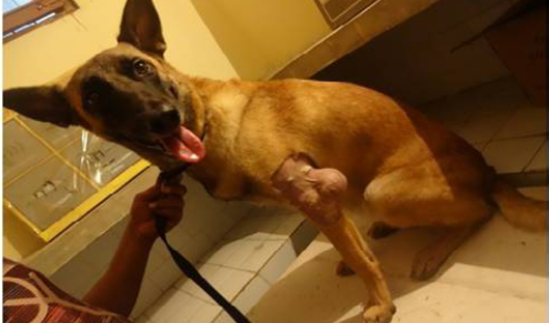
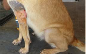
Olecranon bursitis is characterized by a movable swelling over the olecranon tuberosity. Olecranon bursitis or capped elbow or hygroma of elbow or elbow seroma is fluid filled cavity surrounded by a dense fibrous connective tissue due to inflammation over the olecranon bursa. It is a pseudocyst rather than a true cyst because it has no epithelial or synovial lining (Bellah et al., 1993). Canine elbow joint is surrounded by olecranon bursa just beneath the skin to facilitate smooth gliding of the skin over the olecranon process. The elbow is entirely surrounded by brachial and antebrachial fasciae along with the superficial antebrachial fascia on the medial aspect (Constantinescu and Constantinescu, 2009). The growth becomes harder with lesser amount of fluid if not removed and remained as such for longer duration (Fossum, 1997). Underlying causative factor for development of capped elbow in dogs is accumulation of fluid due to repeated trauma to soft tissues eventually becoming encapsulated by fibrous tissue lined with a synovial membrane (Nath et al., 2014). Reports suggest that capped elbow is more frequently reported in young dogs of large breeds before a protective callus forms on the bony prominence (Fossum, 1997; Nath et al., 2014). Hygroma also can develop in older animals that present with arthralgia or orthopaedic pain in other joints, causing excessive pressure over the elbow when lying in sternal recumbency (Birnesser et al., 2005).Primary treatment consists of removing the repeated trauma. Small and early hygroma can be treated with repeated aspiration and application of pressure bandage. The contents are aspirated weekly with careful aseptic precaution (Arican et al. 2005; Durmus and Sangliyan, 2008). Success is unlikely if fluid remain after 3 or 4 treatments (Johnston, 1975). A little information is available about the surgical excision of hygroma in dogs. Therefore, the present case report described the surgical management of capped elbow condition on left elbow of an 8-months-old female Belgium Shepherd dog (Figure 1).
A female dog of aged 8 months, was presented to the dept. of Vet. Surgery and Radiology, Ranchi Veterinary College with the history of growth on the left elbow. Haemorrhagic spot was found on the growth. There was no history of recent trauma to the elbow. The dog was treated earlier with intralesionally administration of dexamethasone and hyluronidase after aspiration of transparent fluid without successful outcome. Rectal temperature, heart rate and respiration rate were found to be within normal range. Keeping in view of progressive enlargement of growth which produced difficulty in walking and dog siting posture, surgery was planned.
The site was prepared aseptically. The animal was administered with Atropine sulphate@0.04 mg/kgbwt. After 5 min., xylazine hydrochloride @ 1mg/kgbwt was administered as preanaesthetic. Propofol “to effect” was given after 15 min. of xylazine hydrochloride administration. Intravenous fluid therapy was initiated prior and continuous infusion rate of propofol was maintained during the period of surgical management. Animal was placed on table in lateral recumbency by keeping the left fore limb on upper side. An elliptical incision (Figure 2) was made along the posteriolateral aspects of the point of elbow and over the growth area.
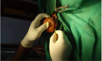
Figure 2: Eliptical incision
Olecranon bursa was then gently separated from its soft tissue attachment by blunt dissections without rupturing it or injuring the joint capsule (Figure 3). Bleeding from major vasculature was checked by ligature with chromic catgut No. 00 whereas, the capillary bleeding was checked by manual pressure with guaze sponge. Hygroma along with

Figure 3: Appearance of bursa
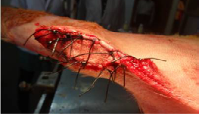
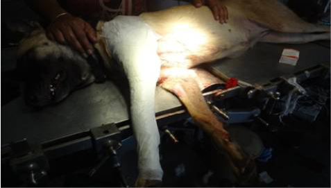

Figure 6: Removed hygroma along with bursa
skin was removed (Figure 6). The fascia and muscles were sutured with chromic catgut No. 0 and skin was sutured with mersilk no. 1 (Figure 4). A thick padding along with pressure bandage was applied over the operative area as well as on full limb (Figure 5). Animal was treated with Ceftriaxone @20 mg/kgbwt intramuscularly for 7 days and Meloxicam @ 0.2 mg/kgbwt for 3 days. Owner was advised to monitor and restrict the movement of the affected limb of his pet for a period of 1 month. External skin sutures were removed 12th day post-treatment with appropriate wound healing without complications (Figure 7).
The dog recovered uneventfully without complications. Treatment of olecranon bursitis at initial stages consisted of eliminating the cause of trauma, pressure bandage, cold hydrotherapy and rest (Arican et al., 2005). In later stage, the treatment of olecranon bursitis has been documented by drainage and aspiration of accumulated fluid and injection of 3-5% carbolic acid (Arican et al., 2005), 4% tincture iodine (Fathy and Radad, 2006) and corticosteroid and penicillin combination (Hayat et al., 2009). In the present case, it was treated with intralesional administration of dexamethasone and hyluronidase, which did not prove to be effective. Johnston (1975) also reported that aspiration and injection of corticosteroid did not prove to be effective in treatment of hygroma as it resulted to infection and skin ulceration.In dogs, the hygroma usually remains sterile unless organisms are introduced through needles or medication. Acute infections have been reported following injection of corticosteroids in to hygromas (Johnston, 1975). The long standing hygroma was treated effectively by surgical removal of hygroma (Nath et al., 2014; Kantia et al., 2015) and application of painrose drain (Johnston, 1975) in dogs. Elbow hygroma in Newfoundland dog was also treated successfully with minimum complications without recurrence with surgical excision and transfer of microvascular free muscle transfer utilizing the rectus abdominis muscle in combination with mesh skin graft and transarticular external fixator (Green et al., 2008). The present case was also long standing and not responded with medical management. Hence it was treated effectively with surgical interventions without complications. The successful outcome of surgery has also been reported by Degreef and Smet (2006), Hayat et al. (2009) and Kumar et al. (2010). In contrast, wound breakdown and ulceration are serious complications of excision of elbow hygromas. The ulcers are rarely healed spontaneously because of repeated trauma to the healing area as reported by Johnston (1975). In present report this problem was controlled by application of thick bandaging for 20 days until complete healing took place.
It is concluded that long standing elbow hygroma not responded with aspiration and intralesional medication can be treated by surgical excision of hygroma.
Acknowledgments
Authors are thankful to Dean, Ranchi Veterinary College for providing necessary facilities to carry out the work.
Conflict of interest
The authors declares that there is no conflict of interest regarding the publication of this paper.
AUTHORS CONTRIBUTION
Arvind Kumar Sharma, Pankaj Kumar, Laxmi Kumari and Lalita Kumari performed the operative procedures and collection of articles related to prevous work and preparation of manuscript whereas, Chandrakala, Madhurndra Kumar Gupta, Sanjit Kumar and Praveen Kumar critically revised the manuscript.
References


