Research Journal for Veterinary Practitioners
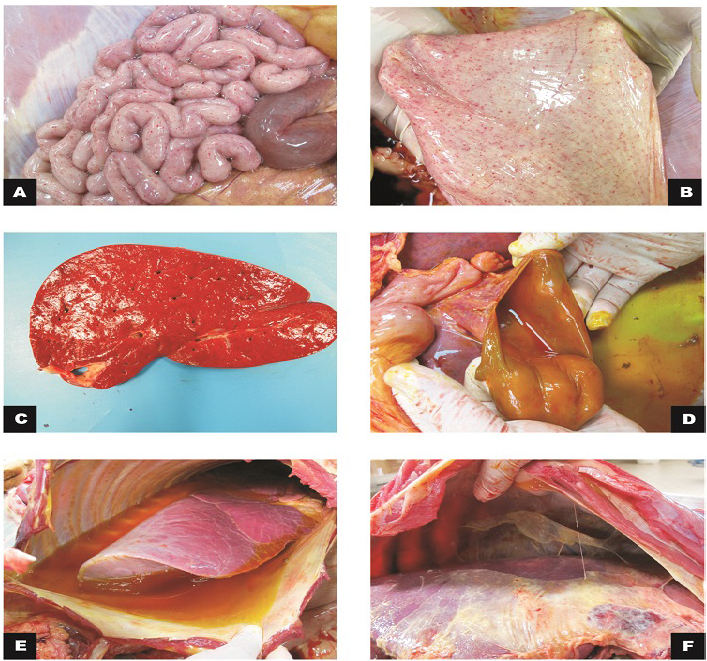
Macroscopic findings in calves
A) Small intestine, jejunum: serosa with marked diffuse petechiae (calf 1); B) Bladder: mucous membrane with marked diffuse petechiae (calf 1); C) Liver: slight increase in volume and orange colour (calf 2); D) Gallbladder: moderate thickening of the wall (calf 2); E) Thoracic cavity and lungs: moderate hydrothorax associated with petechiae and discrete multifocal ecchymosis in the parietal pleura; F) Cranial, middle and right caudal lung lobes not collapsed with reddish cranio-ventral portions (calf 3).
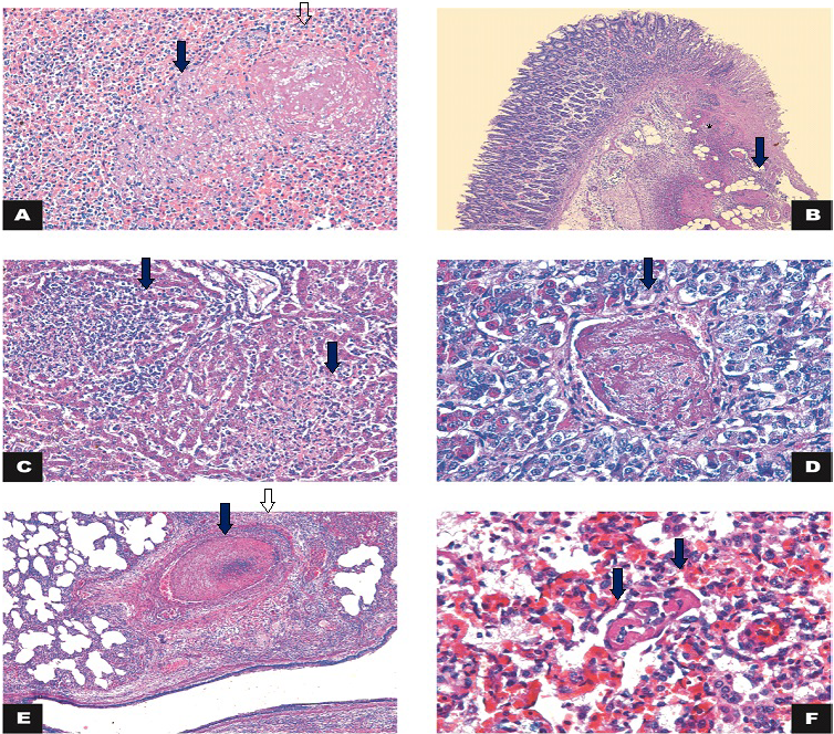
Histology
A) Spleen: necrosis (arrow) associated with thrombosis (open arrow) (calf 3). HE 200x; B) Large intestine: intersection of intact and fibrinonecrotic mucosa associated with multifocal thrombosis (arrow) and inflammatory infiltrate in the lamina propria/submucosa (*) (Calf 1). HE 50x; C) Liver: necrosis and lymphohistiocytic inflammatory infiltrate (arrows) (Calf 2). HE 200x; D) Hypophysis: thrombosis (arrow) (Calf 2). HE 400x; E) Lung: thrombosis (arrow) and vasculitis (arrowhead). HE 50x; F) Lung: capillary thrombosis (arrows) (Calf 4). H&E 400x.


