Research Journal for Veterinary Practitioners
Research Article

Ultrasonographic Identification of Abdominal and Thoracic Lesions Resulting from Foreign Body Syndrome in Buffaloes
Shaimaa Mohamed Gouda
Animal Medicine Department, Faculty of Veterinary Medicine, Zagazig University.
Abstract | Foreign body syndrome or traumatic reticuloperitonitis (TRP) remains the disease of most interest for many clinicians in developing countries. Ultrasonography is remaining the best diagnostic tool for diagnosis of such disease. Therefore, this work was planned to approve the importance of ultrasound in diagnosis and detection of different complications of this disease in buffaloes. Sixty buffaloes with TRP and 20 clinically healthy buffaloes were examined clinically and ultrasonographically. A 3.5 to 5 MHz convex probe was used to scan the area just behind xiphoid cartilage as well as abdominal and thoracic cavities. Peritonitis and abdominal abscesses were detected as abdominal lesions (n=41) while pericarditis, pleurisy and thoracic abscesses were detected as thoracic lesions (no=19) secondary to TRP. The most clinical signs of abdominal affected buffaloes were anorexia, tympany, scanty faeces and decrease of milk production. The thoracic affected buffaloes had the same previous signs in addition to signs of congestive heart failure (no= 12), respiratory signs (no=4) or both (no=3). Signs of pain reaction were observed in 6 cases with abdominal lesions while these signs were detected in all cases had thoracic lesions. The reticulum of healthy buffaloes appeared with half-moon shaped and had 3.95± 0.5 biphasic contractions/ 4 minutes. In contrast, reticulum with TRP appeared corrugated with reduced or absence of reticular motility. Local and diffuse peritonitis were clarified in 26 cases, peri-reticular abscess in 9 cases, hepatic abscesses in 4 cases and splenic abscess in 2 cases. Moreover, pericarditis was detected in 12 cases, pleuropneumonia in 4 cases and thoracic abscess in 3 cases. In conclusion, ultrasonography should be considered as suitable tool for detection the different complications of foreign body syndrome in buffaloes.
Keywords | Ultrasonography, Reticulum, Foreign body, Buffaloes
Editor | Muhammad Abubakar, National Veterinary Laboratories, Islamabad, Pakistan.
Received | May 05, 2015; Revised | May 24, 2015; Accepted | May 25, 2015; Published | May 28, 2015
*Correspondence | Shaimaa Mohamed Gouda, Zagazig University- Egypt; Email: shaimaagouda81@gmail.com
Citation | Gouda SM (2015). Ultrasonographic identification of abdominal and thoracic lesions resulting from foreign body syndrome in buffaloes. Res. J. Vet. Pract. 3(2): 41-46
DOI | http://dx.doi.org/10.14737/journal.rjvp/2015/3.2.41.46
ISSN | 2308-2798
Copyright © 2015 Gouda et al. This is an open access article distributed under the Creative Commons Attribution License, which permits unrestricted use, distribution, and reproduction in any medium, provided the original work is properly cited.
INTRODUCTION
Buffalo (Bubalus bubalis) is a large bovid originating in South, Southeast Asia and China. Today, it is also found in Europe, Australia and in some American countries. Two extant types of water buffalo were recognized based on morphological and behavioural criteria – the river buffalo of South Asia and further west to the Balkans, Egypt and Italy, and the swamp buffalo, found from Assam in the west through Southeast Asia to the Yangtze valley of China in the east (Cockrill, 1977).
Buffaloes as well as cows had unselective feeding habit that results in consumption of foreign objects among their own food. Contractions of the reticulum promote penetration of the wall by the foreign object. Perforation of the wall of the reticulum allows leakage of ingesta and bacteria, which contaminates the peritoneal cavity resulting peritonitis (Andrew et al., 2004). The hardware or foreign body syndrome remains the primary disease of cattle rather than other ruminants. It occurs when pieces of wire, or other sharp metal objects, have been taken by the cattle along with its food (Orpin and Harwood, 2008).
The affected animals generally showed variable clinical signs which depend upon the severity of disease, individual sensitivity and the species of the animal (Abdelaal et al., 2009; Athar et al., 2010).
Diagnosis of reticular affections can be generally achieved using transthoracic ultrasonography, radiography or exploratory laparorumentomy. Ultrasonography is well established in diagnosis of reticular diseases as well as describing the physiological characters of reticulum in cattle. Ultrasonography provides direct views of frequency, duration, speed and amplitude of reticular contractions. The motility of the reticulum has been evaluated by ultrasonography in healthy cows at rest (Braun and Gotz 1994; Kaske et al., 1994; Sleeter and Step, 2007; Braun, 2009), cows and buffaloes with traumatic reticuloperitonitis (Abouelnasr et al., 2012; Aref and Abdel-Hakiem, 2013; Abdelaal and floeck, 2015), reticular abscesses (Braun et al., 1998; Braun, 2005) and vagal indigestion (Braun et al., 2009). Other studies had investigated the effects of atropine, xylazine and scopolamine (Braun et al., 2002) and neostigmine (El-Khodery and Sato, 2008) on reticular motility. The motility of the reticulum during rest, eating, rumination and stress in healthy cows had also been described (Braun and Rauch, 2008).
Buffaloes still require more investigation to clarify the different complications of the foreign bodies after penetration of the reticulum; therefore, following study was carried out.
MATERIALS AND METHODS
Twenty clinically healthy, female buffaloes were selected from the farm of Faculty of Veterinary Medicine- Zagazig University, which were 2.5 to 6.5 years old and weighed 450-650 Kg body weight. Out of 20 buffaloes, 10 were heavy pregnant and 10 were non pregnant. All of the buffaloes were deemed healthy based on the results of a thorough clinical examination, a complete blood cell count, urine analysis and negative faecal examination. Additionally, sixty female buffaloes with the same age and weight (range of healthy control one) were admitted to Veterinary clinic- Zagazig University at 2012-2014. Out of 60 buffaloes, 20 were recently calved, 25 were heavy pregnant and 15 were non pregnant. On admission they were suffered from anorexia, weight loss, tympany and scanty faeces. All buffaloes were submitted to thorough clinical examination, mine detector and ultrasonographic examination and the disease were diagnosed as hardware. Assessment of body temperature, respiratory and heart rates, auscultation of the heart, lungs and rumen were also performed. In addition, examination of the jugular and milk veins and pain tests including; pinching of withers, side sticks were carried out according to Kelly (1984).
Ultrasonographic examinations were carried out on standing, non-sedated animals as described previously by Braun and Gotz (1994) using 3.5- 5.0 MHz convex transducer with penetration depth 20 cm. All healthy control buffaloes were examined after 2 hours from feeding. The probe was placed firstly at the area just behind xiphoid cartilage then moved caudally and laterally to scan the abdominal cavity. Thoracic cavity was also scanned from both sides at intercostal spaces 5th to 12th one. Reticular shape, contour and motility per 4 minutes were assessed.
Rumenotomy was applied as a confirmatory test on 15 buffaloes with hardware disease. Meanwhile, abscesses were confirmed in 16 cases through aspiration under ultrasonographic guidance as described previously by Braun et al (1998). Other 29 cases were not confirmed due to bad prognosis or due to owner’s refusal.
All data were presented as means ± standard deviation (SD) of the means and performed using computer package of Excel, office, 2010.
RESULTS
All apparently healthy buffaloes under investigations showed signs of healthiness including good body condition, alertness, brightness and no external lesion or parasites. Temperature, pulse and respiratory rates/ minute and ruminal motility/2 minutes were 38.46± 0.25, 66.2±1.7, 25.8±1.5 and 3.1±0.5, respectively. On the other hand, all diseased buffaloes were suffered from decreased or absence of appetite, tympany and scanty faeces. Positive pain tests and signs of pain included stretching position, stiffness in gait, abducted elbows and arched back were seen in 6 buffaloes had abdominal lesions and in all buffaloes with thoracic lesions secondary to foreign body invasion (Figure 1). Moreover, the vital parameters as temperature, pulse and respiratory rate/ minute were within control range in 35 cases, elevated in 4 and decreased in 2 cases with abdominal lesions. While all of these parameters were elevated in all buffaloes that suffered from thoracic lesions. Signs of congestive heart failure include, brisket edema and distended jugular veins were recorded in 12 buffaloes with thoracic lesions. Respiratory manifestations as dyspnea, cough and abnormal lung sound either wheezes or crackling or both were observed in 4 buffaloes with thoracic lesions (Figure 2). Both were observed in 3 buffaloes with thoracic involvement.
Ultrasonography of healthy buffaloes revealed a half moon shaped reticulum with 3.95± 0.5 biphasic contractions/ 4 minutes (Figure 3A). It was detected at the area just behind xiphoid cartilage at ventral abdomen and also was detected from left and right para-median until the level of elbows. In all diseased buffaloes, the reticulum appeared corrugated and a tonic or absence of contractions. Basing on ultrasonography the diseased buffaloes were classified into two groups; 41 buffaloes had abdominal lesions (Figure 3B, C and D) included local peritonitis (no=20), diffuse peritonitis (no=6) and abdominal abscess (no=15) and 19 buffaloes had thoracic lesions (Figure 4) included pericarditis (no=12), pleuro-pneumonia (no=4) and thoracic abscess (no=3).
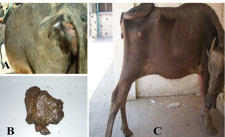
Figure 1: Clinical findings of buffaloes with abdominal lesion due to foreign body syndrome A. tympany; B. scanty feces and C. stretched abdomen due to abdominal pain.
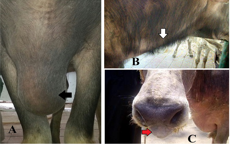
Figure 2: Clinical findings of buffaloes with thoracic lesion due to foreign body syndrome. A. brisket edema (black arrow); B. corded jugular vein (white arrow) and C. mouth breathing due to dyspnea as a respiratory sign (red arrow).
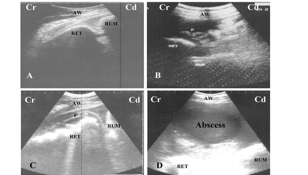
Figure 3: A. Ultrasonogram of normal reticulum during relaxation, it appears a half moon shaped; B and C. Ultrasonogram of reticulum in case of foreign body syndrome, it appears corrugated and surrounded by inflammatory echogenic fibrin (F); D. circumscribed abscess with echogenic capsule and hypoechoic pus content locates between reticulum (RET), rumen (RUM) and abdominal wall (AW); Cr: cranial; Cd: cauadal.
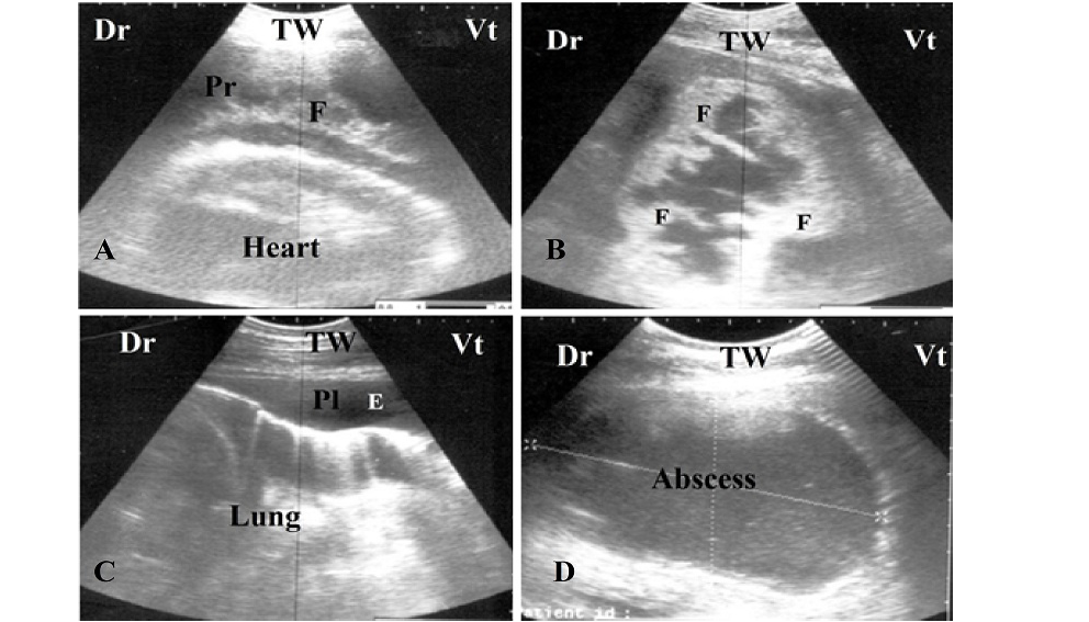
Figure 4: A and B. Ultrasonogram of a buffalo with pericarditis, notice the presence of echogenic fibrin (F) interspersed in pericardial sac (Pr); C. Ultrasonogram of pleurisy, notice the anechoic pleural effusion in pleural sac (Pl) and absence of reverberation artifact of lung; D. Circumscribed abscess with echogenic capsule and hypoechoic pus content locates in mediastina between lung, heart and thoracic wall (TW). Dr: dorsal, Vt: ventral
Peritonitis was described in 26 buffaloes and appeared as hypoechoic materials represented inflammatory exudates which were either localized in 20 buffaloes or diffuse in 6 buffaloes. Abscesses appeared with varying sizes (5-15 cm), as circumscribed masses with echogenic capsule and hypoechoic content and located peri-reticulum (n=9), retro-hepatic (no=4) and retro-splenic (n=2). These masses were confirmed as abscesses by percutaneous aspiration under ultrasonographic guidance.
Pericarditis was seen in 12 cases and appeared as hypoechoic materials with echogenic fibrin interspersed in between the pericardial sac (Figure 4A). Absence of reverberation artifact of normal lung and replaced by hypoechoic structure and echogenic debris with distal comet tail artifact along with anechoic pleural effusion and echogenic fibrin in pleural sac were the features of four pleuropneumonic buffaloes (Figure 4B and C). Large sized thoracic abscesses were described and confirmed in 3 cases (Figure 4D) and had same appearance of abdominal abscesses.
DISCUSSION
Buffaloes as well as cattle are subjected to different abdominal disorders. Most of these disorders in buffaloes are resulted from ingestion of foreign bodies among their own food. The decision of interference depends upon many factors including, whether the foreign body penetrates the reticular wall or not, the location of foreign bodies after reticular and complications that could resulted from the penetrated foreign bodies. All diseased buffaloes under investigation had reduced appetite, ruminal atony with scanty faeces and decreased milk production as has been described by Abdelaal and Floeck (2015). Additionally, Radostitis et al., (2007) stated that the presence of such symptoms is considered as a general sign for indigestion. Signs of pain and systemic reactions were less common in buffaloes with abdominal lesions than those with thoracic lesions. This result indicates that buffaloes with thoracic affection had acute reactions while abdominal lesions mainly chronic in nature. Abdelaal et al. (2009) stated that all buffaloes with traumatic reticuloperitonitis had less signs of pain and systemic reactions more than cattle and attributed this for the natural tolerance of buffaloes for pain. But in the present study the pain and systemic disturbances were observed in buffaloes which had thoracic lesion. Respiratory manifestations were observed in 7 buffaloes with thoracic lesions especially those with pneumonia (n= 4) and thoracic abscess (n= 3). Signs of congestive heart failure were detected in 15 buffaloes with pericarditis (n= 12) and thoracic abscess (n= 3). Both symptoms were observed previously by Abdelaal et al. (2009). From the result of clinical examination of diseases buffaloes, it could be concluded that there is a difficulty to differentiate between different abdominal and thoracic lesions by clinical findings. Hence it is necessary to use additional diagnostic techniques in diagnosis of abdominal and thoracic lesions.
Sonographically, reticulum appeared as a half-moon shaped with 3.95± 0.5 biphasic contractions/4 minutes. These results are nearly similar to that observed by Imran et al, (2012). In the present study, the reticulum was observed in the left and right paramedian behind xiphoid cartilage until the level of elbows. This result was confirmed previously by Abdelaal et al. (2014). Corrugated reticulum with reduction or absence of reticular contractility is considered to be the characteristic ultrasonographic findings of different sequelae of TRP in buffaloes. This result is in consistent with those reported by Braun et al. (1993). The ultrasonographic examination yields precise information about the lesion of foreign body syndrome in buffaloes. The inflammatory reactions appeared as echogenic strands interspersed with anechoic material which was localized at the area of reticulum (local peritonitis, n= 20) or extended to involve the whole abdominal cavity (diffuse peritonitis, n=6). Moreover, abscesses appeared in 15 cases as circumscribed mass with varying sizes (5- 15 cm) and had echogenic capsule with hypoechoic content. This result is in concurrence with those obtained by Braun et al. (1998), Braun (2005) and Mohamed and Oikawa (2007). Additionally, ultrasonography provides us more information about the site of abscesses and suitable site for aspiration of abscesses. Thoracic ultrasonography in buffaloes with foreign body syndrome has proven a good diagnostic procedure for distinguishing traumatic pericarditis from thoracic abscesses and traumatic pleurisy.
CONCLUSION
Ultrasonography is a valuable tool for detecting the lesions resulting from foreign body syndrome in buffaloes, thus it consider a suitable prognostic tool for buffaloes has been affected with this syndrome.
AKNOWLDGEMENT
The author would like to thank the Department of Surgery, Faculty of Veterinary Medicine, Zagazig University, Egypt for their surgical advice.
CONFLICT OF INTEREST
Author declares no conflict of interest
REFERENCES


