Research Journal for Veterinary Practitioners
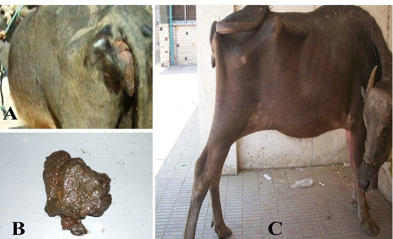
Clinical findings of buffaloes with abdominal lesion due to foreign body syndrome
A. tympany;
B. scanty feces and
C. stretched abdomen due to abdominal pain.
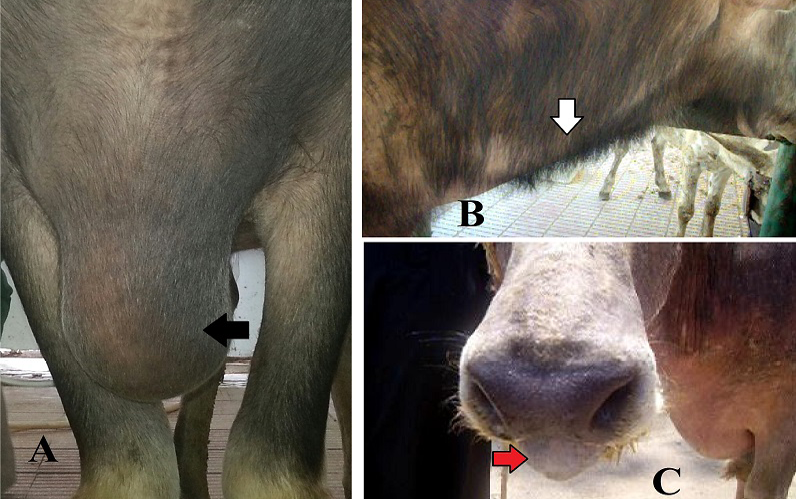
Clinical findings of buffaloes with thoracic lesion due to foreign body syndrome
A. brisket edema (black arrow);
B. corded jugular vein (white arrow) and
C. mouth breathing due to dyspnea as a respiratory sign (red arrow)
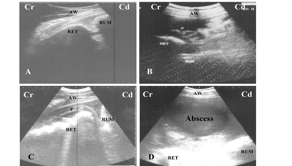
A. Ultrasonogram of normal reticulum during relaxation, it appears a half moon shaped;
B and C. Ultrasonogram of reticulum in case of foreign body syndrome, it appears corrugated and surrounded by inflammatory echogenic fibrin (F);
D. circumscribed abscess with echogenic capsule and hypoechoic pus content locates between reticulum (RET), rumen (RUM) and abdominal wall (AW); Cr: cranial; Cd: cauadal.
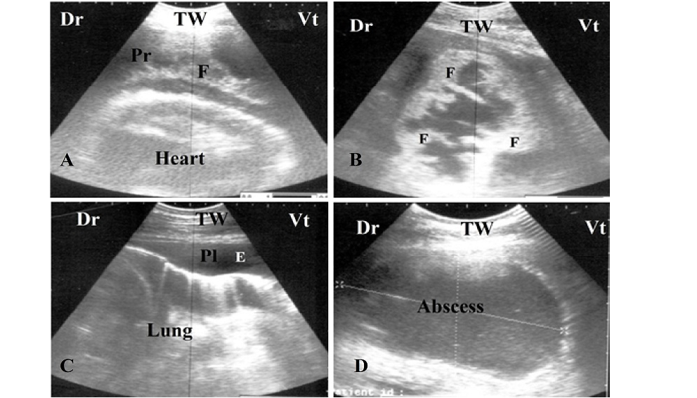
A and B. Ultrasonogram of a buffalo with pericarditis, notice the presence of echogenic fibrin (F) interspersed in pericardial sac (Pr);
C. Ultrasonogram of pleurisy, notice the anechoic pleural effusion in pleural sac (Pl) and absence of reverberation artifact of lung;
D. Circumscribed abscess with echogenic capsule and hypoechoic pus content locates in mediastina between lung, heart and thoracic wall (TW). Dr: dorsal, Vt: ventral


