Research Journal for Veterinary Practitioners
Research Article
Res. j. vet. pract. 3 (2): 36 - 40

Figure 1
Acute tubular necrosis (ATN) and hyaline casts (H&E x 400)
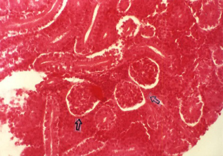
Figure 2
Degenerative tubular changes (H&E x 200)

Figure 3
Foci of hemorrhages in the renal parenchyma (day 15) (H&E x 100)
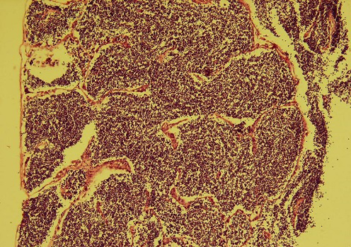
Figure 4
Lymphoid depletion in the spleen (H&E x 200)

Figure 5
Oedema of the wall and hypertrophy of the endothelial cells in arterioles (arrows) and lymphoid depletion in the spleen (H&E x 400)

Figure 6
Cell swelling of the proximal tubules lining epithelium (arrow) (H&E x 400)
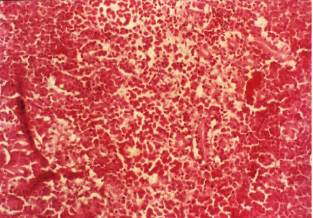
Figure 7
Hemorrhages in the renal parenchyma (H&E x 200)
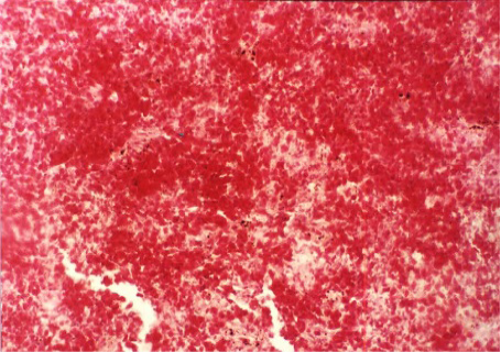
Figure 8
Hemorrhages in the spleen (H&E x 200)


