Research Journal for Veterinary Practitioners
Case Report
Coccidiosis Due to Eimeria arloingi Infection in a Saanen Goat Kid
Aycan Nuriye Gazyagci1*, Tugce Anteplioglu2, Sıla Canpolat2, Hasan Tarik Atmaca2
1Faculty of Veterinary Medicine, Department of Parasitology, Kirikkale University, Kirikkale, Turkey; 2 Faculty of Veterinary Medicine, Department of Pathology, Kirikkale University, Kirikkale, Turkey.
Abstract | Coccidiosis, caused by protozoa of the genus Eimeria, is one of the major parasitic diseases characterized by subclinical contagious enteritis influencing domestic and wild animals. Most often affected are young animals with diarrhoea, poor growth and death. A month of age death in Saanen goat kid was referred to Department of Pathology, Faculty of Veterinary Medicine, Universty of Kırıkkale. At necropsy, severe and diffuse nodular hyperplasia were present on entire small intestine mostly jejunum and ileum. Also intestinal contents were solid and bloody in jejunum and ileum, cecum and large intestinal contents were watery and greenish. Morphological identification of Eimeria arloingi oocysts was carried out based on sporulation techniques and 9900 of oocysts per gram of feces (OPG) were determined by means of the modified McMaster technique. At histo-pathologic examination of intestinal segments were observed whole epithelial layers cells containing oocyst and different numbers of developmental stages of protozoan.
Keywords | Coccidiosis, Eimeria arloingi, Goat, Histopathology, Intestine
Editor | Muhammad Abubakar, National Veterinary Laboratories, Islamabad, Pakistan.
Received | December 08, 2014; Revised | January 15, 2015; Accepted | January 17, 2015; Published | February 07, 2015
*Correspondence | Aycan Nuriye Gazyagci, Kirikkale University, Kirikkale, Turkey; Email: naycani1980@yahoo.com
Citation | Gazyagci AN, Anteplioglu T, Canpolat S, Atmaca HT (2015). Coccidiosis due to Eimeria arloingi infection in a Saanen Goat Kid. Res. J. Vet. Pract. 3(2): 29-32.
DOI | http://dx.doi.org/10.14737/journal.rjvp/2015/3.2.29.32
ISSN | 2308-2798
Copyright © 2015 Gazyagci et al. This is an open access article distributed under the Creative Commons Attribution License, which permits unrestricted use, distribution, and reproduction in any medium, provided the original work is properly cited.
Coccidiosis is a protozoan disease caused by several species of the genus Eimeria, have been observed in worldwide. Eimeria in goats develop in the small and large intestine and causes enteritis in small ruminants and death especially in young animals. Also it leads to growth deficiency and lower productivity in goats which survived from the disease and these animals as a premium play role transmission of clinical coccidiosis (Taylor et al., 2007).
There are 17 Eimeria species have been described in goats (Dinçer and Vatansever, 2001). Dinçer and Vatansever (2001) have defined nine species in Turkey. However, just nine Eimeria species could create an infection and the most pathogenic species in goat is Eimeria arloingi (Taylor et al., 2007). Eimeria species from goat are localized in liver, gallbladder, bile ducts, hepatic and mesenteric lymph nodes as well as small and large intestine (Dik, 2001; Nourani et al., 2006). E. arloingi causes polyp and nodular hyperplasia in intestinal mucosa (Taylor et al., 2007).
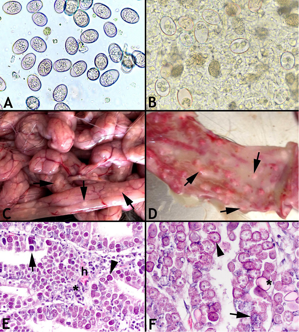
Figure1: A, B, C, D, E and F
A. Unsporulated oocysts in feces detected with flotation examination; B. Sporulated oocysts in feces detected with flotation examination; C. Severe and diffuse noduler hyperplasia (arrows) in intestine serosa; D. Severe and diffuse noduler hyperplasia (arrows) in lumen of duodenum; E. There were macrogamont (arrow head), microgamont (arrow), hyperemi (h), neutrophil and eosinophil infiltration with severe plasmacyte and lymphocyte (asterisk); F. There were macrogamont (arrow head), microgamont (arrow) and oocyst stages of E. arloingi (asterisk) in intestinal epithelium
One month-old goat kid died after showing severe watery with clumps of mucus and yellowish diarrhoea, weakness and anorexia symptoms and was referred Department of Pathology, Faculty of Veterinary Medicine, Universty of Kırıkkale. At necropsy, severe and diffuse nodular hyperplasia were present on jejunum and ileum (Figure 1C and 1D). After collection of faecal samples from the all intestinal segment, native examination and flotation with saturated NaC1 was done for determine number of oocysts (Figure 1A). Fecal examination was performed with the modified McMaster technique, establishing the number of oocysts in 1 g of feces (OPG coefficient) (Thienpont et al., 1986). The intensity of infection in kid was high 9 900 oocysts in 1 g of feaces. Sporulation was performed in wet chamber at 24–26°C in a 2.5% aqueous solution of potassium dichromate (K2Cr2O7) (Pellérdy 1974). The species of Eimeria was determined based on morphological criteria of oocysts such as; shape of oocyst, presence of micropyle, aspect of oocyst wall, polar cap, colour, shape of sporocyst, presence of stieda body, oocystal and sporocystal residues (Coudert, 1992). According to results, E. arloingi was noted (Figure 1B).
Systematically collected tissue samples of the kid were fixed in 10% buffered formalin for 48 hours for histopathologic examination. Then they were embedded in paraffin-wax, sectioned at 4-5 μm thickness and stained with haematoxylin and eosin (H&E). At the histopathologic examination, epithelium cells of almost all layers were detected to be full with E. arloingi. There were macrogamont, microgamont and oocyst stages of E. arloingi in the duodenum, jejunum and ileum epithelium and mild neutrophil and eosinophil infiltration with severe plasmacyte and lymphocyte infiltration (Figure 1E and 1F). Also, hyperemic areas were found.
Eimeria arloingi could result with fatal coccidiosis. Nourani et al., (2006) reported that nodular hyperplastic lesions were seen only in jejunum in Iranian native kid. In this report we observed that the nodular hyperplastic lesions can be seen all segments of intestine especially in jejunum and ileum. Even if the animals survived from the disease, they live as porters. Thus, coccidiosis causes economic losses throughout the global livestock industry. Sharma and Singh (1997) reported that coccidiosis among the whole parasitic diseases has the highest mortality rate. Various studies in Turkey showed that the microscopically determined coccidiosis prevalence was 84% in Middle Anatolia, 86,3% in Ankara and Eskişehir; 100% in Ankara; 53,3% in Ege Region (İzmir, Manisa, Aydın); 94,6% in Elazığ; 69,8% in Van (Göz et al., 2006; Gürler et al., 1990; Güralp and Oğuz, 1967; Merdivenci, 1959; Sayın et al., 1986; Sayın, 1996). Another study showed that the mortality rates due to coccidiosis were 55%, 30% and 24% in Adana, Mersin and Osmaniye, respectively (Özdemir and Çaya, 2012). Around the globe, the prevalence of coccidiosis in goats of varying ages were found between 54-100% and in 36-91% of these cases the causing agent was determined as E. arloingi (Ameh et al., 2000; Balicka-Ramiz et al., 2012; Cavalcante et al., 2011; Ibrahim, 2012; Kahan and Greiner, 2013; Padilla et al., 2009; Silva et al., 2014) One particular study reported from China showed that 83,3% of the 250 Saanen goats were positive for E. arloingi (Zhao et al., 2012).
Saanen goat is a Swiss originated high yielding dairy goat breaded throughout the world. In this case report, we pathologically and parasitologically investigated the dead of the Saanen goat kid that was brought to clinics with manifestations of lack of appetite, inertia and diarrhea. We have determined that the dead was associated with coccidiosis due to E. arloingi and hence the breeder was informed. To best of our knowledge, previously there had been no reported case of Saanen goat coccidiosis due to Eimeria spp. in Turkey. Around the globe economic losses due to coccidiosis in Saanen goat is not to be underestimated. In this case report, coccidiosis due to E. arloingi in Saanen goat is clinically and pathologically well described. Since breeding of Saanen goat is economically a privileged breeding type, and coccidiosis may result in fatal outcome, it is of importance that the breeders should concentrate on preventative measures for protection of Saanen goats from coccidiosis.
CONFLICT OF INTEREST
Author declares no conflict of interest.
REFERENCES


