Research Journal for Veterinary Practitioners
Research Article
Research Journal for Veterinary Practitioners 2 (1S): 11 – 13Special Issue – 1 (2014) (Epidemiology and Occurrence of PPR in Endemic Situation)
Evaluation of Haemagglutination Assay (HA) for the Detection of Peste des Petits Ruminants Virus (PPRV) in Faecal Samples of Recovered Goats
Asma Latif1, Zunaira Akhtar2, Riasat Wasee Ullah1*, Aamer Bin Zahur1, Aman Ullah1, Hamid Irshad1, Adnan Rashid Malik3, Munib Hussain1, Khawar Mahboob4, Shahida Afzal4,
- Animal Health Research Laboratories, Animal Sciences Institute, National Agricultural Research Centre Islamabad
- Department of Clinical Medicine and Surgery, Faculty of Veterinary Sciences, University of Veterinary and Animal Sciences Lahore
- Department of Animal Husbandry, Muzaffarabad, Azad Government of the State of Jammu and Kashmir
- Veterinary Research Institute
, Lahore Cantt, Pakistan
*Corresponding author:riasatwasee252@yahoo.com
ARTICLE CITATION:
Latif A, Akhtar Z, Ullah RW, Zahur AB, Ullah A, Irshad H, Malik AR, Farooq U, Hussain M, Mahboob K, Afzal S (2014). Evaluation of haemagglutination assay (HA) for the detection of peste des petits ruminants virus (PPRV) in faecal samples of recovered goats. Res. J. Vet. Pract. 2 (1S): 11 – 13.
Received: 2014–05–01, Revised: 2014–07–19, Accepted: 2014–08–01
The electronic version of this article is the complete one and can be found online at
(
http://dx.doi.org/10.14737/journal.rjvp/2014/2.1s.11.13
)
which permits unrestricted use, distribution, and reproduction in any medium, provided the original work is properly cited
ABSTRACT
This paper reports the findings of evaluation of Haemagglutination Assay (HA) for detection of Peste des Petits Ruminants (PPR) in faecal samples of sheep and goats persistently infected with PPR. Faecal samples (n=100) collected during an outbreak of PPR were subjected to HA and RT–PCR (gold standard). HA produced more positive results (77/100; 77%) as compared to RT–PCR (29/100; 29%). Kappa analysis indicated no agreement between HA and RT–PCR (kappa = –1.5159). In this study, we found that HA is a non–specific test for detection of PPR Virus (PPRV) in faecal samples of small ruminants, infected with PPRV. Therefore, other sensitive and specific laboratory test should be used for detection of PPRV in faecal samples of persistently infected animals.
INTRODUCTION
Peste des Petits ruminants (PPR) or goat plague is a highly contagious and disastrous viral disease affecting small ruminants specially goats. The disease is caused by Peste des Petits Ruminants Virus (PPRV) that belongs to genus morbillivirus of family Paramyxoviridae (Gibbs et al., 1979). PPR is a Transboundary Animal Disease (TAD) of economic importance. The disease has detrimental effects to whole of the susceptible host population by provoking epidemics and pandemics (FAO, 1999).
Diagnosis of this disease may be carried out using different laboratory techniques like virus isolation, detection of PPRV antigen, detection of genome and nucleic acid sequencing and detection of specific antibodies in serum (Diallo, 2006). PPRV and Measles virus are unique among morbiliviruses as they possess haemagglutination activity. The agglutination of piglet red blood cells (RBCs) with tissue homogenate from PPR affected animals have been reported (Wosu et al., 1985). HA is a simple, quick and cheap diagnostic assay (Ezebie et al., 2004). A previous study used HA to determine the persistence of PPRV in faecal samples and reported persistence of PPRV in faecal samples of goats for 12 weeks (Ezebie et al., 2008). However, no information is available about the specificity and sensitivity of the test for detection of PPRV using faecal samples. Therefore, a study was conducted to evaluate the specificity and sensitivity of HA to detect PPRV in faecal samples.
MATERIALS AND METHODS
PPR Outbreak Profile
An outbreak was reported in an organized goat farm in sub urban area of Lahore, Punjab, Pakistan. The outbreak was confirmed by clinical signs, postmortem findings, RT–PCR and sero–conversion in affected animals using cELISA. The flock consisted of 140 goats with age ranging between 10–18 months. The flock had history of introduction of five new animals from a nearby livestock market. None of the animal had a history of vaccination against PPR. There was no outbreak of PPR in the nearby area/village. The morbidity rate was 100 %. However, 40 of 140 animals died during the outbreak with a mortality rate of 28.5 %.
Antigen
Faecal samples (n=100) were collected directly from rectum of goats twelve weeks post outbreak from recovered goats. The samples were transported to Animal Health Research Laboratories (AHRL) at National Agricultural Research Center (NARC), Islamabad in cold conditions. One gram of each faecal sample was thoroughly mixed with 3 mL of Phosphate Buffer Saline (PBS) having pH 6.8 and incubated at –20oCfor 12 hours. After incubation homogenate was thawed and centrifuged at 15,000 rpm for 15 min at 4oC. Supernatant was collected in cryovials and kept at –20oC for further used as antigen.
Positive Control
A local PPRV isolate (PAK–KP1–06/NARC3) obtained on Vero cell culture was used as positive control for HA test. The isolate was serially passaged 12 times on Vero cells. The HA activity of the PPRV was monitored after each passage using 0.6% chicken RBCs. The highest HA tire (1: 1024) was obtained after 12th passage.
Red Blood Cells
Chicken blood collected in heparinized vacutaniner was washed three times using PBS (pH 6.8). Washed chicken RBCs at 1% concentration were used in HA (Wosu, 1985).
RNA Extraction
RNA for RT–PCR was extracted from homogenate of faecal material using RNeasy kit (Qiagen GmbH, Hilden, Germany) according to manufacturer’s instructions. A negative control was also included during extraction to detect any possible contamination. Extracted RNA was stored at –80oC until further used. The quantity and purity of extracted RNA was determined using Nanodrop (Nano Drop 1000, Thermo scientific Wilmington, DE, USA). PPRV strain Nigeria 75/1 was used as control in RT–PCR to confirm the successful extraction of viral RNA (Balamurugan et al., 2012).
Haemagglutination Test
The HA test was performed as described by Wosu (1985) and Ezeibe et al.,(2004). Briefly, two fold serial dilutions of each faecal sample suspension were made in 50 µL of sterile PBS (pH 7.2) in U bottom microtitration plate. Then 50 µL of chicken RBCs (1%) were added in each well. The positive and negative controls were included in each plate. The plate was incubated at 4oC for 45min and results were noted.
Reverse Transcriptase Polymerase Chain Reaction (RT–PCR)
The amplification of extracted RNA was done using PPRV specific primers based on Nucleoprotein (N) gene(Forsyth and Barrett, 1995). Extracted RNA (2.5 µL) was used for amplification by one step RT–PCR. Each PCR reaction mix (25 µL) tube contained QIAGEN one step 5X RT–PCR buffer with 12.5 mM of MgCl2, 2 µL of enzyme mix, 10mM of each dNTP’s, 5 unit of RNAs inhibitor and 25 pm of forward and reverse primers. The amplification was performed in a 9902 thermal cycler (Applied Biosystems, Courtaboeuf, France). Thermocycler was programmed for an initial reverse transcription for 30 min at 50oC, a PCR activation for 5 min at 95oC, 30 cycles of amplification (1 min at 95oC, 1 min at 50oC, and 2 min at 72oC), and a final extension step at 72oC for 10 min. The amplified PCR products were electrophoresed in 1% agarose gel at 120 V for 15–20 min. The expected band size of the PCR product was 351bp.
Data Analysis
The amount of agreement between the results of HA and RT–PCR was determined using Kappa statistics (Dohoo, 2009). Receiver operating characteristic (ROC) curve of sensitivity was plotted against 1–specificity and area under curve was calculated (Thrusfield, 2005) using STATA 11.2 software.
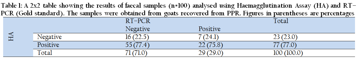
Table 1: ROC curve showing sensitivity and specificity of Haemagglutination Assay (HA) and RT–PCR for detection of PPRV antigen in faecal samples from PPR recovered goats
RESULTS
Of 100 samples tested, both tests agreed on the status of 38 samples; 22 were positive by both tests and 16 were negative by both tests. However, both tests showed a greater disagreement; of 77 samples that were positive by HA, only 22 were positive by RT–PCR. Similarly, HA declared 23 samples as negative; of these 23, only 16 were negative by RT–PCR (Table 1). These findings suggest a poor sensitivity and specificity of HA for detection of PPR antigen in faecal samples as is evident by the area (0.4711) under ROC curve (Figure 1).
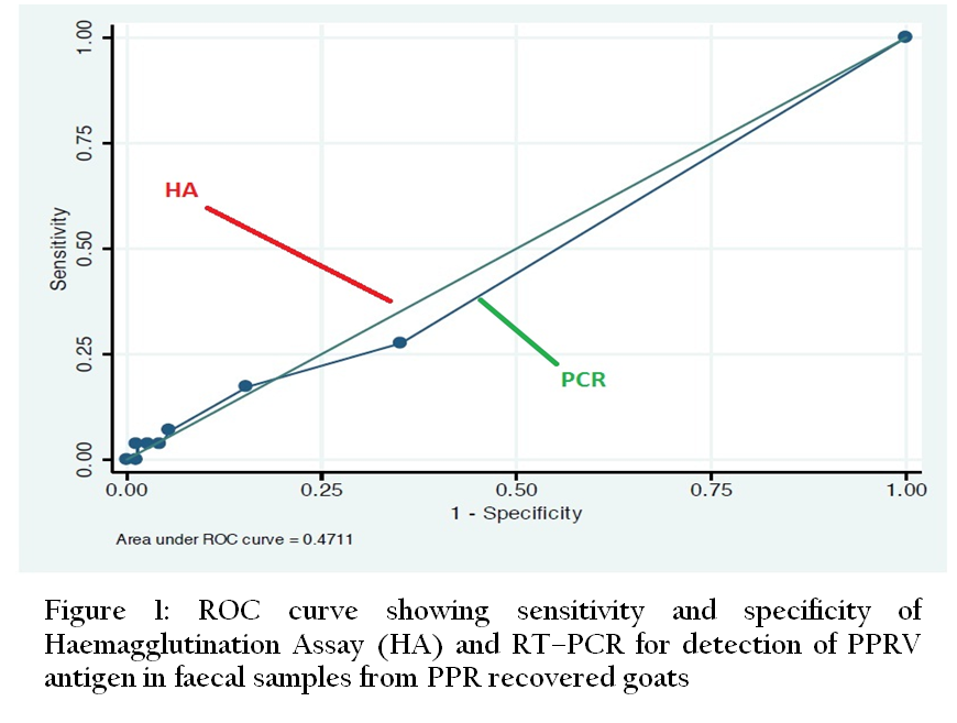
Figure 1: A 2x2 table showing the results of faecal samples (n=100) analysed using Haemagglutination Assay (HA) and RT–PCR (Gold standard). The samples were obtained from goats recovered from PPR. Figures in parentheses are percentages
DISCUSSION
Peste des Petits Ruminants (PPR) is one of the most devastating diseases of small ruminants. It is a highly transmissible and frequently fatal disease of sheep and goats (Gibbs et al., 1979). Various diagnostic tests such as HA, virus isolation, ELISA and RT–PCR are being used for the diagnosis of PPR. Some of these test (ELISA, RT–PCR and isolation) are expensive, require skilled personals and sophisticated laboratory. However, HA is a simple and cheap test and has been used for the diagnosis of PPR (Wosu, 1985; Ezeibe et al., 2004). For example, a study reported HA activity of porcine RBCs with extract from PPR infected mesenteric lymph nodes (Wosu, 1985). However, HA activity with chicken and human ‘O’ RBCs have also been observed (Ezebie et al., 2004). The chicken RBCs were used for HA in this study as they are economical and easily available.
HA test can be performed on samples obtained from live and dead animals. Ocular, nasal, oral discharges and faecal samples obtained from PPR affected live animals have been used for detection of PPR antigen using HA (Manoharan et al., 2005; Abubakar et al., 2012). Therefore, in case of an outbreak samples can be taken from live and dead animals to detect PPR antigen using HA.
To study the persistence of PPR in animals recovered from PPR various studies have used different samples. For example, a recent study where nasal, ocular and oral swabs were analysed using real time PCR, from goats challenged with PPR virus indicated persistence of PPRV for 40 days (Liu et al., 2013). However, a previous study has indicated detection of PPRV in faecal samples of PPR recovered goats for 12 weeks using HA which clearly indicates the persistence of PPRV for longer duration in faecal samples (Ezeibe et al., 2008). Therefore, faecal samples appear to be the sample of choice for persistence studies. However, Ezeibe et al., (2008) did not evaluate the specificity and sensitivity of HA when faecal samples were used to detect presence of PPR antigen. The results of our study (kappa = –1.5159) indicated no agreement between HA and RT–PCR which suggests that HA is a non–specific test when faecal samples are used for detection of PPR antigen. Therefore, HA is not a good choice for detection of PPR antigen in persistence studies and more specific tests such as RT–PCR should be used for this purpose.
ACKNOWLEDGEMENT
Authors are thankful to Agricultural Linkages Program (ALP) of PARC for financial assistance during the study.
REFERENCES
Abubakar M, Arshed MJ, Zahur AB, Ali Q Banyard AC (2012). Natural infection with peste des petitsruminants virus: a pre and post vaccinal assessment following an outbreak scenario. Virus Res. 167(1): 43 – 47.
http://dx.doi.org/10.1016/j.virusres.2012.03.018
PMid:22504337
Balamurugan V, Sen A, Venkatesan G, Yadav V, Bhanot V, Bhanuprakash V, Singh RK (2012). A Rapid and sensitive one step–SYBR green based Semi Quantitative Real Time RT–PCR for the Detection of peste des petits ruminants Virus in the Clinical samples. Virol. Sin. 27 (1):1 – 9
http://dx.doi.org/10.1007/s12250-012-3219-z
PMid:22270801
Barrett T, AC Banyard, Diallo A (2005). Molecular biology of the morbillivruses. In. Rinderpest and Peste des Petits Ruminants, Virus Plagues of Large and Small Ruminants. T. Barrett, P. P. Pastoret and W. P. Taylor (Eds.), Academic Press, Elsevier. 31 – 67.
PMCid:PMC3113918
Diallo A (2006). Control of peste des petits ruminants and poverty alleviation. J. Vet. Med. B. 53: 11 – 13.
http://dx.doi.org/10.1111/j.1439-0450.2006.01012.x
PMid:17123358
Dohoo I, Martin W, Stryhn H (2009) Veterinary Epidemiologic Research, VER Inc.; Canada, 2nd edition.
Ezeibe MCO, Wosu LO, Erumaka IG (2004). Standardisation of haemagglutinationtest for petse des petits ruminant's (PPR). Small Rumin Res. 51: 269 – 272.
http://dx.doi.org/10.1016/S0921-4488(03)00123-8
Ezeibe M, Okoroafor O, Ngene A, Eze J, Eze I, Ugonabo J(2008). Persistent detection of peste de petitsruminants antigen in the faeces of recovered goats. Trop Anim Health Pro. 40: 517 – 519.
http://dx.doi.org/10.1007/s11250-008-9128-3
PMid:18716908
FAO (1999). Report of the FAO– Japan Cooperative Project "Collection of Information on Animal Production and Health" prepared by Baldlock, C., T. Forman, B. Geering and B. Taylor, in collaboration with Infectious Diseases– EMPRES Group. FAO Animal Production and Health Paper 144: Rome, Italy.
Forsyth M, Barrett T (1995). Evaluation of polymerase chain reaction for the detection and characterisation of rinderpest and peste des petitsruminants viruses for epidemiological studies. Virus Res. 39: 151 – 163.
http://dx.doi.org/10.1016/0168-1702(95)00076-3
Gibbs E, Taylor W, Lawman M, Bryant J (1979). Classification of peste des petitsruminantsvirus as the fourth member of the genus Morbillivirus. Intervirology. 11:268 – 274.
http://dx.doi.org/10.1159/000149044
PMid:457363
Liu W, Wu X, Wang Z, Bao J, Li L, Zhao Y, Li J (2013). Virus excretion and antibody dynamics in goats inoculated with a field isolate of peste des petitsruminants virus. Transbound and Emerg Dis. 60: 63 – 68.
http://dx.doi.org/10.1111/tbed.12136
PMid:24589103
Manoharan S, Jayakumar R, Govindarajanand R, Koteeswaran A(2005). Haemagglutination as a confirmatory test for Peste des petitsruminants diagnosis. Small Rum Res. 59: 75 – 78.
http://dx.doi.org/10.1016/j.smallrumres.2004.12.002
Thrusfield M (2005). Veterinary Epidemiology, Blackwell publishing company UK, 3rd edition.
Wosu LO (1985). Agglutination of red blood cells by peste des petits ruminants (PPR) virus. Nigerian Vet. J. 14: 56 – 59.


