Research Journal for Veterinary Practitioners
Case Report
Research Journal for Veterinary Practitioners 2 (6): 111 – 112Surgical Repair of Ruptured Superior Rectus Muscles in a Six Months Old Male Calf
Shongsir Warson Monsang1*, Suvendu Kumar Behera2, Shahjahan Alam3
- College of Veterinary Sciences & A.H., R. K. Nagar, Tripura (W)
- College of Veterinary Sciences & A.H., Selesih, Aizawl, Mizoram
- Khalsa College of Veterinary & Animal Sciences, Amritsar, Punjab, Department of Teaching Veterinary Clinical Complex, Tripura (W)
*Corresponding author:warsonmonsang@gmail.com
ARTICLE CITATION:
Monsang SW, Behera SK, Alam S (2014). Surgical repair of ruptured superior rectus muscles in a six months old male calf. Res. j. vet. pract. 2 (6): 111 – 112.
Received: 2014–05–30, Revised: 2014–07–09, Accepted: 2014–07–10
The electronic version of this article is the complete one and can be found online at
(
http://dx.doi.org/10.14737/journal.rjvp/2014/2.6.111.112
)
which permits unrestricted use, distribution, and reproduction in any medium, provided the original work is properly cited
ABSTRACT
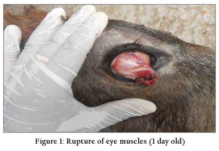
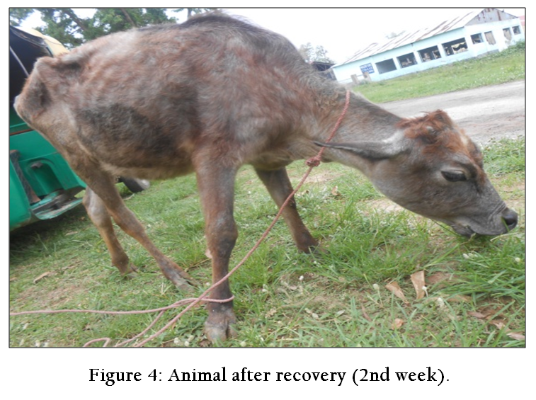
A case of ruptured superior rectus muscles was reported in a 6 months old male calf with the history of having previously fought with an adult horned bull a day before it was brought to clinic for treatment. On examination, the eye muscle was traumatized, soiled and found hanging from just below the upper eyelid covering the entire corneal region. The globe of the eye rotated downward and the calf was nervous and excited to touch. After proper restraint, auriculo-palpebral and retrobulbar nerve blocks were carried out to repair the ruptured eye muscles. The tissue healed completely on the 2nd week post surgery without any complications like wound infections, conjunctivitis, orbital hemorrhage or loss of vision.
Eye is one of the most delicate and sensitive parts of the body. Its front portion is protected by the eyelids and is surrounded by muscles and thick padding of retrobulbar fat within the orbit (Venugopalan, 2007). Muscles of the eye are designed to stabilize and move the eye. Its movements are controlled by muscles innervated by cranial nerves III, IV and VI. Six extraocular muscles that control the rotational movements of the eyeball consists of superior and inferior rectus muscles, medial and lateral muscles and superior and inferior oblique muscles each having different functions for the rotational movements of the eyeball. The superior rectus rotates the eye upwards but is opposed by inferior rectus. Similarly, the functions of medial rectus and superior oblique are opposed by lateral and inferior oblique muscles, respectively. If any damage can occur due to physical traumas like fight, fall or sticks, it might lead to serious complications impairing the rotation of the eyeball in normal directions. If timely approach is not constituted, infections may initiate leading to formation of pus, exudates, and finally loss of vision. Keeping this in mind, prompt surgical repair of ruptured superior rectus muscle which developed due to trauma is discussed in a six months old male local calf.
A case of ruptured superior rectus muscles in a 6 months old male calf was presented at the Khalsa College of Veterinary & Animal Sciences, Amritsar. History revealed that the calf fought with an adult horned bull before a day it was brought to the clinic. On examination, a soiled and bled tissue was seen hanging from just below the upper eyelid which covered the entire corneal region of the eye (Figure 1). The entire eyeball rotated downward with no visible structures of cornea and sclera unless it was manipulated with hand. The animal was nervous and excited to touch and all the clinical parameters were within the acceptable physiological limits. No any other abnormalities were observed.
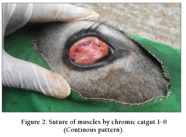
The calf was casted and controlled in lateral recumbency with the affected eye facing upwards. For desensitization of auriculo-palpebral and retrobulbar nerves, 2 ml xylocaine (LOX 2%) was used (Kumar, 2007). After desensitization of eyeball, the affected eye was thoroughly washed with isotonic solution (Normal saline, 0.09%) to remove soiled materials. With the help of mayo scissors, the dead tissue from the ruptured eye muscles were dissected and cleansed with saline solutions. The fresh muscle was sutured with the help of chromic catgut 2-0 (Figure 2) and the eyeball was brought back to the normal anatomical positions. Postoperatively, dexamethasone and gentamycin injections were mixed in 1:1 ratios and injected subconjunctivally to reduce inflammations. From second day, ointment Neosporin was applied over the suture line along with drops Ciprofloxacin daily for 2 weeks. On 2nd week, tissues were completely healed without any complications (Figure 3 & 4).

Eye affections such as conjunctivitis, keratitis, endophthalmitis, uveitis and panophthalmitis are commonly reported among animals (Gelatt, 1981; Slatter, 1981 and Severin, 1984).
Due to lack of awareness among the animal owners and the attending clinicians, the incidences of ocular problems are increasing few folds in the recent times (Tamilmahan et al., 2013). Traumatic eye injuries are not uncommon among domestic and wild animals as they are prone to fight amongst themselves to own their own territory. Small ailments may get worsen if the conditions are not responded and intimidated to proper care and treatment. Chronic eye damage is irreparable and may lead to complete loss of vision which might cause great loss to the owners and society. Therefore, prompt approach followed by accurate diagnosis and successful treatments forms the basis in limiting the economic loss and improving the health conditions of the animals to large extent.
In conclusion, a case of ruptured superior oblique muscles in a male calf is successfully treated by nerve blocks followed by postoperative antibiotics and anti-inflammatory drugs.
REFERENCES
Gelatt KN (1981). Veterinary Ophthalmology, Lea and Febiger, Philadelphia.
PMCid:PMC1903677
Kumar A (2007). Veterinary Surgical Techniques.Vikas Publishers Pvt. Ltd. New Delhi. 107 - 109.
PMid:17565807
Severin AC (1984). Veterinary Ophthalmology Notes, 2ndedn. Colorado State University.
PMCid:PMC498895
Slatter D (1981). Fundamentals of Veterinary Ophthalmology, B.W.Saunders, Philadelphia.
Tamilmahan P, Zama MMS, Rekha P, Muneeswaran NS, Karthik K (2013). A retrospective study of ocular occurrence in domestic animals: 799 cases.Vet. World 6(5): 274 - 276.
http://dx.doi.org/10.5455/vetworld.2013.274-276
Venugopalan A (2007): Essentials of Veterinary Surgery. 8th Edn. Oxford and IBH Publishing Co. Pvt. Ltd.New Delhi.p.280.


