Research Journal for Veterinary Practitioners
Research Article
Research Journal for Veterinary Practitioners 2 (5): 91 – 97Effect of Bovine Collagen Sheet with Fibroblast Cell in full Thickness Skin Wound Healing in Rat Model
Ashok Kumar Balwada, Sandeep Kumar*, Ashok Kumar Sharma, Naveen Kumar, Swapan Kumar Maiti
-
Division of Surgery, Indian Veterinary Research Institute, Izatnagar, Uttar Pradesh, India- 243122.
*Corresponding author:drskgvet@gmail.com
ARTICLE CITATION:
Balwada AK, Kumar S, Sharma AK, Kumar N, Maiti SK (2014). Effect of bovine collagen sheet with fibroblast cell in full thickness skin wound healing in rat model. Res. J. Vet. Pract. 2 (5): 91 – 97.
Received: 2014–05–05, Revised: 2014–05–27, Accepted: 2014–05–28
The electronic version of this article is the complete one and can be found online at
(
http://dx.doi.org/10.14737/journal.rjvp/2014/2.5.91.97
)
which permits unrestricted use, distribution, and reproduction in any medium, provided the original work is properly cited
ABSTRACT
The present study was conducted in eighteen wistar rats of either sex divided into two equal groups (n = 9) to evaluate the healing potential of bovine collagen sheet with fibroblast cell. Full thickness skin wound (20 X 20 mm2) was created on dorsal region of thorax of adult wistar rats. Topical application of paraffin gauze over the wound acted as control (Group -A). The second group (Group - B) animals were treated with collagen sheet and fibroblast cell. Clinical observations, gross observation, exudation, granulation tissue, peripheral swelling and the contraction rate of the wounds were recorded on days 0, 3, 7, 14, 21, 28 post surgery at different interval in both groups. Early granulation tissue formation with reduced exudation and peripheral swelling was observed in treatment group-B and proved better than that of group - A. The percent of wound contraction was similar to gross findings, however, in group - B, complete wound healing was observed on day 17 - 18, but in group - A (control), healing was completed by day 27 - 28. The large wound that cannot be corrected by conventional surgical procedures can be corrected by these cells and graft material to ensure early wound healing. This study will help to heal large skin wound in animals.
INTRODUCTION
Skin wounds are physical injuries that result in an opening or breaking of the skin. Disruption of skin generally leads to increase fluid loss, infection, hypothermia, scarring and compromised immunity (Wysocki, 1999). Wound healing occurs in multiple stages over several days by increased migration and proliferation of cells, as well as by formation and contraction of granulation tissue (Aggarwal et al., 2006). Healing involves migration, infiltration, proliferation, and differentiation of several cell types like keratinocytes, fibroblasts, endothelial cells, macrophages, and platelets which were culminate in an inflammatory response, the formation of new tissue and wound closure (Barrientos et al., 2008).
Collagen is an abundant structural protein in all animals. The mechanisms by which heterologous collagen matrices assist wound healing include hemostatic, spa¬tial, nutritional, and chemotactic effects. Denatured bovine collagen contains more fibronectin binding sites, and at least in-vitro, increased fibronectin binding stimulates migration of fibroblasts (Leipziger et al., 1985). Large number of natural polymers including fibrin, hyaluronic acid, fibrinogen and collagen have been used as biologically superior dermal substitutes with collagen showing the most potential (Powell and Boyce, 2006).
Fibroblasts not only produce and organize the extracellular matrix of dermis but also communicate with each other playing a crucial role in regulating skin physiology (Sorrell and Caplan, 2004). Circulating fibrocytes represent an important source of fibroblasts, particularly during healing of extensive burn wounds where it may be difficult for fibroblasts to migrate from the edges of the wound (Yang et al., 2002). With this background, the present study was designed with the objective to evaluate healing potential of bovine collagen sheet with fibroblast cell for full thickness skin wounds in wistar rats.
MATERIALS AND METHODS
Eighteen clinically healthy adult wistar rats of either sex were used in this study All the animals were procured from Laboratory Animals Research (LAR) section of Indian Veterinary Research Institute, Izatnagar, Uttar Pardesh, India.
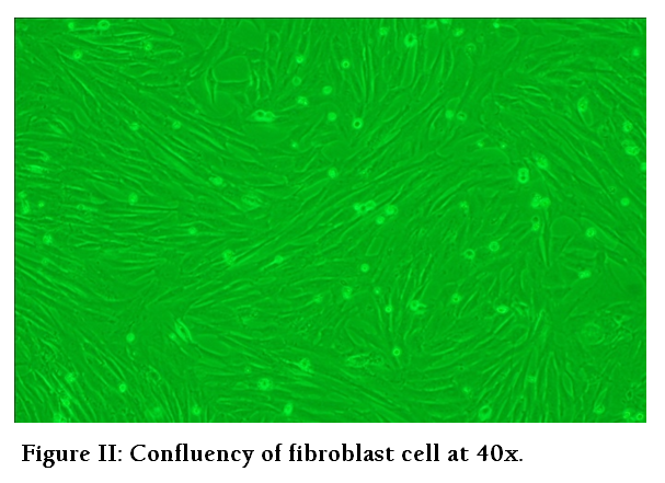
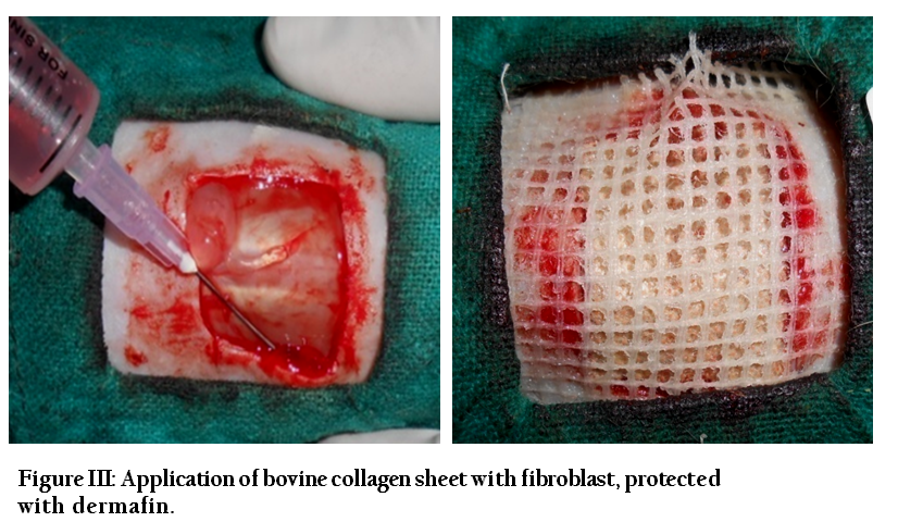
Each animal was caged individually and provided free access to water and a standard diet. All the animals were maintained under standard conditions. All the animals were acclimatized to approaching, handling and animal house conditions for a period of 10 - 15 days prior to the study. They were randomly divided into two groups, viz , Group A and Group B of nine animals each (table 1). Preparation of primary mouse embryo fibroblasts culture was done by pregnant female mouse embryo which was sacrificed at 14th day of gestation (figure 2). Bovine collagen sheet (Skin Temptm) is available in market to heal wound of various type which has no immunological property.
Surgical Procedure
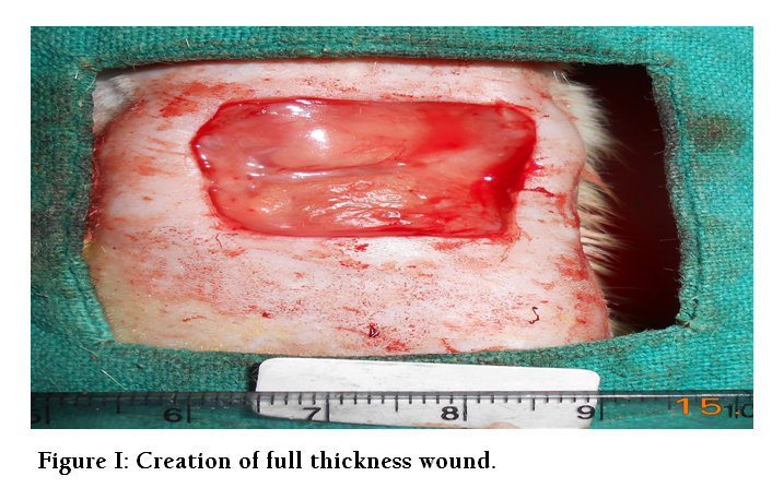
The rats were anaesthetized using xylazine at dose rate of 6 mg/kg body weight and ketamine at dose rate of 60 mg/kg body weight, administered intramuscularly (Gangwar et al., 2013). The animals were placed in sternal position on the operative table. The animal was aseptically prepared starting from the caudal part of the shoulder on the dorsum to the caudal part of the last rib. On the prepared area, a 20x20 mm2 full thickness skin defect was created aseptically (figure 1). The animals in each group were treated as mentioned in Table 1. The wounds were observed for 28 days or till complete healing of wound.
The efficacy of the wound healing was measured on the basis of following parameters.
Clinical Observations
General behavioral changes: Feeding pattern and general behavioral changes in all the animals was observed daily during the observation period
Rectal temperature: Rectal temperature was recorded daily upto 7 post-operative days in all the animals.
Gross observations
The wound site was examined grossly on days 0, 3, 7, 14, 21 and 28 or till completion of healing for the evaluation of following parameters:
Measurement of wound area: Wound area was measured at day 0, 3, 7, 14, 21 and 28 or till completion of healing.
Wound contracture: Wound contraction was measured on 0, 3, 7, 14, 21, and 28 postoperative day as a percentage reduction in wound area. Progressive decrease in the wound area was monitored periodically by tracing the wound margins on a tracing paper and the area was assessed by using a graph paper. The mean wound area and the mean percentage of wound contraction for each interval were calculated for each group (Bohling et al., 2004).
| % contraction = 100 - |
Total wound area on dayn |
X 100 |
| Original wound area on dayo |
Where n is: 0,3,7,14,21 and 28 day
Exudation: The degree of exudation at the site of repair was graded on 1-4 scale as per standard method (Bigbie et al., 1991). 1= None (Apparently dry wound); 2 = Mild exudates (Wound is moist, no oozing on pressing the wound); 3 = Moderate exudates (Wound is moist, slight oozing on pressing the wound); 4= Extreme exudates (Exudates is visible and pressure lead to extensive exudation).
Evaluation of granulation tissue: Granulation tissue evaluation was graded on 1-4 scale as per standard method (Bigbie et al., 1991) (1= Granulation tissue depressed below the skin edge; 2= Granulation tissue proliferated to the level of skin edge; 3= Granulation tissue elevated above skin edges; 4= Granulation tissue elevated above skin edges, projecting over the border of epithelium).
Colour of granulation tissue: Colour of the wound depicts the status of healing and was scored as: 1=Pale yellow; 2=Pale red and 3=Pink.
Time of appearance of granulation tissue: It was recorded as the first day when the granulation tissue was observed
Histopathological Observations
The biopsy specimens from the site were collected on days 7, 14, and 28 for the histo-pathological evaluation. The sections were stained with Hematoxylin and Eosin for evaluation of inflammation, epithelialization and neovascularization. The H&E sections were evaluated microscopically by using histological scoring system (Ghamsari et al., 1996). Special staining for collagen fibers was done by using Masson’s Trichrome stain.
Statistical Analysis
The data was analysed using the suitable Statistical Program for Social analysis (SPSS) software version 16.0 for windows (Snecador and Cochran, 1989). One way ANOVA (Analysis of Variance) and Duncan multiple range test (DMRT) were used to compare the means at different time intervals among different group. Student paired‘t’ test was used to compare the mean value at different time interval with their base value in each group. The subjective data generated from the scoring of various parameters were analysed using Kruskal Wallis test.
Ethical Permission
Necessary permission was undertaken from the Institute Animal Ethics Committee (IAEC), Indian Veterinary Research Institute, Izatnagar (U.P) India to conduct this research work on small laboratory animals.
RESULTS
The animals of both groups remained dull on first postoperative day and assumed a hunched back posture, while resting in their cages. They started taking feed and water partially within 24h after surgery. In all the animals feed and water intake became normal by 3rd postoperative day. The animals of group B started resting on dorsal recumbency from day 12, whereas, in group A it was from day 21.
Significant (P < 0.05) increase in rectal temperature for first 3 days post surgery was recorded in both groups. The hyperthermia recorded in the early postoperative period started returning to normal as soon as the animals recovered from surgical stress. On day 3, temperature was significantly increased within group A.
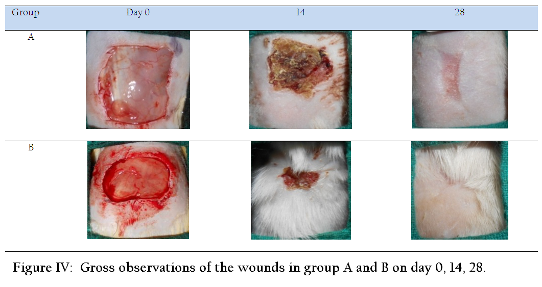
Wound area decreased gradually as the healing progressed. Control wound healed completely by 27- 28 days leaving a large scar indicating the existence of severe contraction. Group B took 17-18 days for complete healing and it was with minimum contraction leaving a little scar than control group (figure 4). Mean ± SE of the total wound area (mm2) of the skin wounds at different time interval are presented in table 2.
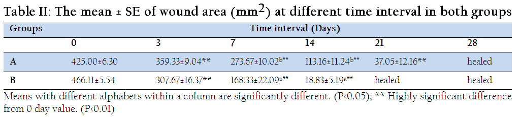
On day 7 and 14, wound areas were significantly (P < 0.05) decreased in group B as compared to group A. Original wound was created as 2 cm X 2 cm dimensions, however, almost all the wounds expanded to various extents and had an area greater than the 4cm2. Significant (P < 0.05) increase in percent of contraction was observed in both the groups during the observation period. On day 7 and 14, wound area was significantly (P < 0.05) contracted in group B as compared to group A. Mean ± SE of the total wound area contraction at different time interval are presented in table 3.

Table 3: The mean± SE of the percentage contraction of the wound area (mm2) in both groups at different time interval
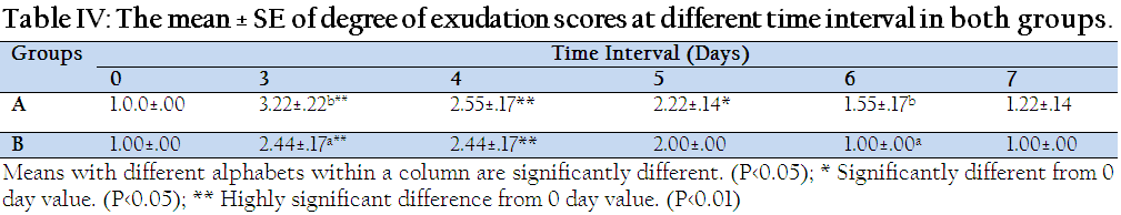
Moderate exudation at the site was observed upto day 3-4 in all the treated as well as control wounds. As the healing progressed the inflammation subsided gradually and therefore, no exudation was observed on day 7 and onwards except in group A. But in animals of group A showed a significantly higher (P < 0.001) values of exudation on day 3 and 6 respectively as compared to group B (table no 4).
Granulation tissue was first observed in group B on day 2-3 and in group A on day 4. On day 13-14 granulation tissue was almost fully covered the wound in group B, whereas, wound area was covered by the granulation tissue in the control group (A) on day 21-22. Animals of group B showed significantly higher granulation tissue values (P < 0.05) on day 14 and day 21 as compared to group A (Table 5). The time (day) of appearance of granulation tissues seen grossly was significantly (P < 0.05) less in group B than group A (table 6).
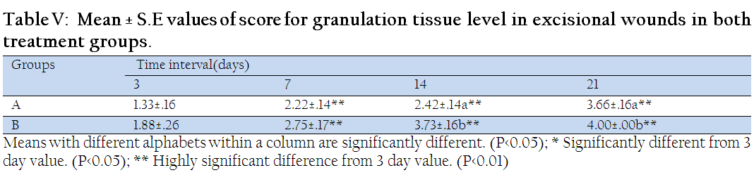
Table 5: Mean ± S.E values of score for granulation tissue level in excisional wounds in both treatment groups
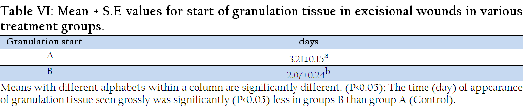
Table 6: Mean ± S.E values for start of granulation tissue in excisional wounds in various treatment groups
Creation of excisional wounds resulted in variable extent of bleeding and formation of clot. The clot was dried and formed a cover over the wounds, which rendered the evaluation of colour of granulation tissue difficult in some of the animals. The scab, which covered the underlying granulation tissue, was detached in majority of the wounds before 18 postoperative days in group B. At this junction the classical ‘shinny, beefy, red’ to pink granulation tissue indicating healthy healing progress was evident. The scar became paler with passage of time, which indicated stage of maturation. The maturation was much earlier in group B than group A
Histopathological observation
Group A: On day 7, the necrosed surface was detached and the edges of the wound had partial epitheliazation. The granulation tissue (fibroplasia and neovascularization) was severe and has less dense, thin and worst arranged collagen fibers and presence of mainly mononuclear cells (lymphocytes and macrophages) and less number of neutrophils. On day 14, severe proliferation of fibroblasts and neovascularization was observed. On day 28, a high degree of collagen deposition was observed with complete epitheliazation but arrangement of collagen fiber in the wound area was however unorganized. (figure 5 and 6)
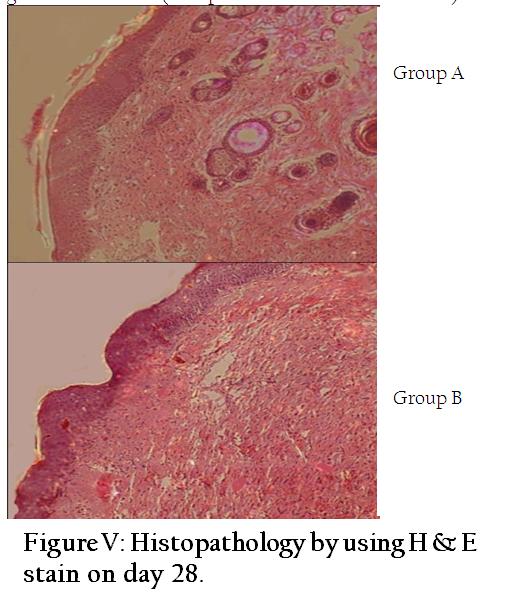

Group B: By day 7, fibroblast proliferation became more prominent, the necrosed surface was detached and the edges of the wound had partial epitheliazation. The granulation tissue (fibroplasia and neovascularization) was mild and had very less dense, thin and worst arranged collagen fibers and presence of mainly mononuclear cells (lymphocytes and macrophages) and less number of neutrophils shows excessive inflammation. By day 14, epitheliazation was partial, inflammation decreased, denser, thick and better arranged collagen fibers. By day 28, epitheliazation was almost completed covering the granulation tissue. Hair follicles were also observed with arranged collagen fiber.
DISCUSSION
In the present study, one 20x20 mm2 square full-thickness skin (dorsal thoracic region) wounds were created on each of 18 adult wistar rats of either sex under standard anaesthetic protocol to evaluate the wound healing potential of bovine collagen sheet and fibroblast cell in full thickness wounds in rats. This study can help in future to heal large skin wounds in human and animal also.
The animals of group B started resting on dorsal recumbency from day 12, whereas, in group A, it was from day 21.These results were in accordance with other workers (Kaarthick, 2011). It indicates the normalcy and healing was progressing well in the experimental group. Dullness, depression and partial anorexia observed in the immediate post operative period (1-2 days) may be attributed to surgical trauma (pain) and inflammation at the site of reconstruction (Gangwar et al., 2006).
Significant (P < 0.05) increase in temperature for first 3 days post surgery was recorded in both groups which may be due to foreign body reaction, surgical trauma and stress to the animals following skin wound. Pyrexia of variable degree in postoperative days has also been reported after the repair of full- thickness skin defects with different materials in rabbits (Gangwar et al., 2006; Purohit, 2008) and rats (Kaarthick, 2011). The increase in temperature may also be attributed to the action of endogenous or leukocytic pyrogen produced by granulocytes, monocytes and macrophages (Atkins, 1960).
Wound contraction has been used to monitor wound healing. Wound area decreased gradually as the healing progressed. The most important cell, the fibroblast attained the peak approximately on day 7 from injury and is responsible for initiating the angiogenesis, epithelialisation and collagen formation. Control wound healed completely by 27 - 28 days leaving a large scar indicating the existence of severe contraction. Group- B took 17 - 18 days for complete healing, but it was with minimum contraction leaving a little scar than control group. Wound contraction is the centripetal displacement of the wound edges that facilitates its closure after trauma. This process is carried out by myofibroblasts that contain α-actin from smooth muscle and is mediated by contractile forces produced by granulation tissue from wound (Neagos et al., 2006). Wound healing rate is defined as the gross epithelialisation of the wound bed. Wound contraction was assessed by percent retention of the original wound area (Schalleberger et al., 2008).
Moderate exudation at the site was observed upto day 3 - 5 in both groups. Exudation may be due to inflammatory reaction at the site in response to surgical trauma. As the healing progressed the inflammation subsided gradually and therefore, no exudation was observed on day 7 and onwards except in group A (control). A significant decrease in exudation after full-thickness wounds treated with small intestinal sub mucosa compared to untreated wounds in rat model was reported by (Kim et al., 2005). According to (Wang et al., 2007), a reverse correlation was detected between the survival area of the skin graft and the degree of exudation of the graft bed.
Granulation tissue was first observed in group B on day 2-3 and in group A on day 4 post surgery. On day 13 - 14, granulation tissue was almost fully covered the wound area in group B, whereas, wound area was covered by the granulation tissue in the control group (A) on day 21 - 22. Granulating tissue can generally be divided into two types, healthy and unhealthy granulating tissues. It is well known that healthy granulating tissue develops only in the absence of foreign bodies such as bacteria, debris, and so forth. Formation of healthy granulating tissue which is closely related to angiogenesis is a very important factor in wound healing (Clark and Denver, 1985). Evidence also sug¬gests that bovine collagen supplies some early nutri¬tional needs of granulation tissue (Motta et al., 1983). Similar finding was also found in this study in group B.
The color of the wound changed from white to dark brown and finally dark on subsequent time intervals. There was not so much difference in wound area of group A and B from day 0 to 3. On day 7; a layer of scar was present on both groups. There was drastic reduction in wound area from day 7 to day 14 in group B in comparison to group A. On day 17 - 18 healing was complete in group B leaving a very little scar, whereas, in control group (A) healing was complete in 27 - 28 days with abundant scar. Similar findings also have been reported after the repair of full thickness skin defects in rabbits (Purohit, 2008) and in rats (Kaarthick, 2011).
In the present study the histopathological samples collected on day 7, 14, and 28 were subjected to Hematoxylin and eosin staining. Masson’s Trichrome staining was also done to assess the collagen formation. On day 7, post implantation, moderate to severe inflammation was present in all the groups; however, it was less in groups- B. The early control of inflammation, as in case of groups-B might facilitate the progress to the next phase of wound healing. On day 14, epithelialisation and neovascularization was faster in group-B as compared to groups-A. On day 28, the collagen fiber arrangement was almost similar to normal skin in group-B. In group-B, hair follicle and skin glands could also be seen as in case of normal skin. Similar findings were also observed by Perme et al. (2008).
Full thickness skin wound healing occurs by granulation tissue formation, contraction and epithelialisation (Fossum et al., 2007). Epithelialisation occurs by migration of undamaged epidermal cells from the wound margins across the granulation bed (Swaim and Henderson, 1990). Exogenous collagen supplementation enabled faster migration of cells that are involved in cutaneous wound healing. Since the exogenous collagen is molecular in nature (Nithya et al., 2003) and supplies endogenous collagen in-vivo, it readily integrates with the wound tissue and facilitates the attachment, migration and proliferation of cells on the wound site (Judith et al., 2010). Ishikawa et al. (1997) demonstrated that fibroblasts can be used to produce a thin three dimensional sheet of ECM material in-vitro. The presence of living fibroblasts in the dermal substitute leads to rapid wound healing as compared to acellular matrix alone (Mirza et al., 2009).
We are thankful to Dr.V.P.Vadodaria, Dean and Principal, College of Veterinary Science and AH, SDAU, Sardarkrushinagar and Dr. R.K.Singh, Head and Station–In–Charge, IVRI, Mukteswar for providing necessary facilities for present work. Help and cooperation provided by field veterinarians at the time of collection of clinical and post mortem samples are highly acknowledged.
CONCLUSION
Bovine collagen sheet with fibroblast cell had better healing potential in comparison to standard dressing material (dermafin) for repair of full thickness skin wounds in rat model.
ACKNOWLEDGEMENT
Authors are highly thankful to the Director, Indian Veterinary Research Institute, Izatnagar-243122, Uttar Pradesh, India for providing necessary facilities.
CONFLIC OF INTEREST
The authors declare that they have no conflicts of interests with respect to their authorship or the publication of this article.
REFERENCES
Aggarwal C, Britton ZT, Alaseirlis DA, Li Y, Wang JHC (2006). Healing and normal fibroblasts exhibit differential proliferation, collagen production, α-SMA expression, and contraction. Ann. Biomed. Eng. 34: 653 – 9.
http://dx.doi.org/10.1007/s10439-006-9090-z
PMid:16568347
Atkins E (1960). Pathogenesis of fever: Physiol. Rev. 40: 58 – 646.
Barrientos S, Stojadinovic O, Golinko MS, Brem H, Tomic-Canic M (2008). Growth factors and cytokines in wound healing. Wound Repair Regen. 16: 585 – 601.
http://dx.doi.org/10.1111/j.1524-475X.2008.00410.x
PMid:19128254
Bigbie RB, Schumacker J, Swaim SF, Purohit KC, Wright JC (1991). Effect of amnion and yeast cell derivate on second intention healing in horses. Am. J. Vet. Res., 52: 1376.
PMid:1928923
Bohling MW, Henderson RA, Swaim SF, Kincaid SV, Wright JC (2004). Cutaneous wound healing in the cat: A Macroscopic description and comparison with cutaneous wound healing in the dog. Vet. Surg. 33: 579 – 587.
http://dx.doi.org/10.1111/j.1532-950X.2004.04081.x
PMid:15659012
Clark RAF, Denver MD (1985). Cutaneous tissue repair: Basic biologic considera¬tions. J. Am. Acad. Dermatol. 13: 701 - 725.
http://dx.doi.org/10.1016/S0190-9622(85)70213-7
Fossum TW, Edlund CS, Johnson AL, Schulz KS, Seim HB, Wasard MD, Baer A, Carroll GL (2007). Surgery of Integumentary System. Manual of Small Animal Surgery Mosby. pp. 159 - 175.
Gangwar AK, Sharma AK, Kumar N, Maiti SK, Gupta OP, Goswami TK, Singh R (2006). Acellular dermal graft for repair of abdominal wall defects in rabbits. J. S. African Vet. Assoc. 77: 79 - 85.
http://dx.doi.org/10.4102/jsava.v77i2.349
PMid:17120624
Gangwar AK, Naveen Kumar, Sharma AK, Devi S, Negi M, Shrivastava S, Mathew D, Remya V, Soanal, Arundeep PS, Maiti SK, Kumar V, Kaarthick DT, Kurade NP, Singh R (2013). Bioengineered acellular dermal matrix for the repair of full thickness skin wounds in rats. Trends Biomater. Artif. Organs. 27: 67 - 80.
Ghamsari SM, Acorda JA, Taguchi K, Abe N, Yamada H (1996). Evaluation of wound healing of the teat with and without low level laser therapy in dairy cattle by laser doppler flow metry in comparison with histopathology, tensiometry and hydroxyproline analysis. Br. Vet. J. 152: 583-591.
http://dx.doi.org/10.1016/S0007-1935(96)80010-8
Ishikawa O, Konodo A, Okada K, Miyachi M, Furumura M (1997). Morphological and biochemical analyses on fibroblast and self produced collagens in a novel 3-D culture. Br. J. Dermatol. 136: 6 - 11.
http://dx.doi.org/10.1111/j.1365-2133.1997.tb08738.x
http://dx.doi.org/10.1046/j.1365-2133.1997.d01-1134.x
PMid:9039287
Judith R, Nithya M, Rose C, Mandal AB (2010). Application of a PDGF-containing novel gel for cutaneous wound healing. Life Sci. 87: 1- 8.
http://dx.doi.org/10.1016/j.lfs.2010.05.003
PMid:20470785
Kaarthick DT (2011). Repair of cutaneous wounds using acellular diaphragm and pericardium of buffalo origin seeded with in-vitro cultured mouse embryonic fibroblasts cells in rat model. MVSc. Thesis submitted to Deemed University, I.V.R.I., Izatnagar, Bareilly (UP). 243 -122.
Kim MS, Hong KD, Kim SH, Kim SH, Lee MS, Jang WY, Khang G, Lee HB (2005). Preparation of porcine small intestinal sub mucosa sponge and their application as a wound dressing in full-thickness skin defect of rat. Intl. J. Biol. Macromole. 36: 54 – 60.
http://dx.doi.org/10.1016/j.ijbiomac.2005.03.013
PMid:15939465
Leipziger LS, Glushko V, DiBernardo B (1985). Dermal wound re¬pair: role of collagen matrix implants and synthetic polymer dress¬ings. J. Am. Acad. Dermatol. 12: 409 - 19.
http://dx.doi.org/10.1016/S0190-9622(85)80004-9
Mirza R, Dipietro LA, Koh TJ (2009). Selective and specific macrophage ablation is detrimental to wound healing in mice. Am. J. Pathol. 175: 2454 - 2462.
http://dx.doi.org/10.2353/ajpath.2009.090248
PMid:19850888 PMCid:PMC2789630
Motta G, Ratto GB, DeBarbieri A (1983). Can heterologous collagen enhance the granulation tissue growth? An experimental study. J. Surg. Sci. 13: 101 – 8.
Neagos D, Mitran V, Chiraku GR, Ciurab Lanku C, Stan C, Cimpean A, Iordachescu D (2006). Skin wound healing in a free floating fibroblast populated collagen lattice model. Romanian J. Biophy. 16: 157 - 168.
Nithya M, Suguna L and Rose C(2003). The effect of nerve growth factor on the early responses during the process of wound healing. Biochemica et Biophysica acta., 1620: 25-31.
Perme H, Sharma AK, Kumar N, Singh H, Dewangan R and Maiti SK (2008). In-vitro biocompatibility evaluation of cross-linked cellular and acellular bovine pericardium. Trends Biomater. Artif. Organs. 23(2): 66-75.
Powell HM, Boyce ST (2006). EDC crosslinking improves skin substitute strength and stability. Biomaterials, 27: 5821 - 5827.
http://dx.doi.org/10.1016/j.biomaterials.2006.07.030
PMid:16919327
Purohit S (2008). Biocompatibility testing of acellular dermal grafts in a rabbit model: An in-vitro and In-vivo study. Ph.D. Thesis submitted to Deemed University, I.V.R.I., Izatnagar, Bareilly (UP). 243 - 122.
Schalleberger SP, Stanley BJ, Hauptman JG, Steficek BA (2008). Effect of porcine small intestinal submucosa on acute full-thickness wounds in dogs. Vet. Surg. 37: 515 – 524.
http://dx.doi.org/10.1111/j.1532-950X.2008.00398.x
PMid:19134100
Snedecor GW, Cochran WG (1989). Statistical Methods, 8th Ed. Oxford and IBH Publishing Company, New Delhi.
Sorrell JM, Caplan AI (2004). Fibroblast heterogenicity; more than skin deep. J. Cell Sci., 117: 667 - 675.
http://dx.doi.org/10.1242/jcs.01005
PMid:14754903
Swaim SF, Henderson RA (1990). Small Animal Wound Management, Lea and Febiger, Philadelphia, London. pp. 1 - 33.
Wang Y, Chen X, Armstrong MA, Gang L (2007). Survival of bone marrow derived mesenchymal stem cells in a xeno-transplantation model. J. Ortho. Res. 25: 926 - 932.
http://dx.doi.org/10.1002/jor.20385
PMid:17415789
Wysocki AB (1999). Skin anatomy, physiology, and pathophysiology. Nurs. Clin. N. Amm. 34: 777 - 97.
PMid:10523436
Yang L, Scott PG, Giuffre J, Shankowsky HA, Ghahary A, Tredget EE (2002). Peripheral blood fibrocytes from burn patients: identification and quantification of fibrocytes in adherent cells cultured from peripheral blood mononuclear cells. Lab. Invest. 82: 1183 – 92.
http://dx.doi.org/10.1097/01.LAB.0000027841.50269.61
PMid:12218079


