Research Journal for Veterinary Practitioners
Review Article
Research Journal for Veterinary Practitioners 2 (5): 84 – 90Epidemiology and Management Strategies of Johne’s disease in Endemic Situations
Muhammad Tanveer Munir1, Anum Rafia Munir2, Murtaz ul Hasan1, Muhammad Abubakar3*
- Faculty of Veterinary and Animal Sciences, PMAS–Arid Agriculture University, Shamasabad, Rawalpindi, Pakistan
- Chemistry department, Government College University Lahore, Pakistan
- National Veterinary Laboratory, Park Road, Islamabad, Pakistan
*Corresponding author:mabnvl@gmail.com
ARTICLE CITATION:
Munir MT, Munir AR, Murtaz ul Hassan, Abubakar M (2014). Epidemiology, diagnosis and control options of johne’s disease in endemic situations. Res. J. Vet. Pract. 2 (5): 84 – 90.
Received: 2014–05–12, Revised: 2014–05–31, Accepted: 2014–05–31
The electronic version of this article is the complete one and can be found online at
(
http://dx.doi.org/10.14737/journal.rjvp/2014/2.5.84.90
)
which permits unrestricted use, distribution, and reproduction in any medium, provided the original work is properly cited
ABSTRACT
Mycobacterium paratuberculosis the subspecies of M. avium, effects wide range of animals including domestic cattle, sheep, goats, buffaloes, camelids and wild ruminants resulting in progressive and chronic enteritis known as Johne’s disease (paratuberculosis). Clinically sick animals show emaciation, diarrhea and eventually death but the risk is that mostly they don’t show clinical sign still can shed bacteria in feces and milk. Organism spread in the animal body through blood and lymph nodes to multiple internal organs. It is economically very important disease in livestock because effected livestock is recommended to be culled due to high treatment costs. Etiology, host range, immunology, epidemiology, stages/ forms, clinical signs, diagnostic tools and treatment have been discussed with special reference to endemic situations. Strategies to control this disease include improved management practices, testing and culling and vaccination. Modifications in management practices is not an easy job and so is the case with testing and culling; vaccine on the other hand is the simple practice but it is not usually practiced by farmers because lack of knowledge/awareness in herdsmen and availability of vaccine.
INTRODUCTION
Johne’s disease (paratuberculosis) is a wasting, chronic granulomatous enteritis (Rosseels and Huygen, 2008) affecting domestic cattle, buffalo, goats, sheep, camels, wild ruminants, some mono–gastric animals and birds (Beard et al., 2001; Bennett et al., 2012). Mycobacterium paratuberculosis, the causative agent of paratuberculosis or Johne’s disease (JD) is a subspecies of Mycobacterium avium, Gram positive, slow growing acid fast bacillus (Gwozdz, 2010; Ayele et al., 2001). After first report of JD in 1895, this organism was isolated in 1910 (Twort, 1910). Strains of this microbe are: I (sheep), II (cattle) and III (intermittent) (Gwozdz, 2010). This progressive and chronic infection is mostly unresponsive to treatment (Ansari et al., 2013).
Affected animals have normal appetite but are weak, diarrhea (bubbly and greenish) is evident in some species and eventually death occurs (Gwozdz, 2010). Effected animals shed organism in milk and feces, following ingestion, this organism spread through blood and lymph vessels affecting visceral organs including male and female reproductive organs (Ayele et al., 2001). Economic effect of disease is considerable as there are losses to livestock industry. Relation of JD with Crohn’s, juvenile sarcoidosis (Blau syndrome), autoimmune thyroiditis, autoimmune diabetes, and multiple sclerosis have caused important issue of public safety (Dow, 2012).
This article tends to review the epidemiology and diagnosis of Mycobacterium paratuberculosis in endemic regions along with adopted control strategies and possible preventive measures in national and international scenario.
Epidemiology
The disease has been reported worldwide and is becoming more common, increasing range of animal species (Vansnick, 2004); but still there are parts of world where it is not endemic (Okuni, 2013). Some Australian states and Sweden are proven to be free of this disease. In ruminants, dairy cattle are most prone to disease; in USA herd prevalence has been reported 91.1% (Lombard et al., 2013), in Chile 28–100% (Kruze et al., 2013).
Co–infection of paratuberculosis with other diseases has been reported, e.g., brucellosis (Singh et al., 2013a). Prevalence of Johne’s disease in goats has been reported from all over the world with prevalence of 7.9% in Republic of Cyprus (Liapi et al., 2011), 76.9% USA (Manning et al., 2002), 74.3%Chile (Salgado et al., 2007), 62.9% France (Mercier et al., 2010), 79.4 %India (Singh et al., 2013) and 44.1% Argentina (Fiorentino et al., 2012).
There are many reports of paratuberculosis from Pakistan. Sikandar et al., (2011) reported 11.19% (Cattle: 6.67%, Buffaloes: 12.5%) confirmed cases for paratuberculosis in 134 suspected samples. It has also been seen in ovine species (Sikandar et al., 2013). Abbas et al. (2011) tested samples in 3 semen production units in Punjab, Pakistan and found almost 20% positive breeding bulls and almost 33 % positive teaser bulls.
Host Species
Mycobacterium avian sub-specie paratuberculosis (MAP) effect wide range of animals mostly ruminants. Cattle, buffaloes, sheep, and goats are the most effected specie of domestic animals (Rosseels and Huygen, 2008; Singh et al., 2013; Khan et al., 2010). In wild animals almost all ruminants get infected including giraffe, deer (de Lisle and Collins 1995) and wild goats. Camels are also prone to this disease (Al–Ghamdi, 2013). In non–ruminants it have seen to cause disease in horse, badgers, bears, rabbit (Greiget al.,1999), cats, armadillos, opossums, mouse, rats, macaques, stoats, pigs, weasels, crow and fox (Beard et al., 2001; Hutchings et al., 2010). Johne’s disease have got attention due to relation with Crohn’s disease (Behr and Kapur, 2008; Over et al., 2011; Ayele et al., 2001). Calves get infection in their early six months of age or in–utero. Young animals are more susceptible, most probably because they have immature cellular immunity. Age relation with MP infestation has been proven in some studies (Windsor and Whittington, 2010; Thakur et al., 2013).
Transmission
Mycobacterium paratuberculosis is a contagious infection. Affected animals shed organism in feces and milk (Hines et al., 2007; Seyyedin et al., 2010; Hasonova et al., 2009). Contaminated food, water sources, vehicles and other equipment may be a source of transmission from one herd to other. Male animals may carry MP in accessory reproductive organs and to some extent in semen. Embryo from infected animals may carry infection and will transmit it when transplanted in other animals. Calves may get infected by the colostrums they getting from effected cow (Stabel, 2008); calves have been reported to shed microbe in feces at 5 months of age (Hasonova et al., 2009). Humans may get MP from raw milk, meat and contact with animals (Eltholth et al., 2009; Alluwaimi, 2007).
Immunology
Mycobacterium paratuberculosis is an intracellular pathogen. Following the infection the body responds by opposing it with T helper 1 (Th1) (Wu et al., 2007; Wadhwa et al., 2013) that produce interferon gamma and IgG2 (Nielsen, 2008; Begg et al., 2011). In later stages of infection Th2 (humoral) response (Wadhwa et al., 2013) may be present but it doesn’t prove sufficient to check infection (Stabel, 2000). Lybeck et al., (2011) reported shedding of MP in feces in effected goats before interferon gamma which usually preceded humoral immune response.
Stages and Forms
In cattle, paratuberculosis is classified into three stages I (early infection), II (subclinical), and III (clinical) (Wadhwa et al., 2013). At stage I, infection progresses without shedding adequate bacteria in feces. In stage II, the number of bacteria increases in intestinal mucosa and fecal shedding is intermittent. At stage III, which is a terminal stage bacterial load increases and clinical signs appear; animal suffer from chronic diarrhea, weight loss decreased production and anemia (Vansnick, 2004).
Clinical Signs
As paratuberculosis is a chronic disease so, mild and progressive signs are seen in animals. Milk production decreases in all lactating animals and body condition becomes poor. Weight loss and emaciation becomes evident depending upon stage of infection. Diarrhea is reported in some animals that is intermittent at initial stages but tend to persist in later stages. Reduced ruminal motility is also reported in effected goats (Lybeck et al., 2011)
Blood Parameters
Lybeck et al., (2011) reported decrease in hematochrit, hemoglobin and albumin levels in effected goats. Almujalli and Al–Ghamdi (2012) reported increase in creatinine, blood urea nitrogen, magnesium, AST and ALT in diseased camels.
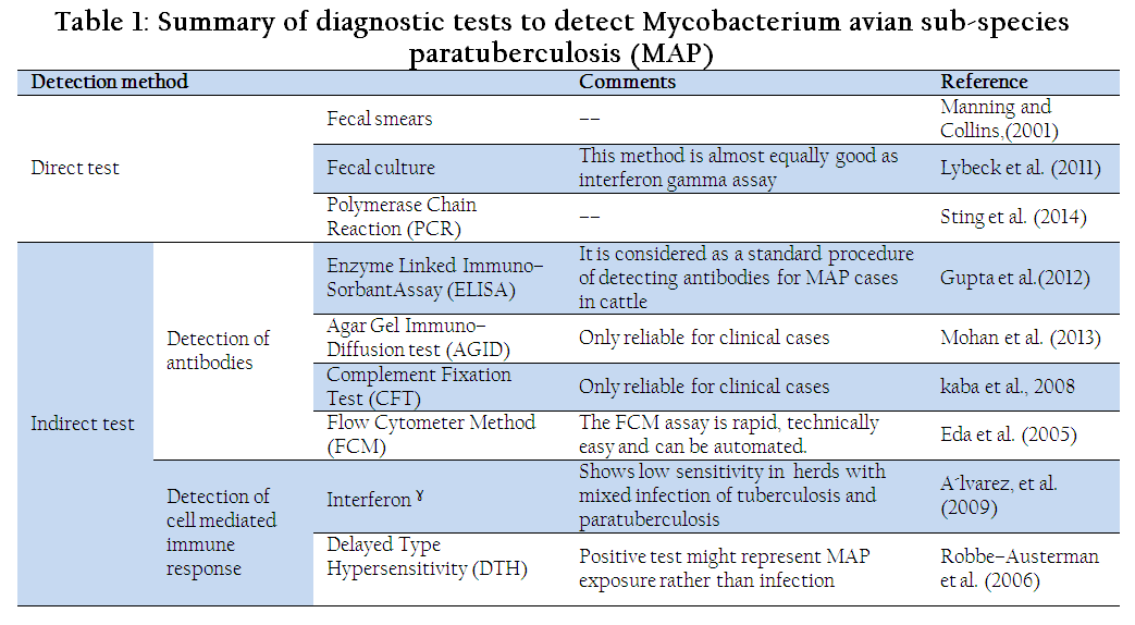
Table 1: Summary of diagnostic tests to detect Mycobacterium avian sub-species paratuberculosis (MAP)
Pathology
In early stages of disease, lesions may not be evident but in clinical cases they can be seen. Lybeck et al., (2011) reported lesions in goats affected with MAP; enlargement of lymph nodes in jejunum was evident with yellowish necrotic foci on cortex. Enteritis is usually seen with ulcerated intestinal mucosa and MAP can be isolated from lesions in intestine and draining lymph nodes of clinically effected animals. Lesions in intestine are seen in jejunum and extend to rectum in advanced stages of disease. Edema and fluid may be found in body cavities. Histopathological examinations exhibit diffused granulomatous enteritis, accumulation of epithelioid giant cells and macrophage in submucosa and mucosa of intestine (Almujalli and Al–Ghamdi, 2012).
Diagnosis
Detection and diagnosis of Mycobacterium paratuberculosis is difficult due to long incubation period (4 months to 15 years) and other reason is the lack of accurate tests which can predict the infection (Nielsen, 2008). Diagnosis is based on clinical signs, postmortem lesions, histopathology and diagnostic tests including direct test e.g. fecal smears, fecal culture and polymerase chain reaction (PCR) and indirect tests e.g. delayed–type hypersensitivity (DTH), interferon Assay, enzyme linked immuno–sorbent assay (ELISA), agar gel immunodiffusion (AGID), complement fixation test (CFT). Differential diagnosis includes kidney failure, gastrointestinal parasitism, renal amyloidosis, peritonitis, chronic salmonellosis, lymphosarcoma and other chronic infectious diseases, copper deficiency and starvation.
DIRECT TESTS
Fecal Smears
As the diseased animals shed pathogen in feces so they can be observed in feces or in pathological lesions from intestine. It is the simple and easy method for detection of etiological agent. Acid fast stain i.e. Ziel–Nelson or Wright's stain is used to highlight the pathogen in smear and observations under oil immersion (X 1000) are positive when clumps of 3–4 acid fast MAP are seen (Manning and Collins, 2001). Sensitivity of this test is very low if used in preclinical stages of paratuberculosis but it is helpful when clinical phase starts (Ansari et al., 2013).
Fecal Culturing
First isolation of MAP was reported in 1910 (Twort, 1910) and complete method of isolation was described in 1912 (Twort and Ingram, 1912). Many authorities consider it most specific and sensitive method of MAP detection. Initially MAP has been grown on egg based medium (
Twort, 1910; Twort and Ingram, 1912), then egg yolk was used instead of whole egg because egg white retard microbial growth (Herrold, 1931), but later antiformin and malachite green were used for decontamination.
Polymerase Chain Reaction (PCR)
It is better and advanced technique (Chaudhary et al., 2009) able to detect MAP and distinguish it from other species and subspecies of Mycobacteria. IS900 and IS901 insertion element is considered unique to MAP and can be used in a PCR gene amplification technique for diagnosis (Slana et al., 2009). Detection limit of PCR in MAP cases is 104 organisms/gm, it is also a limitation of its use. Addition of pretreatment of fecal sample using silica membrane mini–columns and magnetic particles can enhance the detection rate of MAP by PCR (Sting et al., 2014).
INDIRECT TESTS
Detection of Antibodies
Detection of serum antibodies seems to be satisfactory method for screening at mass level but there are many problems related with the detection of antibodies against MAP and its interpretation. Antibodies against paratuberculosis are lately formed because it is a chronic disease and have long incubation period and they are difficult to be detected in preclinical stage sometimes even at clinical stages animal fails to develop antibodies against MAP. Cross reactivity of MAP with other organisms can make antibody interpretation difficult.
Enzyme Linked Immuno–Sorbent Assay (ELISA)
It is considered as a standard procedure of detecting antibodies for MAP cases in cattle (Gupta et al., 2012). Sensitivity of ELISA in present condition changes from low (where minor shedding is present) to high (clinical stage is reached); so we can say that sensitivity of ELISA in MAP case increases with disease progression (Donat et al., 2014). This method can also detect antibodies in milk and blood (Gupta et al., 2012; Collins et al., 2005).
Agar Gel Immunodiffusion (AGID) Test
This test is based on descriptions of precipitation lines formed between antigen used and serum samples. This test is economical and mostly reliable in small ruminants. AGID test in unsatisfactory for subclinical cases due to low specificity and sensitivity (Ferreira et al, 2002), but it gives reliable results in clinical cases (Mohan et al., 2013; Robbe–Austerman et al., 2006).
Complement Fixation Test (CFT)
This test can be used for mass screening of infected animals but interpretation is reliable only at clinical stage (kaba et al., 2008; Slana et al., 2008). Many types of antigens and protocols are being practiced in different countries and laboratories so elucidation of results lacks clarity.
Flow Cytometry Method (FCM)
This method is capable of distinguishing MAP–infected from MAP–non–infected cattle as well as MAP from M. avium subsp. Avium and M. scrofulaceum (Eda et al., 2005). The FCM assay is rapid (completed in less than 4 hours), technically easy and can be automated for handling large numbers of samples (Eda et al., 2005).
Interferon–∂Assay
This method is successfully used for detection of cytokines for the indication of CMI to check exposure of animal to MAP (Nielsen and Toft, 2008). Buffy coat (leukocytes) is collected from heparinized blood and exposed to antigen to measure CMI by the release of gamma interferon (Manning and Collins, 2001). This test shows low sensitivity when used to detect infection in a herd with mixed infection of tuberculosis and paratuberculosis (A´lvarez, et al., 2009; A´lvarez, et al., 2008).
Delayed Type Hypersensitivity (DTH) Reaction
This is a similar test performed in animals for detection of tuberculosis. Delayed type hypersensitivity is measured by injecting intradermal antigens (Johnin or Avian purified protein derivative) in skin to detect cell mediated immunity (CMI). Reaction is allowed to occur and after 72 hours thickness of 2mm will give the indication of positive result (Robbe–Austerman et al., 2006; Manning and Collins, 2001). This is not reliable because MAP antigens are already present in environment (Whittington et al., 2003).
Treatment
There is no treatment for JD to give satisfactory results. Combination of different drugs has been practiced as treatment measure, mostly with isoniazid, clofazimine and rifampin (Borody et al., 2007; St–Jean and Jernigan, 1991). Monensin is also used with the aim of prevention in calves and to reduce shedding in cattle (Fecteau and Whitlock, 2011). Click (2011) concluded from an experiment that Dietzia prebiotic can successfully treat the bovine paratuberculosis and can prevent JD development in MAP infected calves. Recently, lactic acid bacteria (LABATCC 334) have been used as probiotics for treatment of experimentally induced JD in mice (Cooney et al., 2014).
Economic Impact
Paratuberculosis results in economic losses that are primarily associated with decreased milk production, decreased weaning weights in young calves, increased replacement costs, decreased slaughter value (Lombard, 2011) and early culling (Vázquez et al., 2012; Hasnova and Pavlik, 2006). Economic losses to dairy industry are significant (Lu et al., 2013a; Hasnova and Pavlik, 2006; Vidić et al., 2013); estimated losses to US dairy industry cost $200–250 million annually (Cho et al., 2012; Ottet et al., 1999). Pillars et al. (2009) reported averaged $79/cow/year with a median of $66/cow/year annual losses due to paratuberculosis. Raizman et al. (2009) reported less milk production in JD positive cows Indirect economic loss related with JD is trade restriction.
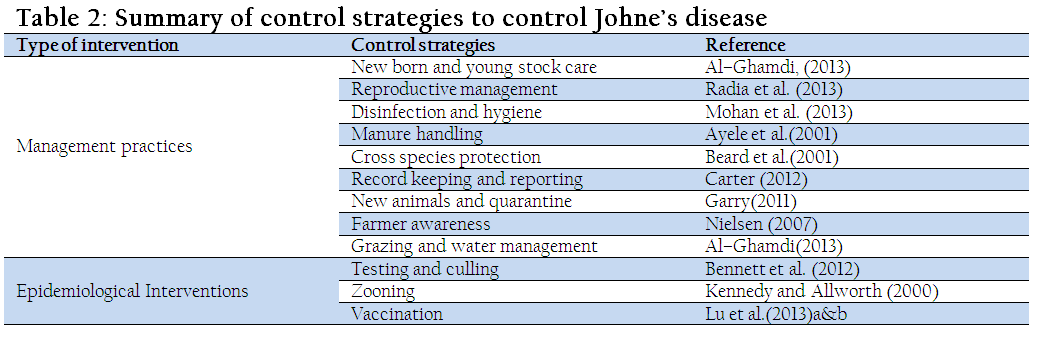
Control Strategies
Countries in different regions of world adopt different control strategies for Johne’s disease depending upon the epidemiology. Basic focus of control strategies is management modification, test, culling and vaccination (Bastida and Juste, 2011; Cho et al., 2012; Al–Ghamdi, 2013; Khol and Baumgartner, 2012; Bennett et al., 2012). Pillars et al. (2009) reported that implementation of JD control programs cost average $30/cow/year (median of $24/cow/year). While, annual losses due to JD averaged $79/cow/year (median of $66/cow/year). This study clearly showed that investment in JD control program is cost effective.
MANAGEMENT PRACTICES
Newborn and Young Stock Management
Calf rearing practices has been proved to be very helpful for JD control (Ridge et al., 2010). JD infests animals at younger age so they should be kept under proper management practices. Parturition should be in clean and manure free area to avoid contact of newborn at early with MAP. Calves should be kept in separate pens to avoid contact with adults possibly carrying MAP (Al–Ghamdi, 2013).
Reproductive Management
Semen may contain MAP having potential of transmitting and causing disease to inseminated animals and newborns. So breeding bulls should be tested for MAP and semen samples must undergo laboratory examination. Periparturient period management is also an important task to be considered to avoid transmission of MAP from dam to calf. Radia et al., (2013) investigated the impact of specific peri–parturient management practices on within–herd. They concluded that management practices aiming to limit the fecal–oral transmission are effective than aiming to limit MAP transmission via colostrums and milk.
Disinfection of Area (Hygiene)
Better hygienic practices in farm management help to control JD (Mohan et al., 2013). MAP can survive many disinfectants exposure, but 5% formalin, 2% calcium hypo–chloride and 2.5% phenol can kill the pathogen. Presence of organic matter may reduce effectiveness of disinfectant and detergents can be used on feces to allow penetration by disinfectants.
Manure Handling
Manure may harbor the MAP (Seyyedin, 2008). Good manure management and disposal techniques are also important. Manure build–up should be prevented, and surfaces should be kept clean (Ayele et al., 2001). Grewal et al., (2006) observed that thermophilic composting is more effective than pack storage in reducing MAP in dairy manure in pathogen sensitive environments.
Cross Specie Transmission
Cross–species transmission of the MAP strains can occur, but seems to be relatively uncommon (Sohal et al., 2010). Therefore, the greatest risk of infection for cattle appears to be from other cattle, and for sheep from other sheep.
Quarantine the New Animals
Farmers with uninfected herds should buy replacement animals from test–negative herds with good records and management practices. All animals should be quarantined and tested before mixing them to the herd.
Farmer Awareness about Disease
Farmers must be educated about the benefits of JD control program and losses related with paratuberculosis; such practices have helped to lower the prevalence of disease (Nielsen, 2007; Carter, 2012).
Grazing and Water Management
Animals shed plenty of MAP on grazing area in feces that is a potential source of disease transmission. Effected pastures should be reseeded and preferably not used for grazing by unaffected animals because MAP may persist on grass and soil for 1–4 years and remains viable to find the host. Water facilities and sources must be uncontaminated because MAP survives in ponds and rivers for five months (Al–Ghamdi, 2013).
Test and Culling
This program consists of periodic testing of herd and positive animals are culled or separated (Garry, 2011). Testing of animals can be performed by tests mentioned in diagnostic portion; single or multiple tests in combination can be used. Control of paratuberculosis would be easier and enhance the efficiency of overall control program if we remove animals that are shedding large numbers of organisms (Garry, 2011; Bennett et al., 2012).
Zoning
This is the general approach to diseases control in animals, where zones are made on basis of severity and prevalence of disease and movement is restricted in them (Kennedy and Allworth, 2000). Disease free, protected and control areas are managed accordingly. But zoning may be a barrier in trading and marketing (Ayele et al., 2001).
Vaccination
Routine vaccination in herds provide partial protection for susceptible calves but its efficacy decreases with the progress of disease (Lu et al., 2013a and 2013b); so, there is no efficient vaccine available and not practically possible (Wadhwa et al., 2013). Still vaccination practice are helpful for delaying the onset of shedding, slowing progression from low shedding to high shedding, reducing infectiousness of shedders, extending latent period of infected animals, and reducing clinical disease (Lu et al., 2013b; Wadhwa et al., 2013; Rosseels and Huygen, 2008; Kumar et al., 2014). Singh et al., (2013b), successfully used vaccination in a calf in preclinical stage and noted reduced severity of disease in an adult cow having clinical signs, by using ‘Indian Bison Type’ biotype of MAP (strain S 5) of goat origin. Singh et al., (2011) vaccinated goats and found recovery rate of 85% under optimal conditions of nutrition while 15 % could not recover because of clinical stage. Thakur et al., (2013) resulted that an appropriate age of vaccination should be considered in vaccination protocols. Both live (attenuated and non–attenuated) and killed whole cell vaccines have been used against paratuberculosis (Bastida and Juste, 2011; Rosseels and Huygen, 2008; Knust et al., 2013). In a few cases, subunit vaccines consisting of sonicated bacteria, bacterial cell fractions or recombinant MAP antigens have been used but they have shown a much lower degree of protection (Kathaperumal et al., 2009; Koets et al., 2006). DNA vaccines have also been practiced with better success rates (Park et al., 2008).
CONCLUSION
Paratuberculosis is the progressive glumerulo entritis effecting wide range of animals. Control of JD is challenge for veterinarians and famers because of nature of organism and lack of policies to control. Most studies focus on management related control because it is very much effective. Second strategy is testing and culling of positive animals. Wide range of tests are available mostly test show positive results when animals starts shedding of MAP in feces and milk. Third control strategy for JD is vaccination, it is recommended in calves at early ages but limitation of vaccination is that it gives false positive results with tuberculosis testing.
REFERENCES
A’lvarez J, De Juan L, Bezos J, Romero B, Sa’ez JL, Gordejo R, Briones V, Moreno MA, Mateos A, Domı’nguez L, Aranaz A (2008). Interference of paratuberculosis with the diagnosis of tuberculosis in a goat flock with a natural mixed infection. Vet Microbiol. 128:72–80.
http://dx.doi.org/10.1016/j.vetmic.2007.08.034
PMid:17954015
A’lvarez J, de Juan L, Bezos J, Romero B, Sa’ez JL, Marque’s S, Domı’nguez C, Mı’nguez O, Ferna’ndez–Mardomingo B, Mateos A, Domı’nguez L, Aranaz A (2009). Effect of paratuberculosis on the diagnosis of bovine tuberculosis in a cattle herd with a mixed infection using interferon–gamma detection assay. Vet Microbiol. 135: 389–393
http://dx.doi.org/10.1016/j.vetmic.2008.09.060
PMid:18986776
Abbas M, Munir M, Khaliq SA, Ikram Ul Haq M, Khan MT, Qureshi ZA(2011). Detection of Paratuberculosis in Breeding Bulls at Pakistani Semen Production Units: A Continuous Source of Threat. ISRN Veterinary Science, vol. 2011, Article ID 501235. doi:10.5402/2011/501235.
http://dx.doi.org/10.5402/2011/501235
Al–Ghamdi GM (2013). Mycobacterium avium subspecies paratuberculosis in Camels; Clinical Aspects and Control Suggestions. Egypt. Acad. J. Biolog. Sci. 5(1):11–15.
Alluwaimi AM (2007). The etiology of mycobacterium avium subspecies paratuberculosis in Crohn's disease. Saudi Med J. 28(10): 1479–1484.
PMid:17914503
Almujalli AM, Al–Ghamdi GM (2012). Clinicopathological Findings of Partuberclosis in Camels Possible Steps for Control Strategy. Res. J. Biolog. Sci. 7 (3):128–131.
http://dx.doi.org/10.3923/rjbsci.2012.128.131
Ansari MM, Peer FU and Wani SA (2013). Studies on clinic-therapeutic management of paratuberculosis in bovine. Blue Cross Book. 29: 60-61.
Ayele WY, Machackova M, Pavlik I (2001). The transmission and impact of paratuberculosis infection in domestic and wild ruminants. Vet. Med. Czech. 46(7–8): 205–224.
Bastida F, Juste RA (2011). Paratuberculosis control: a review with a focus on vaccination. J. Immune Based Ther. Vaccines. 9:8.doi:10.1186/1476–8518–9–8.
http://dx.doi.org/10.1186/1476-8518-9-8
Beard PM, Daniels MJ, Henderson D, Pirie A, Rudge K, Buxton D, Rhind S, Greig A, Hutchings MR, McKendrick I, Stevenson K, Sharp JM (2001). Paratuberculosis infection of nonruminant wildlife in Scotland. J. Clin. Microbiol. 39:1517–21.
http://dx.doi.org/10.1128/JCM.39.4.1517-1521.2001
PMid:11283080 PMCid:PMC87963
Begg DJ, De Silva K, Carter N, Plain KM, Purdie A, Whittington RJ (2011). Does a Th1 over Th2 dominancy really exist in the early stages of Mycobacterium avium subspecies paratuberculosis infections? Immunobiology. 216: 840–846.
http://dx.doi.org/10.1016/j.imbio.2010.12.004
PMid:21281979
Behr MA, Kapur V (2008). The evidence for mycobacterium paratuberculosis in Crohn's disease. Curr. Opin. Gastroenterol. 24:17–21.
http://dx.doi.org/10.1097/MOG.0b013e3282f1dcc4
PMid:18043227
Bennett R, McClement I, McFarlane I (2012). Modelling of Johne's disease control options in beef cattle: A decision support approach. Livestock Sci. 146(2–3): 149–159.
http://dx.doi.org/10.1016/j.livsci.2012.03.002
Borody TJ, BilkeyS, Wettstein AR, Leis S, Pang G, Tye S (2007). Anti–mycobacterial therapy in Crohn's disease heals mucosa with longitudinal scars. Dig. Liver Dis. 39(5):438–44.
http://dx.doi.org/10.1016/j.dld.2007.01.008
PMid:17369114
Carter MA (2012). Prevalence and Prevention of Paratuberculosis in North America. Japanese J Vet Res. 60(Supplement): S9–S18, 2012.
PMid:22458196
Chaudhry ZI, Khan FA, Badar S, Shahid M (2009). Detection of Mycobacterium avium subsp. paratuberculosis in Domestic Ruminants in Lahore, Pakistan. Pak J Zool.41(2):160.
Cho J, Tauer LW, Schukken YH, Gómez MI, Smith RL, Lu Z, Grohn YT (2012). Economic analysis of Mycobacterium avium subspecies paratuberculosis vaccines in dairy herds. J. Dairy Sci. 95(4):1855–72. doi: 10.3168/jds.2011–4787.
http://dx.doi.org/10.3168/jds.2011-4787
Click RE (2011). Successful treatment of asymptomatic or clinically terminal bovine Mycobacterium avium subspecies paratuberculosis infection (Johne's disease) with the bacterium Dietzia used as a probiotic alone or in combination with dexamethasone: Adaption to chronic human diarrheal diseases. Virulence. 2(2): 131–143. Doi:10.4161/viru.2.2.15647.
http://dx.doi.org/10.4161/viru.2.2.15647
Collins MT, Wells SJ, Petrini KR, Collins JE, Schultz RD, Whitlock RH (2005). Evaluation of Five Antibody Detection Tests for Diagnosis of Bovine Paratuberculosis. Clin Diag Immunol. 12(6): 685–692. 1071–412X/05/$08.000 doi:10.1128/CDLI.12.6.685–692.
Cooney MA, Steele JL, Steinberg H, Talaat AM (2014). A murine oral model for Mycobacterium avium subsp. paratuberculosis infection and immunomodulation with Lactobacillus casei ATCC 334. Front. Cell. Infect. Microbiol. 05 February 2014 | doi: 10.3389/fcimb.2014.00011
http://dx.doi.org/10.3389/fcimb.2014.00011
de Lisle GW, Collins DM (1995). Johne's disease in deer. Vet. Rec. 136:336.
http://dx.doi.org/10.1136/vr.136.13.336-a
PMid:7604515
Donat K, Schlotter K, Erhardt G, Brandt HR (2014). Prevalence of paratuberculosis in cattle and control measures within the herd influence the performance of ELISA tests. Vet. Rec. 174(5):119. doi: 10.1136/vr.101533.
http://dx.doi.org/10.1136/vr.101533
Dow CT(2012). M. paratuberculosis Heat Shock Protein 65 and Human Diseases: Bridging Infection and Autoimmunity.Autoimmune Diseases,Volume 2012, Article ID 150824, Hindawi Pub Corp. doi:10.1155/2012/150824.
http://dx.doi.org/10.1155/2012/150824
Eda S, Elliott B, Scott MC, Waters WR, Bannantine JP, Whitlock RH, Speer CA (2005). New Method of Serological Testing for Mycobacterium avium subsp. paratuberculosis (Johne's Disease) by Flow Cytometry. In: Foodborne Pathogens and Disease. Fall 2005. 2(3): 250–262. doi:10.1089/fpd.2005.2.250.
http://dx.doi.org/10.1089/fpd.2005.2.250
Eltholth MM, Marsh VR, Winden SV, Guitian FJ (2009). Contamination of food products with Mycobacterium avium paratuberculosis: a systematic review. J of Appl Microbiol. 107: 1061–1071. ISSN 1364–5072. doi:10.1111/j.1365–2672.2009.04286.x
Eppleston J, Begg DJ, Dhand N, Watt B, Whittington RJ (2014). Environmental Survival of Mycobacterium avium subsp. paratuberculosis in Different Climatic Zones of Eastern Australia. Appl. Environ. Microbiol. AEM 03630–13. doi:10.1128/AEM.03630–13.
Fecteau ME, Whitlock RH (2011). Treatment and Chemoprophylaxis for Paratuberculosis. Vet. Clin. Food Anim. 27:547–557.
http://dx.doi.org/10.1016/j.cvfa.2011.07.002
PMid:22023833
Ferreira R, Fonseca LS, Lilenbaum W (2002). Agar gel immunodiffusion test (AGID) evaluation for detection of bovine paratuberculosis in Rio de Janeiro, Brazil. Lett.Appl.Microbiol.35(3):173–5.
http://dx.doi.org/10.1046/j.1472-765X.2002.01149.x
PMid:12180935
Fiorentino MA, Gioffré A, Cirone KC, Morsella, Alonso B, Delgado F, Paolicchi F(2012). First isolation of Mycobacterium aviumsubsp. paratuberculosis in a dairy goat in Argentina: Pathology and molecular characterization. Small Rum Res. 108: 133– 136.
http://dx.doi.org/10.1016/j.smallrumres.2012.06.010
Garrido JM, Vazquez P, Molina E, Plazaola JM, Sevilla IA, Geijo MV, Alonso–Hearn M, Juste RA (2013). Paratuberculosis vaccination causes only limited cross–reactivity in the skin test for diagnosis of bovine tuberculosis. PLoS One. 8(11):e80985.
http://dx.doi.org/10.1371/journal.pone.0080985
PMid:24303029 PMCid:PMC3841166
Garry F (2011). Control of Paratuberculosis in Dairy Herds. Vet. Clin. Food Anim. 27: 599–607.
http://dx.doi.org/10.1016/j.cvfa.2011.07.006
PMid:22023838
Greig A, Stevenson K, Henderson D, Perez V, Hughes V, Pavlik I, Hines ME, McKendrick I, Sharp JM (1999). Epidemiological study of paratuberculosis in wild rabbits in Scotland. J of Clin Microbiol. 37:1746–1751.
PMid:10325318 PMCid:PMC84940
Grewal SK, Rajeev S, Sreevatsan S, Michel FC (2006). Persistence of Mycobacterium aviumsubsp. paratuberculosis and other zoonotic pathogens during simulated composting, manure packing, and liquid storage of dairy manure. Appl Environ Microbiol. 72(1):565–74.
http://dx.doi.org/10.1128/AEM.72.1.565-574.2006
PMid:16391093 PMCid:PMC1352242
Gupta A, Rani SM, Agrawal P, Gupta PK (2012). Sero–Prevalence of Paratuberculosis (Johne's Disease) in Cattle Population of South–Western Bangalore Using ELISA Kit. Open J Vet Med. 2: 196–200.doi:10.4236/ojvm.2012.24031.
http://dx.doi.org/10.4236/ojvm.2012.24031
Gwozdz JM (2010). Paratuberculosis (Johne's Disease). Australian and New Zealand Standard Diagnostic Procedures August 2010.
Hasnova L, Pavlik I (2006). Economic impact of paratuberculosis in dairy cattle herds: a review. Vet Med.51 (5): 193–211.
Hasonova L, Trcka I, Babak V, Rozsypalova Z, Pribylova R, Pavlik I(2009). Distribution of Mycobacterium avium subsp. Paratuberculosis in tissues of naturally infected cattle as affected by age. Vet Med. 54(6): 257–269.
Herrold RD (1931). Egg yolk agar medium for the growth of organisms would be M. paratuberculosis strain 18 or tubercle bacilli. J. Infect. Dis. 48:236–241.
http://dx.doi.org/10.1093/infdis/48.2.236
Hines ME II, Stabel JR, Sweeney RW, Griffin F, Talaat AM, Bakker D, Benedictus G, Davis WC, de Lisle GW, Gardner IA, Juste RA, Kapur V, Koets A, McNair J, Pruitt G, Whitlock RH (2007). Experimental challenge models for Johne's disease: A review and proposed international guidelines. Vet Microbiol. 122: 197–222.
http://dx.doi.org/10.1016/j.vetmic.2007.03.009
PMid:17467201
Hutchings MR, Stevenson K, Greig A, Davidson RS, Marion G, Judge J (2010). Infection of Non–ruminant Wildlife by Mycobacterium avium subsp. paratuberculosis. In Paratuberculosis; Organism, Disease, Control. Edited by Behr MA, Collins DM. Wallingford: CAB International; 2010:188–200.
http://dx.doi.org/10.1079/9781845936136.0188
Kaba J, Gerlach GF, Nowicki M, Rypuła K (2008). Agreement between ELISA and complement fixation test used for diagnosing of paratuberculosis in goats. Pol. J. Vet. Sci. 11(3):209–12.
PMid:18942542
Kathaperumal K, Kumanan V, McDonough S, Chen LH, Park SU, Moreira MA, Akey B, Huntley J, Chang CF, Chang YF (2009). Evaluation of immune responses and protective efficacy in a goat model following immunization with a coctail of recombinant antigens and a polyprotein of Mycobacterium aviumsubsp. paratuberculosis.Vaccine.27:123–135.
http://dx.doi.org/10.1016/j.vaccine.2008.10.019
PMid:18955101
Kennedy DJ, Allworth MB (2000). Progress in national control and assurance programs for bovine Johne's disease in Aus.Vet. Microbiol.77(3–4):443–51.
http://dx.doi.org/10.1016/S0378-1135(00)00329-1
Khan FA, Chaudhry ZI, Ali MI, Khan S, Mumtaz N, Ahmad (2010). Detection of Mycobacterium aviumsubsp. Paratuberculosis in tissue samples of cattle and buffaloes. Trop. Anim Health Prod. 42:633–638. DOI 10.1007/s11250–009–9467–8.
http://dx.doi.org/10.1007/s11250-009-9467-8
Khol JL, Baumgartner W (2012). Examples and suggestions for the control of paratuberculosis in European cattle. Jpn. J. Vet. Res. 60: 1–7.
Knust B, Patton E, Ribeiro–Lima J, BohnJF, Wells SJ (2013). Evaluation of the effects of a killed whole–cell vaccine against Mycobacterium aviumsubsp. paratuberculosis in 3 herds of dairy cattle with natural exposure to the organism. J the Amer Vet Med Assoc. 242(5): 663–669.doi: 10.2460/javma.242.5.663.
http://dx.doi.org/10.2460/javma.242.5.663
Koets A, Hoek A, Langelaar M, Overdijk M, Santema W, Franken P, Eden W, Rutten V(2006). Mycobacterial 70 kD heat–shock protein is an effective subunit vaccine against bovine paratuberculosis.Vaccine.24:2550–2559.
http://dx.doi.org/10.1016/j.vaccine.2005.12.019
PMid:16417949
Kruze J, Monti G, Schulze F, Mella A, Leiva S (2013). Herd–level prevalence of Map infection in dairy herds of southern Chile determined by culture of environmental fecal samples and bulk–tank milk qPCR. Prev. Vet. Med. 111(3–4):319–24.
http://dx.doi.org/10.1016/j.prevetmed.2013.05.011
PMid:23791126
Kumar A, Singh SV, Srivastava AK, Gangwar NK, Singh PK, Gupta S, Chaubey KK, Tiwari R, Chakraborty S, Dhama K(2014). Comparative Evaluation of 'Indigenous' and Commercial Vaccines in Double Challenge Model for the Control of Caprine Paratuberculosis in India. J Biol Sci. 14(3):169–182.
http://dx.doi.org/10.3923/jbs.2014.169.182
Liapi M, Leontides L, Kostoulas P, Botsaris G, Iacovou Y, Rees C, Georgiou K, Smith GC, Naseby DC (2011). Bayesian estimation of the true prevalence of Mycobacterium aviumsubsp. paratuberculosis infection in Cypriot dairy sheep and goat flocks. Small Rumin. Res. 95(2–3): 174–178.
http://dx.doi.org/10.1016/j.smallrumres.2010.09.010
Lombard JE (2011). Epidemiology and economics of paratuberculosis. Vet. Clin. North. Am. Food Anim. Pract. 27(3):525–35. doi: 10.1016/j.cvfa.2011.07.012.
http://dx.doi.org/10.1016/j.cvfa.2011.07.012
Lombard JE, Gardner IA, Jafarzadeh SR, Fossler CP, Harris B, Capsel RT (2013). Herd–level prevalence of Mycobacterium aviumsubsp. paratuberculosis infection in United States dairy herds in 2007. Prev. Vet. Med. 108: 234–238. doi: 10.1016/j.prevetmed.2012.08.006
http://dx.doi.org/10.1016/j.prevetmed.2012.08.006
Lu Z, Schukken YH, Smith RL, Gröhn YT (2013a). Using vaccination to prevent the invasion of Mycobacterium aviumsubsp. paratuberculosis in dairy herds: a stochastic simulation study. Prev. Vet. Med.110(3–4):335–45.
http://dx.doi.org/10.1016/j.prevetmed.2013.01.006
PMid:23419983
Lu Z, Schukken YH, Smith RL, Gröhn YT (2013b). Impact of imperfect Mycobacterium avium subsp. paratuberculosis vaccines in dairy herds: A mathematical modeling approach. Prev Vet Med. 108(2–3): 148–158.
http://dx.doi.org/10.1016/j.prevetmed.2012.08.001
PMid:22921715
Lybeck KR, Storset AK, Djønne B, Valheim M, Olsen I (2011). Faecal shedding detected earlier than immune responses in goats naturally infected with Mycobacterium aviumsubsp. Paratuberculosis. Res Vet Sci. 91: 32–39.
http://dx.doi.org/10.1016/j.rvsc.2010.08.012
PMid:20869736
Manning JBE, Collins MT (2001). Mycobacteriumaviumsubsp.paratuberculosis: pathogen, pathogenesis and diagnosis. Rev. Sci. Tech. Off. Int. Epiz. 20(1): 133–150.
Manning JBE, Steinberg H, Krebs V, Collins MT (2002). Diagnostic testing patterns of natural Mycobacterium paratuberculosis infection in pygmy goats. Can. J. Vet. Res. 67: 213–218.
Martin SW, Meek AH, Willeberg P (1987). Veterinary Epidemiology. Iowa State University Press, Ames, IA, USA, pp. 245–258 (Chapter 10).
Mercier P, Baudry C, Beaudeau F, Seegers H, Malher X (2010). Estimated prevalence of Mycobacterium aviumsubspeciesparatuberculosis infection in herds of dairy goats in France. Vet. Rec. 167(11): 412–416.
http://dx.doi.org/10.1136/vr.c4454
PMid:20834001
Mohan A, Das P, Kushwaha N, Karthik K, Niranjan AK (2013). Investigation on the status of Johne's disease based on agar gel immunodiffusion, Ziehl–Neelsen staining and nested PCR approach in two cattle farm. Vet World. 6(10): 778–784.
http://dx.doi.org/10.14202/vetworld.2013.778-784
Nielsen SS (2007). Danish control program for bovine paratuberculosis. BCVA Cattle Practice 15(2): 161–168.
Nielsen SS (2008). Transitions in diagnostic tests used for detection of Mycobacterium aviumsubsp. Paratuberculosis infections in cattle. Vet Microbiol. 132: 274–282.
http://dx.doi.org/10.1016/j.vetmic.2008.05.018
PMid:18599225
Nielsen SS, Toft N (2008). Ante mortem diagnosis of paratuberculosis: a review of accuracies of ELISA, interferon–g assay and faecal culture techniques. Vet. Microbiol. 129: 217–235.
http://dx.doi.org/10.1016/j.vetmic.2007.12.011
PMid:18255239
Okuni JB (2013). Occurence of Paratuberculosis in African Countries: a Review. J. Vet. Adv. 3(1): 1–8.
Ott SL, Wells SJ, Wagner BA (1999). Herd–level economic losses associated withJohne's disease on US dairy operations. Prev. Vet. Med. 40:179–92.
http://dx.doi.org/10.1016/S0167-5877(99)00037-9
Over K, Crandall PG, O'Bryan CA, Ricke SC(2011). Current perspectives on Mycobacterium aviumsubsp. paratuberculosis, Johne's disease, and Crohn's disease: a Review. Critical Rev in Microbiol. 37(2): 141–156. ISSN 1040–841X print/ISSN 1549–7828, DOI: 10.3109/1040841X.2010.532480.
http://dx.doi.org/10.3109/1040841X.2010.532480
Park SU, Kathaperumal K, McDonough S, Akey B, Huntley J, Bannantine JP, Chang YF (2008). Immunization with a DNA vaccine cocktail induces a Th1 response and protects mice against Mycobacterium aviumsubsp. paratuberculosis challenge. Vaccine. 26:4329–4337.
http://dx.doi.org/10.1016/j.vaccine.2008.06.016
PMid:18582521
Pillars RB, Grooms DL, Wolf CA, Kaneene JB (2009). Economic evaluation of Johne's disease control programs implemented on six Michigan dairy farms. Prev. Vet. Med. 90(3–4):223–32. doi: 10.1016/j.prevetmed.2009.04.009.
http://dx.doi.org/10.1016/j.prevetmed.2009.04.009
Radia D, Bond K, Limon G, van Winden S, Guitian J (2013). Relationship between periparturient management, prevalence of MAP and preventable economic losses in UK dairy herds. Vet Rec. 173(14):343. doi: 10.1136/vr.101408.
http://dx.doi.org/10.1136/vr.101408
Raizman EA, Fetrow JP, Wells SJ (2009). Loss of income from cows shedding Mycobacterium aviumsubspecies paratuberculosis prior to calving compared with cows not shedding the organism on two Minnesota dairy farms. J. Dairy. Sci. 92(10):4929–36. doi: 10.3168/jds.2009–2133.
http://dx.doi.org/10.3168/jds.2009-2133
Ridge SE, Heuer C, Cogger N, Heck A, Moor S, Baker IM, Vaughan S (2010). Herd management practices and the transmission of Johne's disease within infected dairy herds in Victoria, Australia. Prev. Vet. Med. 95(3–4):186–97. doi: 10.1016/j.prevetmed.2010.05.001.
http://dx.doi.org/10.1016/j.prevetmed.2010.05.001
Robbe–Austerman S, Gardner IA, Thomsen BV, Morrical DG, Martin BM, Palmer MV, Thoen CO, Ewing C (2006). Sensitivity and specificity of the agar–gel– immunodiffusion test, ELISA and the skin test for detection of paratuberculosis in United States Midwest sheep populations. Vet. Res. 37 (2006): 553–564. DOI: 10.1051/vetres:2006018.
http://dx.doi.org/10.1051/vetres:2006018
Rosseels V, Huygen K (2008). Vaccination against paratuberculosis. Expert rev of Vac. 7(6): 817–832. DOI 10.1586/14760584.7.6.817.
Salgado M, Kruze J, Collins MT (2007). Diagnosis of paratuberculosis by fecal culture and ELISA on milk and serum samples in two types of Chilean dairy goat herds. J. Vet. Diagn. Invest. 19: 99–102.
http://dx.doi.org/10.1177/104063870701900117
PMid:17459841
Seyyedin M, Slaeh TZ, Tadjbakhsh H, Najafi MF, Rabbani M (2008). Isolation and Identification of Mycobacterium aviumsubsp. Paratuberculosis from Milk, Manure and Fecal Samples of Holstein–Fresian Dairy Cattle. Pak J of Biol Sci. 11(24): 2639–2645.
http://dx.doi.org/10.3923/pjbs.2008.2639.2645
Seyyedin M, Zahraei T, Najafi MF (2010). Comparison of isolation frequency of Mycobacterium aviumsubspeciesparatuberculosis from different types of samples. Pak. Vet. J. 30(3): 143–149.
Sikandar A, Cheema AH, Adil M, Younus M, Zaneb H, Zaman MA, Tipu MY, Masood S (2013). Ovine paratuberculosis– A histopathological study from Pakaistan. The J of Ani & Pl Sci. 23(3): 749–753. ISSN: 1018–7081.
Sikandar A, Cheema AH, Younus M, Aslam A, Zaman MA, Rehman T (2011). Histopathological and serological studies on paratuberculosis in cattle and buffalos. Intern Workshop on Dairy Sci Park, Peshawar, Pakistan. 21–23/11/2011.
Singh SV, Chaubey KK, Gupta S, Gupta VK, Agrawal ND, Kumar N (2013a). Co–infection of Mycobacterium aviumsubspecies paratuberculosis and Brucellamelitensisin a sirohi breed of goats in India. Adv. Anim. Vet. Sci. 1 (6): 188 – 190.
Singh SV, Gupta S, Singh PK, Singh AV, Sohal JS, Kumar N, Kumar A, Kumar K, Singh S (2013b). Therapeutic Management of Clinical Bovine Johne's disease Using Goat Based 'Indigenous Vaccine' in Native Hariana Cattle: Case Reports. Adv. Anim. Vet. Sci. 1(1S):23–28.
Singh SV, Singh PK, Singh MK, Singh AV, Sohal JS (2011). Therapeutic potential of Johne's disease vaccine: A follow up post vaccination study in a goatherd of endangered Jamunapari breed, naturally infected with Mycobacterium aviumsubspeciesparatuberculosis. Int J livestock Prod. 2(13): 192–204.
Slana I, BartošM, RoubalP, Babák V, Pavlík I (2009). Mycobacterium aviumsubsp. paratuberculosisand M. a. aviumDetected by Culture, Is900 and Is901 Highly Sensitive Pcr in Bulk Tank Milk from Dairy Herds in the Czech Republic between 2002 and 2004. Czech J. Food Sci. 27(5): 372–378. Sohal JS, Singh SV, Singh AV, Singh PK (2010). Strain diversity within mycobacterium aviumsubspecies paratuberculosis– A review. Indian J. Exp. Biol. 48: 7–16.
Slana I, Paolicchi F, Janstova B, Navratilova P, Pavlik I (2008). Detection methods for Mycobacterium aviumsubsp.paratuberculosis in milk and milk products: a review. Vet Med. 53(6): 283–306.
Stabel JR (2000). Transitions in immune responses to Mycobacterium paratuberculosis. Vet. Microbiol. 77: 465–473.
http://dx.doi.org/10.1016/S0378-1135(00)00331-X
Stabel JR (2008). Pasteurization of colostrum reduces the incidence of paratuberculosis in neonatal dairy calves. J Dairy Sci. 91(9):3600–6. doi: 10.3168/jds.2008–1107.
http://dx.doi.org/10.3168/jds.2008-1107
Sting R, Hrubenja M, Mandl J, Seemann G, Salditt A, Waibel S (2014). Detection of Mycobacterium aviumsubsp. paratuberculosis in faeces using different procedures of pre–treatment for real–time PCR in comparison to culture. Vet. J. 199(1):138–42. doi: 10.1016/j.tvjl.2013.08.033.
http://dx.doi.org/10.1016/j.tvjl.2013.08.033
St–Jean G, Jernigan AD (1991). Treatment of Mycobacterium paratuberculosis infection in ruminants. Vet. Clin. North. Am. Food. Anim. Pract. 7(3):793–804.
PMid:1760762
Thakur A, Aagaard C, Stockmarr A, Ersen P, Jungersen G (2013). Cell–Mediated and Humoral Immune Responses after Immunization of Calves with a Recombinant Multiantigenic Mycobacterium aviumsubsp. Paratuberculosis Subunit Vaccine at Different Ages. Clin Vac Immunol.20(4): 551–558.
http://dx.doi.org/10.1128/CVI.05574-11
PMid:23389934 PMCid:PMC3623413
Tkachuk VL, Krause DO, McAllister TA, Buckley KE, Reuter T, Hendrick S, Ominski KH (2013). Assessing the Inactivation of Mycobacterium aviumsubsp. paratuberculosis during Composting of Livestock Carcasses. Appl Environ Microbiol. 79(10): 3215–3224.
http://dx.doi.org/10.1128/AEM.03768-12
PMid:23503307 PMCid:PMC3685258
Twort FW (1910). A method for isolating and growing the Leprabacillus of man (preliminary note). Proc. R. Soc. Lond. Ser. B.83:156–158.
http://dx.doi.org/10.1098/rspb.1910.0074
Twort FW, Ingram GLY (1912). A method for isolating and cultivating Mycobacterium enteritischronic pseudotuberculosisbovis, Johne, and some experiments on the preparation of a diagnostic vaccine for pseudotuberculosis enteritis of bovines. Proc. R. Soc. Lond. Ser. B.84:517–542.
http://dx.doi.org/10.1098/rspb.1912.0011
Vansnick E (2004). Johne's disease in zoo animals: development of molecular tools for the detection and characterization of Mycobacterium aviumsubspecies paratuberculosis. Doctorial thesis: Instituutvoor Tropische Geneeskunde Departement Diergeneeskunde.
Vázquez P, Garrido JM, Juste RA (2012). Effects of paratuberculosis on Friesian cattle carcass weight and age at culling. Spanish Journal of Agricultural Research. 10(3): 662–670. doi: 10.5424/sjar/2012103–2728.
http://dx.doi.org/10.5424/sjar/2012103-2728
Vidić B, Savić S, Vidić V, Jovičin M, Prica N (2013). Economic impact of paratuberculosis on milk production. Biotech in Ani Husb. 29(2): 183–191. ISSN 1450–9156 DOI: 10.2298/BAH1302183V.
http://dx.doi.org/10.2298/BAH1302183V
Wadhwa A, Kumar N, Velasco–Villa A, Eda S (2013). Overview of Johne's disease immunology. Vet World. 6(11): 901–904.
http://dx.doi.org/10.14202/vetworld.2013.901-904
Whipple DL, Callihan DR, Jarnagin JL (1991). Cultivation of Mycobacterium Paratuberculosis from Bovine Fecal Specimens and a Suggested Standardized Procedure. J. Vet. Diagn. Invest. 3: 368–373
http://dx.doi.org/10.1177/104063879100300424
PMid:1760476
Whittington RJ, Marsh IB, Taylor PJ, Marshall DJ, Taragel C, Reddacliff LA (2003). Isolation of Mycobacterium aviumsubsp. paratuberculosis from environmental samples collected from farms before and after destocking sheep with paratuberculosis. Aust. Vet. J. 81(9): 559–563.
http://dx.doi.org/10.1111/j.1751-0813.2003.tb12887.x
PMid:15086096
Windsor PA, Whittington RJ (2010). Evidence for age susceptibility of cattle to Johne's disease. Vet J. 184(1):37–44. doi: 10.1016/j.tvjl.2009.01.007.
http://dx.doi.org/10.1016/j.tvjl.2009.01.007
Wu CW, Livesey M, Schmoller SK, Manning EJ, Steinberg H, Davis WC, Hamilton MJ, Talaat AM (2007). Invasion and persistence of Mycobacterium aviumsubsp. paratuberculosis during early stages of Johne's disease in calves. Infect. Immun.75(5):2110–9.
http://dx.doi.org/10.1128/IAI.01739-06
PMid:17296749 PMCid:PMC1865790


