Research Journal for Veterinary Practitioners
Case Report
Research Journal for Veterinary Practitioners 2 (4): 67 – 69Surgical Management of Concurrent Umbilical Hernia and Intestinal Fecolith in a white Yorkshire Piglet; Case Report
Shongsir Warson Monsang1*, Saumen Kanti Pal2, Mritunjay Kumar1, Joyabrata Roy1
- Departement of Teaching Veterinary Clinical Complex, College of Veterinary Science & A.H., R.K.Nagar , Agartala-799008, Tripura (W), India
- Department of Pharmacology & Toxicology, College of Veterinary Science & A.H., R.K.Nagar , Agartala-799008, Tripura (W), India
*Corresponding author:warsonmonsang@gmail.com
ARTICLE CITATION:
Monsang SW, Pal SK, Kumar M, Roy J (2014). Surgical management of concurrent umbilical hernia and intestinal fecolith in a white Yorkshire piglet; Case report. Res. J. Vet. Pract. 2 (4): 67 – 69.
Received: 2014–02–28, Revised: 2014–03–28, Accepted: 2014–03–29
The electronic version of this article is the complete one and can be found online at
(
http://dx.doi.org/10.14737/journal.rjvp/2014/2.4.67.69
)
which permits unrestricted use, distribution, and reproduction in any medium, provided the original work is properly cited
ABSTRACT
An intact 3 months old male white Yorkshire pig was presented with the complaint of swelling at the umbilical region since one month which later became larger as the pig gradually grows. Appetite of the animal was reported to be normal though scanty faeces were passed out at regular intervals. On palpation, the swelling was hard, non–painful and irreducible. No hernial ring could be palpated which could confirm hernia. Exploratory surgery confirmed a fecolith obstructing the hernial ring. Hence, surgery was done under diazepam and ketamine anesthesia to correct the cause. The animal recovered uneventfully at 10 days post surgery without any complications.
Umbilical hernia is one of the most common developmental defects seen in swine (Searcy–Bernal et al., 1995; Edwards and Mulley, 1999) and is a subject of concern for people where pig rearing is largely practiced to prevent the lost it might suffer It occurs due to weaken supportive muscles around the umbilical stump or navel area of the pig that result in protrusion of abdominal contents into the overlying subcutis. Intestines in direct contact with skin stimulate formation of adhesions that can interfere with normal digestion if it is not corrected at appropriate time. The size of hernia varies depending on the extent of the umbilical defect and the amount of abdominal contents contained within it. The etiology in both large and small animals is likely to have a genetic component; however, cutting the umbilical cord too close to the abdominal wall is also considered as other possible cause.
Diagnosis is usually straightforward, especially if the hernia is manually reducible. Umbilical hernia and umbilical abscess often are seen together, especially in cattle and swine. Exploratory puncture, such as via fine–needle biopsy with cytopathology, may be required for confirmation in such cases. In the present paper, a case of umbilical hernia and enterotomy for removal of fecolith is discussed.
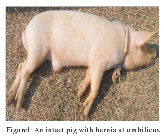
An intact 3 months old male white Yorkshire pig was brought to the Teaching Veterinary Clinical Complex, R.K.Nagar, Tripura, with a long standing swelling present at the ventral abdominal wall at the point of umbilicus (Figure 1). Anamnesis suggested that the swelling tends to increase in size as the pig gradually grows. First hand palpation revealed a hard mass about 6cm, non–painful and irreducible structure with no signs of palpable hernial ring. Fine needle aspiration was done to differentiate it from abscess or tumor. Although the appetite and water intake was reported to be normal, only scanty faeces were voided out at some point of time. Clinical parameters like heart rate, respiratory rate and rectal temperature were within the normal physiological limits. As accurate diagnosis could not be confirmed, exploratory surgery was planned without much delay to overcome any complications.
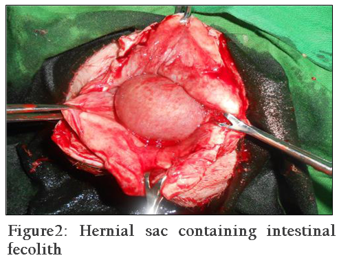
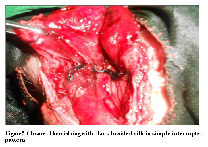
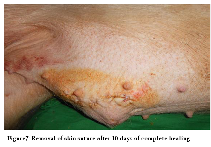
After 12 hours of fasting, the pig was tranquilized with Sequil (Triflupromazine HCl @ 2mg/kg, IM) to control and restrain without much stress. Premedication with Diazepam @2mg/kg, IV, was followed by injection of ketamine HCl (@ 5mg/kg, IV) (Hall et al., 2001; Thurmon et al., 1996) after 10 minutes to achieve a surgical state of anesthesia. Maintenance was done with diazepam–ketamine (1:2) combinations throughout the surgical procedure. After aseptic preparation, animal was placed in dorsoventral position and surgical incision was made over the skin of the herniated mass to cut through the hernial sac and expose the hernial contents. The hernial content consisted of intestines filled with fecolith which sealed the hernial ring (Figure 2). The affected intestine was exteriorized and isolated and incision was given at the antimesenteric border to extract the fecoliths (Figure 3). Thereafter, the opening was closed by Continous Lembert’s suture with 2/0 Chromic catgut (Figure 4). Subsequently, the herniated intestines were slowly reposed back with the help of blunt–end forceps and fingers avoiding any hemorrhage and over–manipulation to prevent any complications. Herniorrhaphy was done using black braided silk in simple interrupted pattern after the ring was freshened (Figure 5) and the muscles and skin closed in usual manner (Figure 6). Post–surgically, antibiotics (Inj. Intacef 500mg bid, IM) and analgesics (Inj. Flunimeg @ 1.1 mg/kg, IM) were administered for a minimum period of 5 days along with regular antiseptic dressings (Scavon cream) and spray (Topicure). Liquid diets for first few weeks followed by semi–solid to solid foods were also advised accordingly. No serious complications were observed during the observation period and the wounds heal completely at 10th day post surgery (Figure 7).

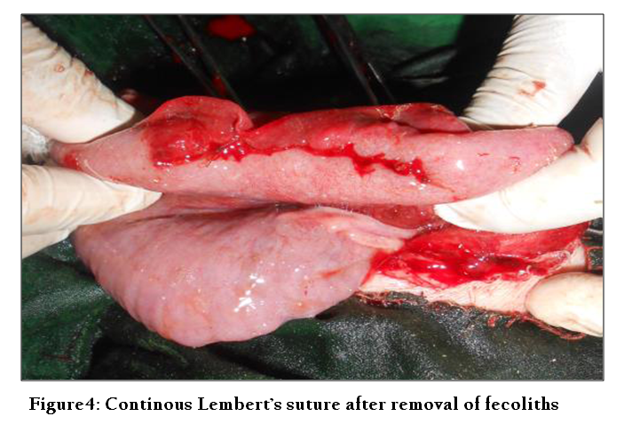
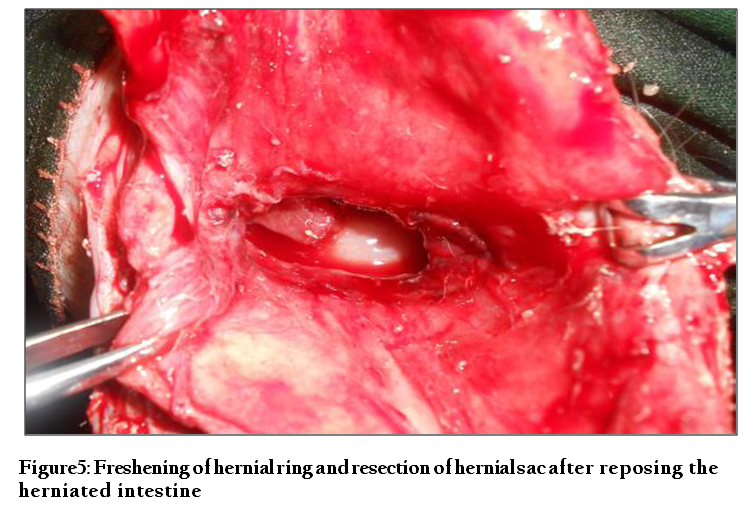
Figure 5: Freshening of hernial ring and resection of hernial sac after reposing the herniated intestine
The exact incidence and cause of umbilical hernia is still unknown. A “familial” cause has been suggested and a few specific genes have been recently shown to associate with this condition (Zhao et al., 2008). Environmental conditions may also play a role in the incidence of this defect as it is thought that environmental compromises such as navel infections early in life may be linked to the incidence of this condition (Ronald and Barbara, 2008). Small umbilical hernias are not serious and sometimes close by themselves as the animal grows. Large umbilical hernias can strangulate when a loop of intestine or portion of another body organ, get pinched off within it. Such cases need to be surgically removed as it involves life threatening. In veterinary practice, concurrent reports of both hernia and intestinal fecolith as a hernial content is rarely reported. The animal may be at risk when such fecolith is present as it might disturb the whole length of digestive system. If prompt diagnosis and treatment is not initiated, the conditions may lead to possible complications like adhesions and hydrocele of the hernial sac, incarcerations and torsions (Venugopal, A. 2007) and abscess as reported in goats (Al–Sobayil and Ahmed, 2007). However, these signs were not observed in our present case except for presence of a fecolith. In the present case, hernia of this kind may be due improper management of umbilicus during the first few weeks after birth. Genetic factors may also contribute to such factor as most of the cases reported are without proper history. Enterotomy was done after packing off the intestines with sterilized clothe to avoid any leakage of the intestinal contents into the abdominal cavity thereby to avoid chances of peritonitis. Herniorrhaphy and enterotomy attempted in this case proved successful and the animal recovered uneventfully without any complications at 10th day post surgery.
In conclusion, umbilical hernia and intestinal fecolith was treated successfully with herniorhhaphy and enterotomy, respectively, in a white Yorkshire piglet. Prompt surgical intervention is the only treatment of choice for corrections of these defects to prolong the life of the patient. Delayed response and ineffective treatment may lead to serious complications which may ultimately lead to the death of the animal.
REFERENCES
Al–Sobayil, F.A., and Ahmed, AF (2007). Surgical treatment for different forms of hernias in sheep and goats. J. Vet. Sci, 8: 185–191.
http://dx.doi.org/10.4142/jvs.2007.8.2.185
PMid:17519574 PMCid:PMC2872719
Edwards M.J. and Mulley, RC (1999). Genetic, developmental and neoplastic diseases. In: Straw BE, D'Allaire S, Mengeling WL, Taylor DJ, eds. Diseases of Swine. 8th ed. Ames, Iowa: Iowa State University Press; 1999; 704–705.
PMid:10476024
Hall LW, Clarke KW and Trim CM (2001). Veterinary Anaesthesia, 10th edn. Philadelphia, W. B. Saunders
Ronald, O. Bates (State Swine Specialist, Department of Animal Science, Michigan State University) and Barbara Straw (State Extension Swine Veterinarian, College of Veterinary Medicine, Michigan State University) and published in the Michigan State University Pork Quarterly, 2008, Volume 13.
Searcy–Bernal, R., Gardner, I.A. and Hird, D.W. 1994. Effects of and factors associated with umbilical hernias in a swine herd. JAVMA. 204:1660–1664.
PMid:8050950
Thurmon, J.C., Tranqilli, W.J. and Benson, G.J. (Eds) 1996. Lumb and Jones' Veterinary Anaesthesia, 3rd edn. Philadelphia, Lea & Febiger.
Venugopalan, A. (2007): Essentials of Veterinary Surgery. 8th Edn. Oxford and IBH Publishing Co. Pvt. Ltd. New Delhi.p.280.
Zhao, X, Du, Z–Q., Vukasinovic, N. V., Rodriguez, F. R., Clutter, A. C. and Rothchild, M.F. 2008. Candidate gene association for hernia and cryptorchidism in commercial lines of pigs. J. Anim. Sci. 86 (Suppl. 2). Abstr.


