Research Journal for Veterinary Practitioners
Research Article
Research Journal for Veterinary Practitioners 2 (4): 58 – 62Investigation of Hepatitis–Hydropericardium Syndrome Caused by Fowl Adenovirus (FAdV) Infection in Broiler Chicken Flocks in Northern Parts of India
Munuswamy Palanivelu*, Shambhu Dayal Singh, Mariappan Asok Kumar, Rajamani Barathidasan, Berin P. Varghese, Shyama N. Prabhu, Kuldeep Dhama
-
Avian Diseases Section, Division of Pathology, Indian Veterinary Research Institute, Izatnagar, (Uttar Pradesh) – 243 122, India
*Corresponding author:drpalvet@gmail.com
ARTICLE CITATION:
Palanivelu M, Singh SD, Kumar MA, Barathidasan R, Varghese BP, Prabhu SN, Dhama K (2014). Investigation of hepatitis–hydropericardium syndrome caused by fowl adenovirus (FAdV) infection in broiler chicken flocks in northern parts of India. Res. J. Vet. Pract. 2 (4): 58 – 62.
Received: 2014–03–07, Revised: 2014–03–21, Accepted: 2014–03–21
The electronic version of this article is the complete one and can be found online at
(
http://dx.doi.org/10.14737/journal.rjvp/2014/2.4.58.62
)
which permits unrestricted use, distribution, and reproduction in any medium, provided the original work is properly cited
ABSTRACT
The present study describes disease investigation of outbreaks of hepatitis–hydropericardium syndrome (HPS) in three different commercial broiler flocks each with a flock size of 5000, 10000, and 20000 birds respectively, of different age groups ranging from 3 to 5 weeks in western Uttar Pradesh and Srinagar region of India. Representative broiler birds (n= 15) from these flocks were brought for postmortem investigation and diagnosis. History obtained from the flock owners revealed a large number of clinically sick birds and varying mortality rates (10–15% (10% in one farm and 15% in two farms). In all the three flock in spite of routine vaccination against major viral diseases (Marek’s disease and infectious bursal diseases). Necropsy examination of the dead and sacrificed birds showed enlarged, pale, friable liver; swollen kidneys with alternating areas of pale and hemorrhagic parenchyma; and varying degrees of hydropericardium. Representative tissue samples of liver, spleen and kidney were collected for histopathology and molecular detection of the HPS causative virus. Histopathologically, there were varying degrees of hepatitis, nephrosis to nephritis, hemorrhages in hepatic and renal parenchyma. Moderate number of hepatocytes showed the presence of large basophilic intranuclear inclusion bodies. Polymerase chain reaction (PCR) testing of the clinical tissue samples, using Hexon protein gene specific primers, yielded virus specific amplicon of 897 bp size confirming the presence of fowl adenovirus (FAdV). The recurrent outbreaks of infectious hepatitis – hydropericardium syndrome in broiler industry in different parts of India need to be investigated further to assess the economic losses due to this disease as well as to adapt suitable prevention and control measures.
INTRODUCTION
Hepatitis–hydropericardium syndrome is one of the important pathological manifestations most commonly associated with adenoviral infection in birds (Balamurugan and Kataria, 2004; Kataria et al., 2013). Adenoviral infections are frequently reported among birds including domestic poultry and wild species of birds but majority of these viruses do not produce apparent clinical signs as a primary etiological agent. However, a few adenoviruses are directly associated with disease conditions and symptoms such as hydropericardium syndrome (HPS), inclusion body hepatitis (IBH), gizzard erosions, respiratory illness, reduced egg production, enteritis, reduced feed conversion, and retarded growth depending upon the serotypes or genotypes involved (Rahul et al., 2005; Domanska–Blicharz et al., 2011; Asthana et al., 2013). The adenoviruses that is associated with these afore mentioned lesions are grouped under the genus Aviadenovirus. The aviadenovirus is further subdivided into five distinct genotypes (A–E) with 12 different serotypes. These viruses are shed in all secretions and excretions, particularly in the feces, which act as the main source of infection to other healthy birds of a flock (Benjamin et al., 2013). The fowl adenoviruses (FAdVs) are diagnosed routinely by isolation of the virus in embryonated chicken eggs or in cell culture, demonstration of virus particles by electron microscopy, or by detection of viral genome by polymerase chain reaction (PCR) (Lim et al., 2012). The virus has affected poultry industry worldwide (Asthana et al., 2013; Kataria et al., 2013) and in past two decades and during recent years many disease outbreaks have been reported from different parts of India (Dahiya et al., 2002; Memon et al., 2006; Gowthaman et al., 2012).
The present paper describes the disease outbreaks of hepatitis–hydropericardium syndrome associated with aviadenovirus infection in broiler chicken flocks in northern states of India and the investigations carried out based on clinical, post–mortem, histopathological examination of affected poultry birds and the molecular detection of the causative virus. The study adds to the epidemiological data available with regard to this virus, seeing other recent reports of disease outbreaks of FAdVs/HPS in the country appropriate prevention and control measures are suggested to alleviate the economic losses to the poultry producers from this economically important virus/disease.
MATERIALS AND METHODS
Source of Samples
Representative clinical tissue samples from liver, spleen and kidneys of each dead and sacrificed birds (n=5) of the affected farms collected during outbreaks of hepatitis–hydropericardium syndrome (HPS) in three different commercial broiler flocks of different age groups (3 to 5 weeks) in western Uttar Pradesh (2 farms) and Srinagar region (1 farm) of India, have constituted as the study materials for disease investigation based on histopathological examination and polymerase chain reaction (PCR) testing for the presence of fowl adenovirus (FAdV).
Histopathology
Tissue samples of 2–3 mm thickness from liver, spleen and kidneys were fixed in 10% neutral buffered formalin and processed for histological examination as per routine paraffin embedding technique (Luna et al., 1968). Tissue sections of 4–5µ thickness were cut and stained with Hematoxylin and Eosin for histo–pathological evaluation.
PCR Testing for Detection of FAV
Viral DNA was extracted from the infected liver, spleen and kidney samples using DNeasy Tissue Kit (Qiagen, Vienna, Austria) as per the manufacturer's protocol. For conventional PCR, 1 μl of DNA was amplified using 10 pmol of each primer (Forward, 5'– CAA RTT CAG RCA GAC GGT –3'; Reverse, 5'– TAG TGA TGM CGS GAC ATC AT –3'). The gene encoding L1 region of the hexon protein of fowl adenovirus group–I was chosen to select primers (Thakor et al., 2012). The reaction mix was amplified in thermocycler (Quanta Biotech, United Kingdom) by initial denaturation at 95 ºC for 3 min followed by 30 cycles of denaturation at 94 ºC for 30 seconds; annealing at 57 ºC for 30 seconds; elongation at 72 ºC for 1 minute followed by final elongation at 72 ºC for 4 minutes. The PCR amplified products were separated by electrophoresis in a submerged 1% agarose gel and visualized under ultraviolet light in a Gel Documentation System.
RESULTS
History, Clinical Signs and Gross Pathology
History from the flock owners revealed that the flock size varied from 5,000 to 20,000 birds and the mortality was relatively high in birds aged between 3–5 weeks than in the other age groups in spite of good management practices.
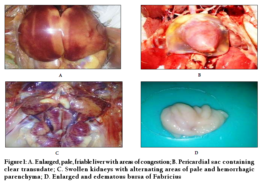
The clinical signs in the affected broiler birds consisted of depression, reduced feed and water intake, reluctance to move, ruffled feathers, huddling, prostration followed by death (10–15%). The necropsy lesions included, pale, friable liver with areas of congestion; varying degrees of hydropericardium containing clear, straw colored fluid; and swollen kidneys with alternating areas of pale and hemorrhagic parenchyma; the bursa of Fabricius was enlarged and edematous (Figure 1).
Histopathology
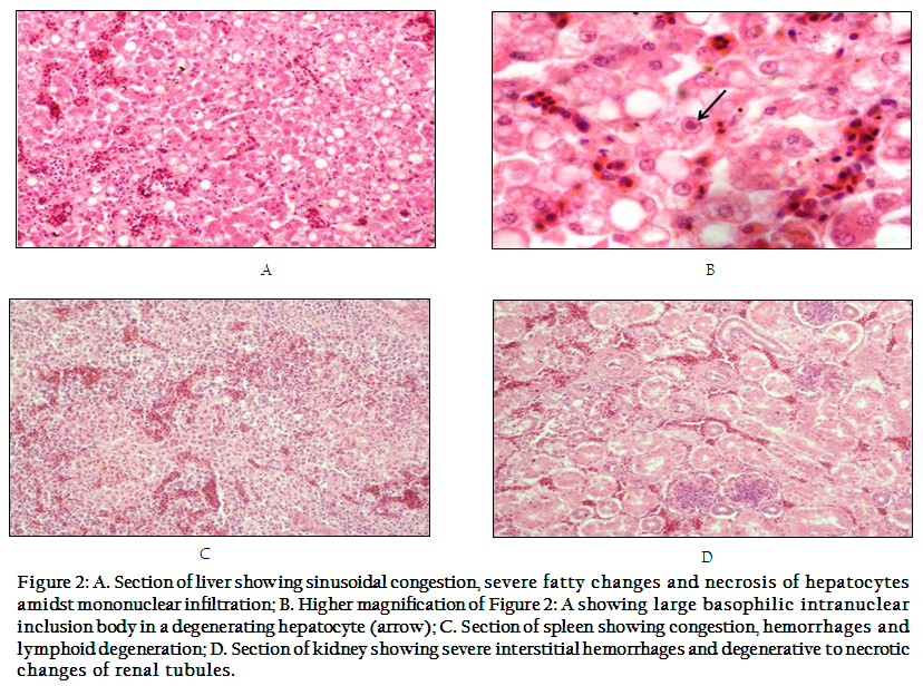
Histopathological findings encountered in the liver were congestion, areas of focal hemorrhages and hepatitis. The hepatic cords in majority of the cases were dissociated and moderate number of hepatocytes showed varying degrees of fatty changes and necrotic changes (Figure 1A) amidst focal collections of leucocytes predominated by mononuclear cells. In a few hepatocytes, the nuclei had disappeared completely, leaving a ghost cell consisting of one or more large vacuoles. Varying degrees of pyknosis, karyorrhexis and karyolysis were evident in the majority of the hepatic cells. In some hepatocytes, karyomegaly and margination of the chromatin was apparent. A few number of the degenerating hepatocytes showed large basophilic intranuclear inclusion bodies which were surrounded by a clear halo with an irregular in outline (Figure 2B). Sections of spleen were characterized by sub–capsular hemorrhages and congestion amidst hemorrhages of parenchyma (Figure 2C). Kidneys showed severe congestion and interstitial hemorrhages along with degenerative and necrotic changes in tubular epithelia (Figure 2D). Bursa of Fabricius from few infected birds revealed inter–follicular edema and degenerative changes.
PCR Detection of FAV in Clinical Samples
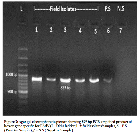
The PCR products visualized in agarose gel electrophoresis showed the presence of amplified DNA products of 897 bp in all the amplified DNA samples from tested tissues (liver, spleen and kidney), confirming the presence of hexon protein gene specific for fowl adenovirus (Figure 3).
DISCUSSION
Viral diseases in birds are major threat to the poultry industry as they cause heavy mortality and severe economic loss in terms of production and productivity. These losses may result either due to primary infections by the potentially pathogenic viruses or by secondary opportunistic pathogens. There are many serotypes of fowl adenoviruses (FAdV) that infects chicken but some serotype produces a clinical form of disease, while infection with other serotype may not result in overt disease particularly in healthy birds. The inclusion body hepatitis–hydropericardium syndrome (IBH–HPS) associated with FAdV has been reported to occur in broilers as well as layers throughout the world (Balamurugan and Kataria, 2004; Asthana et al., 2013). In the past two decades several disease outbreaks have been recorded from poultry flocks of different stares and regions of India, causing severe economic losses to the poultry owners and the industry (Dahiya et al., 2002; Gowthaman et al., 2012; Sawale et al., 2012; Kataria et al., 2013; Kumar et al., 2013). The presence of immunosuppressive disease conditions like infectious bursal disease (IBD), chicken infectious anemia (CIA) and mycotoxins prior to infection with FAdV are believed to be the predisposing factor in the pathogenicity of IBH–HPS syndrome (Toro et al., 1999; Toro et al., 2000; Shivachandra et al., 2003; Gowthaman et al., 2012; Hussain et al., 2012). However, several cases of IBH have been reported without any obvious immunosuppressive diseases (Hafez, 2011).
Gross pathological lesions reported in IBH–HPS include an enlarged pale and friable liver sometimes with necrotic foci, ecchymotic hemorrhages, and less consistently ecchymotic hemorrhages can be observed in leg and breast muscles (Meenakshi–Bal et al., 2005). The heart may appear flabby with varying degrees of hydropericardium characterized by presence of a straw–coloured transudate in the pericardial sac. In addition, nephritis, enlarged spleens and thymus atrophy may also be observed in most dead birds (Deepak et al., 2004; Goyal et al., 2009; Hafez, 2011). Most of these gross lesions were consistently found in all the birds examined from the three outbreaks attended during the present disease investigation study. The histopathological lesions observed in the present disease outbreaks were similar and consistently in line with the previous reports by various workers (Kumar et al., 1997; Nakamura et al., 1999; Venkatesha et al., 2005; Amit et al., 2010; Domanska–Blicharz et al., 2011; Hafez, 2011).
The molecular tool of PCR is being widely used to amplify and detect fowl adenoviruses (FAdVs) specific gene in clinical samples for confirmatory diagnosis of diseases associated with these viruses (Ganesh et al., 2002; Parthiban et al., 2004; Rahul et al., 2004; Kataria et al., 2005; Kataria et al., 2013). The primers most commonly used to detect FAdV genome in recent time are set of primers that amplify gene specific for Hexon protein with a product size of 897 bp (Meulemans et al., 2001). The present study demonstrated amplification of gene specific for Hexon protein in all the clinical samples tested by PCR, representing birds of the three different flocks of the two sates confirming the presence of FAdV infection in broilers with apparent hepatitis – hydropericardium syndrome. To conclude, frequent screening and investigation of poultry flocks for fowl adenovirus infection and molecular characterization of FAdV isolates is warranted to identify the subtype(s) of FAdV associated with hepatitis – hydropericardium syndrome in poultry flocks in different parts of India, which would help design appropriate disease prevention and control strategies to counter this virus and its disease outbreaks for alleviating economic losses being suffered by poultry owners and the industry.
ACKNOWLEDGEMENTS
The authors are highly thankful to the Director, Indian Veterinary Research Institute, Izatnagar, Uttar Pradesh, for providing facilities to carry out this disease investigation and the study.
REFERENCES
Amit G, Hitesh P, Pal JK and Prajapati KS. (2010). Isolation, identification and molecular characterization of Inclusion Body Hepatitis virus. Vet. World., 3: 415–417.
Asthana M, Chandra R and Kumar, R. (2013). Hydropericardium syndrome: current state and future developments. Arch. Virol., 158(5): 921–931.
http://dx.doi.org/10.1007/s00705-012-1570-x
PMid:23242777
Balamurugan V and Kataria JM (2004). The hydropericardium syndrome in poultry–A current scenario. Vet. Res. Commun., 28: 127–148.
http://dx.doi.org/10.1023/B:VERC.0000012115.86894.1e
PMid:14992243
Benjamin S, Ferdinand S, Brigitte B, Michaela A, Robert F, Giovanni C, Calogero T, Isabella M, Richard JWC and Philipp O (2013). Adenoviral gizzard erosion in broiler chickens in Germany. Avian Dis., 57: 159–163.
http://dx.doi.org/10.1637/10330-082312-Case.1
Dahiya S, Ravinder N, Srivastava RN, Hess M, Baldev R and Gulati BR (2002). Fowl adenovirus serotype 4 associated with outbreaks of infectious hydropericardium in Haryana, India. Avian Dis., 46(1): 230–233.
http://dx.doi.org/10.1637/0005-2086(2002)046[0230:FASAWO]2.0.CO;2
Deepak JN, Kataria JM., Dhama K and Verma KC (2004). Pathogenicity of fowl adenovirus serotype–4 isolated from case of hydropericardium syndrome. Indian J. Comp. Microbiol. Immunol. Infect. Dis., 25(1): 7–13.
Domanska–Blicharz K, Tomczyk G, Smietanka K, Kozaczynski W and Minta Z (2011). Molecular characterization of fowl adenoviruses isolated from chickens with gizzard erosions. Poult. Sci., 90: 983–989.
http://dx.doi.org/10.3382/ps.2010-01214
PMid:21489943
Ganesh K, Suryanarayana VV and Raghavan R (2002). Detection of fowl adenovirus associated with hydropericardium hepatitis syndrome by a polymerase chain reaction. Vet. Res. Commun., 26: 73–80.
http://dx.doi.org/10.1023/A:1013361906791
PMid:11862998
Gowthaman V, Singh SD, Dhama K, Barathidasan R, Kumar MA., Desingu PA, Mahajan NK and Ramakrishnan MA (2012). Fowl adenovirus (FAdV) in India: evidence for emerging role as primary respiratory pathogen in chickens. Pak. J. Biol. Sci., 15(18): 900–903.
http://dx.doi.org/10.3923/pjbs.2012.900.903
PMid:24205761
Goyal D, Singh A, Sood N, Gupta K and Sood NK (2009). Pathological changes in naturally occurring inclusion body hepatitis and hydropericardium syndrome in poultry. Ind. J. Vet. Pathol., 33(1): 105–106.
Hafez HM (2011). Avian adenoviruses infections with special attention to inclusion body hepatitis/hydropericardium syndrome and egg drop syndrome. Pak. Vet. J., 31: 85–92.
Hussain I, Mahmood M.S, Arshad MI, Akhtar M, Mahmood F and Rafique A (2012). Immune system dysfunction in broiler chickens experimentally inoculated with fowl adenovirus serotype–4 associated with inclusion body hepatitis hydropericardium syndrome. Turk. J. Vet. Anim. Sci., 36: 223–230.
Kataria JM, Mohan MC, Dey S, Dash BB and Dhama K (2005). Diagnosis and immunoprophylaxis of economically important poultry diseases: a review. Indian J. Anim Sci., 75(5): 555–567.
Kataria JM, Dhama K, Nagarajan S, Chaktaborty S, Kaushal A and Deb R (2013). Fowl adenoviruses causing hydropericardium syndrome in poultry. Adv. Anim. Vet. Sci., 1(4S): 5–13.
Kumar R, Chandra R, Shukla SK, Agrawal DK and Kumar M (1997). Hydropericardium syndrome in India: a preliminary study on the causative agent and control of the disease by inactivated autogenous vaccine. Trop. Anim. Hlth. Prod., 29: 158–164.
http://dx.doi.org/10.1007/BF02633014
PMid:9316232
Kumar V, Kumar R, Chandra R, Bhatt P and Dhama K (2013). Outbreaks of inclusion body hepatitis (IBH) in chickens: pathological studies and isolation of fowl adenovirus. Adv. Anim. Vet. Sci., 1(3S): 21–24.
Lim TH, Kim BY, Kim MS, Jang JH, Lee DH, Kwon YK, Lee JB, Park SY, Choi IS and Song CS (2012). Outbreak of gizzard erosion associated with fowl adenovirus infection in Korea. Poult. Sci., 91: 1113–1117.
http://dx.doi.org/10.3382/ps.2011-02050
PMid:22499868
Luna LG. (1968). Manual of Histologic Staining Methods of the Armed Forces Institute of Pathology. McGraw Hill, New York, USA
Meulemans G, Boschmans M, Van den Berg TP and Decaesstecker M (2001). Polymerase chain reaction combined with restriction enzyme analysis for detection and differentiation of fowl adenoviruses. Avian Pathol., 30: 655–660.
http://dx.doi.org/10.1080/03079450120092143
PMid:19184959
Meenakshi–Bal MS, Kumar H and Sandhu KS (2005). Pathology of hydro–pericardium syndrome in an outbreak in broilers. Ind. J. Vet. Pathol., 29: 46–47.
Memon ZN, Gachal GS, Yusuf M and Arian MA (2006). Incidence of hydropericardium syndrom disease in broilers of Hyderabad, Sindh. Int. J. Poultry Sci., 5(7): 673–676.
http://dx.doi.org/10.3923/ijps.2006.673.676
Nakamura K, Mase M, Yamaguchi S, Shiobahara T and Yuasa N (1999). Pathologic study of specific pathogen free chicks and hens inoculated with adenovirus isolated from hydropericardium syndrome. Avian Dis., 43: 414–423.
http://dx.doi.org/10.2307/1592638
PMid:10494409
Parthiban M, Manoharan S, Aruni AW., Prabhakar TG, Chandran NDJ and Koteeswaran A (2004). Usefulness of polymerase chain reaction in early detection and tissue tropism of fowl adenovirus in experimentally infected chicken. Vet. Res. Commun., 28: 617–622.
http://dx.doi.org/10.1023/B:VERC.0000042873.56448.bd
PMid:15563109
Rahul S, Kataria JM, Senthilkumar N, Dhama K, Uma R, Sylvester SA and Satheesh CC (2004). Polymerase chain reaction based differentiation of various fowl adenovirus serotypes causing inclusion body hepatitis – hydropercardium syndrome (IBH–HPS) of poultry in India. Indian J. Comp. Microbiol. Immunol. Infect. Dis., 25(1): 1–6.
Rahul S, Kataria JM, Senthilkumar N and Dhama K (2005). Association of fowl adenovirus serotype–12 with hydropericardium syndrome of poultry in India. Acta Virologica, 49: 139–143.
PMid:16047743
Sawale GK, Gupta SC, Srivastava PK, Sabale SS, Ingole KH, Pawale NH and MoreBK (2012). Inclusion body hepatitis–hydropericardium syndrome in commercial broiler chickens. Ind. J. Vet Pathol., 36(2): 255–257.
Shivachandra SB, Sah RL, Singh SD, Kataria JM and Manimaran K (2003). Immunosuppression in broiler chicks fed aflatoxin and inoculated with fowl adenovirus serotype–4 (FAV–4) associated with hydropericardium syndrome. Vet. Res. Comm., 27: 39–51.
http://dx.doi.org/10.1023/A:1022058623634
PMid:12625402
Thakor KB, Dave CJ, Prajapati KS, Fefar DT and Jivani BM (2012). Molecular characterization of avian adenovirus causing Inclusion Body Hepatitis–Hydropericardium syndrome in broiler chickens in Anand, Gujarat, India. Vet. World, 5: 178–182.
http://dx.doi.org/10.5455/vetworld.2012.178-182
Toro H, Pruse R, Raue R, Cerda L, Geissea C and Hess M (1999). Characterization of fowl adenoviruses from outbreaks of inclusion body hepatitis/hydropericardium syndrome in Chile. Avian Dis.43: 262–270.
http://dx.doi.org/10.2307/1592616
PMid:10396639
Toro H, Gonzalez C, Cerda L, Hess M, Reyes E and Geissea C (2000). Chicken anemia virus and fowl adenoviruses: association to induce the inclusion body hepatitis/hydropericardium syndrome. Avian Dis., 44: 54–58.
http://dx.doi.org/10.2307/1592507
Venkatesha V, Gowda RNS, Vijayasarathi SK and Rao S (2005). Histopathology of hydropericardium syndrome (HPS) in poultry. Ind. J. Anim. Sci., 75: 271–273.


