Research Journal for Veterinary Practitioners
Case Report

Intussusception of Proventriculus in a Blue & Gold Macaw- A Case Report
S. Ramesh1*, N. Pazhanivel1, S. Meignanalakshmi1,S. Subapriya1, R. Sokkalingam2
1Madras Veterinary College, Tamil Nadu Veterinary & Animal Sciences University, Chennai-600 007, Tamil Nadu, India; 2Society for Prevention of Cruelty to Animals (SPCA), Chennai-600 007, Tamil Nadu, India.
Abstract | A pet blue gold macaw was brought to the local Pet Clinic, Chennai, Tamil Nadu, and India with a history of inappetence for more than 7 days for diagnosis and treatment. The bird was treated for the symptoms of dehydration followed by antibiotics for suspected bacterial infection. After treatment with rehydrating fluids and antibiotics, the bird showed improvement; however, it died after two days of therapy following which a detailed necropsy was conducted. Postmortem examination revealed intussusception of proventriculus with no foreign bodies or parasites detected either in the proventriculus or gizzard. Intestines showed scarce contents with no apparent signs of endoparasites. Histopathological examination of the visceral organs including liver, spleen, pancreas, kidney, lungs, proventriculus, ventriculus and intestine revealed congestion. In addition to congestion, spleen revealed haemorrhages while intestine showed mononuclear infiltration and necrosis on microscopic examination. Based on the gross and histopathological findings, it was concluded that the bird suffered from intussusception of proventriculus which caused the death of the bird.
Keywords | Blue Gold Macaw, Intussusception of proventriculus, Pathology
Received | April 23, 2021; Accepted | May 11, 2021; Published | August 01, 2021
*Correspondence | Ramesh Shunmugam, Madras Veterinary College, Tamil Nadu Veterinary & Animal Sciences University, Chennai-600 007, Tamil Nadu, India; Email: rameshlibya2010@gmail.com
Citation | S. Ramesh, N. Pazhanivel, S. Meignanalakshmi, S. Subapriya, R. Sokkalingam (2021). Intussusception of proventriculus in a blue &gold macaw- a case report. Res J. Vet. Pract. 9(3): 21-23.
DOI | http://dx.doi.org/10.17582/journal.rjvp/2021/9.3.21.23
ISSN | 2308-2798
Copyright © 2021 Shunmugam et al. This is an open access article distributed under the Creative Commons Attribution License, which permits unrestricted use, distribution, and reproduction in any medium, provided the original work is properly cited.
INTRODUCTION
The blue and gold macaws (Ara ararauna), native to south and central America characterized by gorgeous blue body, dark lemon-yellow chest and gradient hues of green on top of its head are probably the most popular large psittacine birds kept as pets because of their striking colors, intelligence, sociability, vocal abilities and close bonding to humans. Like other psittacine birds, these macaws are also prone to a variety of ailments. Though intestinal intussusception, telescoping of one part of intestine into an adjacent segment has been recorded in domestic animals, the reports on an incidence of proventricular intussusception in birds especially in macaws are very scanty. Hence the present paper reports on an incidence of intussusceptions of proventriculus in a male blue- and gold macaw.
MATERIALS AND METHODS
A blue &gold macaw was presented at local pet clinic, Chennai, India with a history of lethargy and inappetence. The bird was housed in a cage alone and fed with commercial feed, fruits and vegetables. The bird was reported active and alert with normal feeding habits 7 days before presentation. Clinical examination was carried out on ailing bird and the bird was treated for the symptoms of dehydration first followed by antibiotics for suspected bacterial infection (Ringer’s Lactate and 5% Dextrose in equal parts @ 50 ml /kg/ day IV Enrofloxacin @ 10 mg/Kg IM and Febendazole @ 15 mg/Kg orally (Luis, 2007). A detailed necropsy was carried on the dead bird and the gross lesions were recorded. Intestinal contents were processed and subjected to microscopic examination for presence of endoparasites. Heart blood and liver impression smears were stained by Giemsa stain and examined under microscope for the presence of bipolar organisms as well as other hemoparasites. Tissue samples namely proventriculus, gizzard, liver, kidney, spleen, lungs, pancreas and intestine were collected in 10 % formalin and processed by routine paraffin embedding method. The tissue sections were cut into 4-6 µm thickness and stained by hematoxylin and eosin for histopathological studies.
RESULTS AND DISCUSSION
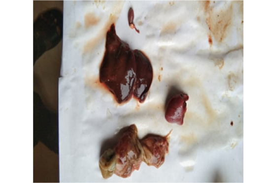
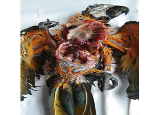
On clinical examination, the ailing bird was found to be dull, severely dehydrated and unable to move. External examination revealed no abnormalities. The bird weighed 700g and the body temperature recorded was 107°F. Despite treatment, the condition of the bird deteriorated and the bird died after two days of therapy. Carcass showed severe emaciation. At necropsy, the sex was identified as male. The mucosal surface of the entire digestive tract revealed diffuse hemorrhages. The predominant finding recorded was in the proventriculus which was seen partly intussuscepted into the gizzard. Liver and spleen showed diffuse congestion. Kidneys were slightly swollen and revealed congestion. Trachea and lungs also showed marked congestion. (Figure 1 & 2) Intestinal contents were watery in consistency and blackish in colour which on microscopic examination revealed no endoparasitic larvae or eggs. No bipolar organisms could be detected in either heart blood smear or liver impression smear. Histopathological examination of the proventriculus, ventriculus, intestine spleen and lungs showed congestion and haemorrhage while liver revealed only congestion. In addition to congestion, proventriculus, ventriculus and intestine revealed multifocal degeneration and necrosis of mucosal epithelium with mononuclear infiltration. Crypts revealed degenerative changes and also necrosis. Pancreas showed congestion, mild acinar cell degeneration and mild mononuclear infiltration. Based on the above findings, diagnosis was confirmed as intussusception of proventriculus.
The present gross findings were in accordance with that of Luis (2007) who also observed similar findings in a 10 years old female Yellow Rosella which died of intussusception of proventriculus . Similar findings were also reported in young synthetic and native lines of chicken (Pradeep and Reddy, 2019).
Nancy et al. (2019) reported proventricular intussusceptions in nineteen laying hens. Hens with proventricular intussusceptions were severely emaciated. Postmortem findings revealed prominent keel, marked muscle atrophy, generalized serous atrophy of fat, no visible proventriculus, esophagus directly entering the ventriculus, and an enlarged, spherical, firm ventriculus, which contained an invaginated, swollen, diffusely ulcerated proventriculus. Histopathological examination showed severe, diffuse necrosis and ulceration of the mucosa of proventriculus. They have attributed the cause to the sporadic emaciation in older laying hens.
Intussusception of intestine had been associated with ulcerative enteritis and coccidiosis in birds (Peckham, 1978). Olasemi and Olatunji (2011) recorded jejunal intussusceptions in a flock of pullets associated with necrotic enteritis of unconfirmed origin and the cause was found to be a shift in gut micro flora which resulted from an imbalance in use of anticoccidial vaccines, anticoccidial drugs and Antibiotic Growth Promoters (AGPs).At necropsy, the intestine was ballooned and the caudal aspect of the evaginating segments revealed a yellowish grey membrane which sloughed off readily showing multifocal haemorrhagic and necrotic ulcers underneath. The intestinal contents were brownish grey in colour.
Okaye (1985) recorded duodenal coccidiosis and intussusceptions of ileum near ileocaecal junction in a flock of 1017 pullets aged 7 weeks. Affected areas were inflamed, necrotic and often gangrenous with sloughing of the surface epithelium. He also observed similar intussusceptions in another farm housed with 328 pullets aged 12-weeks. However examination of intestinal contents revealed negative for coccidiosis. Based on his findings, he opined that starvation might cause intestinal intussusception in fowls and the basic mechanism responsible for the condition appears to be hyperperistalsis and dysperistalsis produced by hunger sensation. However William (1986) opined that intussusception is probably not caused by starvation per se, since it is rarely associated with poultry diseases which cause primary anorexia. He also opined that the coccidiosis or coccidiasis sometimes associated with intussusceptions might develop secondarily following reduction of intake of food and, therefore, of anticoccidial agent, caused by the latter condition.
Palanivelu et al. (2014) reported a case of jejunal intussusception of jejunum in an eight weeks old pullet naturally infected with coccidiosis.Necropsy revealed intussusception within the jejunal segment, the mesenteric and serosal blood vessels were congested and the serosa showed multiple diffuse petechial hemorrhages and necrotic foci, predominantly, in the upper intestinal segments. The lumen of theintestine at the vicinity of intussusception was characterized by necrotic enteritis often with denuded mucosa. On incision of the intussuscepted segments, around 4 centimeter of the jejunal segment was found to be invaginated into its distal part. The lumen of invaginated portion contained hemorrhagic content amidst necrotic debris. Microscopic examination of intestinal contents revealed the presence of oocysts of Eimeria species
The exact cause of intussusception in the present case could not be ruled out as opined by William (1986) who also could not identify even a single cause in three fowls affected with intussusception.
acknowledgements
No acknowledgement.
conflict of interest
There are no conflict of interest.
authors contribution
All the authors contributed for research and manuscript writing.
REFERENCES


