Research Journal for Veterinary Practitioners
Case Report

An Incidence of Cutaneous Pox in an Amazon Parrot (Amazonaoratrix)
S.Ramesh1*, K. Porteen1, S. Meignanalakshmi1, S.Subapriya1, R.Sokkalingam2
1Madras Veterinary College, Tamil Nadu Veterinary and Animal Sciences University, Chennai, TamilNadu, India; 2Veterinary Officer, Society for Prevention of Cruelty to Animals (SPCA), Chennai, TamilNadu, India.
Abstract | A yellow headed Amazon parrot (Amazonaoratrix) maintained as a pet at Chennai was referred to the local veterinarian for clinical diagnosis and treatment with the history of dullness, inappetance and presence of nodules on the unfeathered areas. Physical examination of the bird revealed numerous lesions which were small, focal, and nodular to grayish scab present on the breast, thigh and toes. Examination of the scab using PCR confirmed the presence of Avipox virus. The ailing bird was treated with enrofloxacin for eight days and the bird became active with normal feeding habits after ten days
Keywords | Amazon parrot, Cutaneous pox, PCR
Received | April 21, 2021; Accepted | May 11, 2021; Published | June 25, 2021
*Correspondence | Ramesh Shunmugam, Madras Veterinary College, Tamil Nadu Veterinary and Animal Sciences University Chennai, TamilNadu, India; Email: rameshlibya2010@gmail.com
Citation | Ramesh S, Porteen K, Meignanalakshmi S, Subapriya S, Sokkalingam R (2021). An incidence of cutaneous pox in an amazon parrot (amazonaoratrix). Res J. Vet. Pract. 9(2): 18-20.
DOI | http://dx.doi.org/10.17582/journal.rjvp/2021/9.2.18.20
ISSN | 2308-2798
Copyright © 2021 Shunmugam et al. This is an open access article distributed under the Creative Commons Attribution License, which permits unrestricted use, distribution, and reproduction in any medium, provided the original work is properly cited.
INTRODUCTION
A wide variety of birds gets affected with Avipox viruses (APVs), the largest group of slowly spreading viruses belonging to the subfamily Chordopoxvirinae and family Poxviridae. More than 232 avian species including both domestic as well as free ranging birds viz., crow, sparrow, pigeons, partridges, mynahs, peafowls, eagle, hawk, bustards, doves, pigeons etc. are affected with pox worldwide (Samour, 2008, Joshi et al., 2012, Ramesh et al., 2018).The disease is transmitted either by direct contact or indirect contact through fomites. In addition, mosquitoes play an important role in transmission of this disease. The disease occurs in three different clinical forms namely cutaneous form (dry pox), diphtheritic form (wet pox) and septicemic form(generalized). Of the three forms, cutaneous form is the most common form, characterized by proliferative and nodular lesions on the unfeathered areas while diphtheritic form is characterizedby the appearance of plaques on the mucous membranes of the upper digestive and respiratory tract resulting in difficulty in breathing and feeding. The third form, septicemic form which is very rare form is characterized by generalized lesions resulting in death in majority of cases (Olatunde et al., 2016). Only limited number of cases on avian pox viruses affecting wild birds have been reported (Kyung et al., 2011). Hence the present paper reports an incidence of cutaneous pox in a yellow headed Amazon parrot maintained as a pet in Chennai.
MATERIALS AND METHODS
A yellow headed Amazon parrot maintained as a pet was referred to the local pet clinic, Chennai, India for clinical diagnosis and treatment with the history of dullness, inappetence and presence of nodules on the unfeathered areas. A thorough physical examination was carried out. The cutaneous lesions were collected aseptically and collected in a sterile container and processed for PCR examination.
Droppings were collected in a clean glass container and subjected to microscopic examination.
DNA was extracted from the scab lesions of the infected the birds using Qiagen DNeasy Blood & Tissue kit (Germany) as per manufacturer instructions. The isolated DNA was stored at -20˚C till further use. The PCR amplification was carried out with primers
Forward primer: 5’CAGCAGGTGCTAAACAACAA-3’and
Reverse Primer R: 5’-CGGTAGCTTAACGCCGAATA-3’
optimised in 25 µl PCR reaction mixture using Eppendorf thermal cycler under standardized cycling conditions, Initial denaturation for 5 minutes at 940C, followed by denaturation- 940C for 2 minutes, primer annealing-600C for 1 minute and extension-720C for 1 minute for 35 cycles and final extension at 720C for 2 minutes as per Huw Lee and Huw Lee (1997). The PCR products were subjected to Agarose gel electrophoresis. The amplified product was visualized as a single compact band of expected product size was determined by comparing with the standard molecular weight marker. The gel was visualized in gel doc system and photographed. The droppings were processed by routine sedimentation and floatation techniques and subjected to microscopic examination. The bird was treated parenterally with enrofloxacin for three days @ 5mg/kg b wt and orally @20mg/kg b wt for five days to prevent secondary bacterial infections. Povidone iodine ointment was applied topically over the cutaneous lesions.
RESULTS
The bird was found to be dull, inactive and weak. On physical examination, numerous wart like lesions measuring 1-3 mm in diameter were present scattered on the breast, thigh and the toes (Figure 1). Examination of these lesions using PCR confirmed the presence of Avipox virus. PCR targeting p4b gene revealed that all the three samples viz cutaneous lesions from the breast, toes and thigh were positive for Avipox virus (Figure 2).The droppings were yellowish in colour and watery in consistency which on microscopic examination revealed no endoparasitic larvae or eggs. The bird became active with normal feeding habits after ten days of treatment.
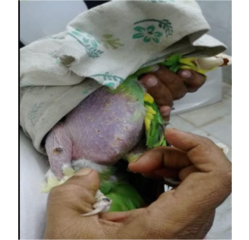
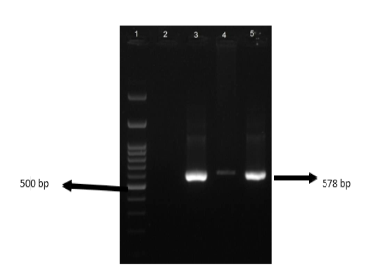
Figure 2: . PCR- 100 bp Ladder (1), negative control (2), lesions over the breast (3), toes (4) and thigh (5)
DISCUSSION
The present findings were in accordance with that of Mohan and Fernandez (2008) who also recorded numerous round rough and firm coalescing masses measuring 0.5- 1.0 cm in diameter on the eyelids, beak, and the mouth of two local pigeons. Wart-like lesions were also recorded on the legs and head around eyelids and beaks of doves (Kyung et al., 2011). Erosions, crust and nodular lesions on the unfeathered areas were also observed in a male jungle crow (Joshi et al., 2012). Ramesh et al. (2018) recorded numerous firm yellowish nodules (1-3 cm indiameter) on the unfeathered areas of skin namely beak, eyelids and feet while no diphtheritic lesions either in the upper respiratory tract or digestive tract in a pigeon found dead on the roadside and affected with avipox. Reddy and Sivajothi (2018) also recorded similar cutaneous lesions in pigeons as observed in the present study which were small, focal, nodular greyish to white scab. Clinical lesions of yellowish crust/nodules on and around beak and eyes and feet were also observed in a scattered group of around 40 pigeons (Audarya et al., 2018)
Avian pox virus was confirmed in Oriental Turtle Doves by PCR using primers specific to the p4b core protein gene of avian pox virus (Kyung et al., 2011) as confirmed in the present case. Molecular diagnosis of avipox was also carried out in pigeons using nodules / scab which were collected aseptically and the virus specific p4b gene (578 bp) was successfully amplified and visualized in 1% agarose gel on electrophoresis (Audarya et al., 2018).
Ramesh et al. (2018) also recorded yellowish watery intestinal contents which on microscopic examination revealed no endoparasites as observed in the present study. Sudhakara Reddy and Sivajothi (2018) reported effective treatment of azithromycin in pigeons infected with cutaneous pox and disappearance of pox lesions by 10 to 12 days of post treatment as recorded in the present case using enrofloxacin.
CONFLICT OF INTEREST
There are no conflicts of interest.
AUTHORS CONTRIBUTION
All the authors contributed for the research and manuscript writing, editing and formatting.
REFERENCES


