Research Journal for Veterinary Practitioners
Research Article

Pregnancy Toxemia in Ewes, and Role of Metabolic Disorder Causing It
Gaadee H. I. M1*, Gehan M. S2
1First Researcher. Biochemistry Unit, Agriculture Research Center (ARC), Animal Health Research Institute;
2First Researcher, Parasitological Unit, Agriculture Research Center (ARC). Animal Health Research Institute.
Abstract | Pregnancy toxemia is a common problem that occurs due to an imbalance between energy requirements in pregnant Ovis aries and the energy provided by the diet consumed. The study was carried on 45 Ovis aries reared under the same living and weather conditions and classified into three groups according to pregnancy and healthy state (group A)., pregnancy with more than on fetus and suffering from clinically pregnancy toxemia, (group B)., pregnant with more than one fetus and clinically healthy and (group C)., non-pregnant and clinically healthy and used as a control. The aim of the study was to evaluate the role of nutritional disorders in initiating the process of pregnancy toxemia in Ovis aries and the associated clinical, biochemical and hematological changes that occur with this disorder, pregnancy toxemia in Ovis aries diagnosed by clinical signs and confirmed by evaluated β-hydroxybutyrate level by chemical and urinalysis. Clinical examination was performed to record clinical changes associated with the disease, two types of blood sample collected for hematological and biochemical studies, where biochemical parameters as glucose, cholesterols, albumin, total protein levels were found statistically lower (P<0.05), in group A than B, C groups, while triglyceride, ALT and AST were found a significant increase in group A than B,C groups. Also MDA as oxidative stress parameter was found significant higher (P<0.05), while cocktails and GSH were considerably decreased, while hematological analysis revealed that erythropenia, oligochromenia, reduced hematocrit, leukocytosis and lymphocytosis in group (A and B).
Keywords | Ovis aries, Sheep, Pregnancy toxemia, Metabolic disorders, Oxidant, Antioxidant response.
Received | December 03, 2020; Accepted | December 29, 2020; Published | March 12, 2021
*Correspondence | Gaadee HIM, First Researcher. Biochemistry Unit, Agriculture Research Center (ARC), Animal Health Research Institute; Email: dr.hodaosman@yahoo.com
Citation | Gaadee HIM, Gehan MS (2021). Pregnancy toxemia in Ovis aries, and role of metabolic disorder cousing it. Res J. Vet. Pract. 9(1): 1-8.
DOI | http://dx.doi.org/10.17582/journal.rjvp/2021/9.1.1.8
ISSN | 2308-2798
Copyright © 2021 Gaadee and Gehan. This is an open access article distributed under the Creative Commons Attribution License, which permits unrestricted use, distribution, and reproduction in any medium, provided the original work is properly cited.
Introduction
Pregnancy toxemia (twin-lamb”disease), is a metabolic disorder of pregnant small ruminants, caused by an abnormal metabolism of carbohydrates and fats, which occurs in the final stage of pregnancy (Brozos et al., 2011). As the energy required during pregnancy is much greater than the energy produced by eating, theses negative energy balance during the late pregnancy is considered key to the development of pregnancy toxemia, (Pough., 2002). Pregnancy toxemia can also be observed in poorly nourished Ovis aries sheep with only a single fetus. (Bani Israel et al, 2008), at any age, breed, over fat or very poor condition (Schlumbohm and Harmeyer.,2004). Risk factors include multiple fetuses, poor quality of ingested energy, decreased dietary energy level, genetic factors, obesity, lack of good body condition, high parasitic load, stress factors and multiple pregnancies (Hefnawy et al., 2011), also Ketosis can occur in obese Ovis aries since fat occupy more space in abdomen resulting a less space for the rumen to hold feed. Additionally, conditions that interrupt feed intake, such as storms, overcrowding, other diseases, can also induce this metabolic disease (Cal-Pereyra et al., 2012).The disease, has a significant economic impact on sheep flock which reflects directly in national income due to mostly loss of Ovis aries sheep and fetuses, (Lima et al., 2012)
Pregnancy toxemia is characterized chemically by hypoglycemia and hyper-ketonemia resulting in the animal being unable to maintain an adequate energy balance, (Cal-Pereyra et al., 2015), Where almost 80% of the fetal growth takes place in the final 6 weeks of pregnancy, with 30–40% of the maternal glucose supply being utilized by the fetal–placental unit (Rook, 2000). If Ovis aries do not receive at least half of the required energy during this period, fat deposits are mobilized in large quantities (Firat and O¨ spinner, 2002).
The clinical symptoms of pregnancy toxemia shows depression, restlessness, grinding of the teeth, sometimes constipation, loss of condition, acetone smell from the mouth, dystocia and neurologic signs include blindness, stiffness, incoordination, tremors in the neck muscles, convulsions and lateral recumbence, (Pough., 2002).
While Laboratory finding of pregnancy toxemia in Ovis aries is associated with a hypoglycemic, hypocalcemia and/or hypomagnesaemia which may complicate the clinical picture (Vania and Plamen, 2017).The liver is important for the blood glucose metabolism, for the glucose’s tissue supply and because it is virtually the only organ where the gluconeogenesis takes place, so it is good to measure liver function tests although there are small contributions from the kidney (Jyothi et al., 2014; Harmeyer and Schlumbohm, 2006).Ketone body can generate superoxide radicals that can then form hydroxyl radicals. These free radicals exert their cytotoxic effect by initiation of membrane phospholipid peroxidation resulting in accumulation of the final products of lipid peroxidation. These products are known to crosslink membrane components causing alterations in membrane permeability and lipid organization as well as cellular dysfunction and membrane damage, while normal cells have the capacity to detoxify superoxide radicals using antioxidant enzymes, as superoxide dismutase (SOD), glutathione peroxidase (GSH-Px), glutathione reductase, and catalase (CAT), which help maintain the intracellular concentration of reduced glutathione and NADPH ,which is necessary for the optimal function of antioxidant defense system (Jain et al., 2006).
The aim of this study was to evaluate the role of nutritional disorders in late stage pregnancy in Ovis aries in initating the process of pregnancy toxemia and its clinical picture, oxidative stress and antioxidant defense system in addition to associated hemato-chemical changes as mirror pregnancy toxemia in the Ovis aries.
Material and methods
Animals
The study was carried on 45 Ovis aries aged (2-7 years) and selected from private farms reared in different villages in Assiut governorate, Egypt, and reared similarly under the same living and weather condition with unsatisfactory standards of animal management and feeding. Ovis aries in the study were at 15-18 weeks of gestation and aged between 2 and 7 years and weighting 40-60 kg, these Ovis aries were subjected to careful clinical and laboratory investigations, according to (Radostits, 2007), and divided into three groups, group (A), included 15 pregnant Ovis aries, showed signs of pregnancy toxemia (pregnant with twins by ultrasound, recumbence, tremor of muscles jaw, depression, restless and blinds) and confirmed by laboratory demonstration of β-Hydroxy butyrate (BHBA) concentrations,Greater than 3.0 mmol/L (Lacetera et al., 2001). (Group B), included 15 healthy pregnant Ovis aries (Pregnant with two or more lambs by ultrasound and in the last month of pregnancy). BHBA concentration > 0.86 mmol /, without any clinical signs of disease (Lacetera et al., 2001). Group C, included 15 non-pregnant Ovis aries, clinical and laboratory healthy and consider as a control group.
Sampling
Blood samples:Two types of blood samples were collected by puncture of the jugular vein using sterile needles and vacationers with and without EDTA. Samples were obtained in the morning before feeding and were stored and transported at 40Ƈ analysis was performed within 24 hours.
1 - 5 ml blood samples from each Ovis aries under the study were collected by the jugular vein puncture in a tube without anticoagulant for subsequent serum preparation and stored until used for chemical analysis. 2 - 5 ml blood with EDTA anticoagulant for hematological analysis.
Fecal sample: A fresh fecal sample was collected from each Ovis aries under the study for parasitological examination, to exclude the parasitic Ovis aries.
Urine sample: Urine sample was collected from each Ovis aries under the study for microscopically and urine stripe examination for detection ketone levels.
Clinical examination
Clinical examinations were conducted according to (Radostits et al., 2007), to record the more clinical signs associated with pregnancy toxemia and excluded the Ovis aries with other diseases.
Parasitological examination
Parasitological examination was carried to exclude the parasitic Ovis aries from the study, according to (Soulsby., 1982), Ovis aries under the study were dosed with Albendazole twice within 15 days, and then the feces are re-examined to ensure that there are no worms.
Urine stripe examination
Ketone urine Strips provide an accurate and quick way to detect pregnancy toxemia in sheep. A range of shades
Table 1: Clinical signs of Ovis aries with pregnancy toxemia, healthy pregnancy and healthy non pregnant.
| Clinical signs | Pregnancy toxemia ewes (group A) | Pregnant healthy ewes (group B) | Non pregnant ewes (group C) |
| Number | 15 | 15 | 15 |
|
Ultrasound examination |
Pregnant with twin (Diseased) | Pregnant with twin (Healthy) | Non pregnant |
| Temperature | 38.8 | 38.9 | 39.6 |
| Fruity odor | Present | ------- | ----- |
| Heart rate (min) | 80- 81 | 78 -79 | 76- 77 |
| Respiratory rate (min) | 24- 29 | 24- 26 | 18- 19 |
| Rumen motility (min) | 9 - 10 | 10 -11 | 10 -12 |
| Depression | + | - | - |
| Mucous membrane | Pale - Icterus | Pale | Rose red |
| Nervous signs | + | - | - |
| Recumbence | + | - | - |
| Apparent Blinds | + | - | - |
| Staggering gait | + | - | - |
gives a semi-quantitative result in about 40 seconds. The urine samples were analyzed using Multistix 10 SG reagent strips (Siemens Healthcare Private Limited, India) for the qualitative determination of ketone bodies, glucose and Protein (Gurdogan et al., 2014).
Briefly. Wash your hands, take a urine sample in a small container, immerse the absorptive end of the strip into the sample for a few seconds, remove strip, wait until strip change its color. Compare the strip with the color chart on the packaging and record the results.
Biochemical analysis
Biochemical parameters were evaluated as the following:
Glucose, Triglyceride, Cholesterol, β-Hydroxybutyrate, Alanine aminotransferase (ALT), Aspartate aminotransferase (AST), albumin and total protein, this parameter were evaluated by spectrophotometry method using commercially available kits supplied by Biomed diagnostics (Egypt), according to manufacturer’s instruction.
Serum oxidant and antioxidant analysis: Oxidant and antioxidant status were evaluated by measuring malondialdehyde (MDA), glutathione transferase (GSH), superoxide dismutase (SOD) and catalase (CAT). All these parameters were determined by spectrophotometry method by using commercial diagnostic kits, bio diagnostic (Egypt).
Hematological analysis
Hematological parameters include, red blood cell count (RBCs), total white blood cell count (TWBCs), Hemoglobin (HB), packet cell volume (PCV) and parameters of differential leukocyte count were determined according to routine hematological procedures.
Statistical analysis
Data were analyzed by using one-way analysis of variance (ANOVA). All data were analyzed using Sigma Stat 3.1, statistical software for data analysis (SPSS Inc., Chicago, IL, USA). Values were represented by means ± standard error (SE). All differences were considered statistically significant at P < 0.05 according to (SAS, 2011).
Results
Clinical examination
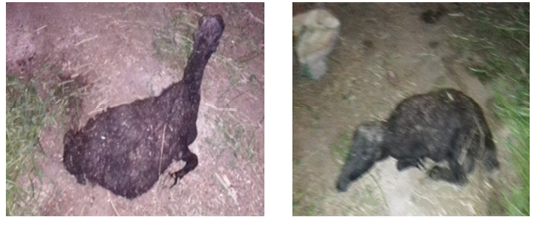
Clinical signs of three groups of Ovis aries sheep are shown in Table 1.Where the clinical signs in group (B,C),pregnant healthy and control group were within the reference ranges while the clinical signs of group (A),pregnant Ovis aries sheep with clinical toxemia revealed reduced appetite, enhanced thirst and depression. In some sheep, locomotor disturbances of progressing nature followed by lying down, tremor of the head and neck. As the disease progressed, ataxia, Sternal recompense, abortion, coma and death were observed (Figure, 1, 2).
Fecal analysis
Fecal examination for all Ovis aries under study to help select the Ovis aries free from parasites and exclude parasitic Ovis aries.
Urine analysis
Urine analysis by urine test strip was carried for all Ovis aries under study, where Urine analysis was remarkable for ketonuria and illustrated in Table (2), (urine ketone bodies
Table 2: Biochemical parameters monitor of pregnancy toxemia Ovis aries compared with healthy pregnant Ovis aries and healthy non pregnant Ovis aries.
|
|
Pregnancy toxemia ewes (group A) |
Healthy pregnant ewes (group B) |
Healthy non pregnant ewes (group C) |
|
BHBA(mmol/l) |
5.02±0.03↑*** |
1.09±0.05↑* |
0.32±0.01 |
|
Glucose (mmol/l) |
1.02±0.03↓*** |
2.91±0.08↓* |
4.78±0.04 |
|
Cholesterol(mg/dl) |
46.45±0.15↓** |
67.05±0.14↓* |
77.33±0.22 |
|
Triglycerides(mg/dl) |
108.46±0.05↑*** |
87.33±0.023↑* |
57.32±1.02 |
|
ALT(u/l) |
88.62±0.49↑** |
39.60±0.83↑* |
26.81±0.73 |
|
AST(u/l) |
171.20±7.46↑*** |
68.21±0.16↑* |
57.26±0.80 |
|
Albumin (g/dl) |
1.82±0.21↓** |
2.33±0.14↓* |
3.38±0.029 |
|
Total Protein(g/dl) |
5.02±0.12↓** |
7.04±0.24↓* |
8.32±0.17 |
|
Urine multistix |
+++ |
+ |
- |
group B ++, in group A, +++., with Bayer
Multistix 10 SG®).

Biochemical analysis
Blood β-Hydroxybutrate acid (BHBA) analysis in a control group of sheep were within the reference range (Table. 2). BHBA were statistically increased in group A and B ((5.02±0.03mmol/l, 1.09±0.05mmol/l), respectively, than the control group (0.32±0.01mmol/l), where statistical significant value (р<0.005).
Statistical significant difference biochemical parameters between three groups were present in Table 2, where glucose, cholesterols, albumin, total protein levels were found statistically lower (P<0.05) than control groups, while, triglyceride, ALT and AST were found significant increases than control groups. In Table 3, MDA was found higher (P<0.05), while catalase and GSH were found highly significant decrease.
Table 3: Serum oxidant and antioxidant parameters in pregnancy toxemia Ovis aries in compared with pregnant healthy Ovis aries and non-pregnant healthy Ovis aries
|
Group |
Pregnancy toxemia ewes (group A) | Pregnant healthy ewes (group B) | Non pregnant ewes (group C) |
| MDA (nmol/ml) | 14.46±0.24* | 11.93±0.18 | 10.21±0.21 |
| SOD (u/ml) | 460±30.84 | 456±28. 16 | 455±26. 27 |
| Catalase (u/l) | 190±6. 24↓** | 210±7. 53↓* | 250±5. 63 |
| GSH (nmol/l) | 1. 57±3. 21↓* | 2. 38±9. 21 | 2. 84±8. 43 |
Hematological analysis
Blood picture in the control group showed that all parameters were within the physiological ranges, while in pregnancy toxemia group, erythrocyte count, hemoglobin and hematocrit showed significantly decreased in comparison with other groups, while insignificant change between pregnant healthy group and control group. Leukocyte count and lymphocyte showed a statistical significant increase in group A in relation to control group (12.70±3.3 х109/l) and (70.63±0.4 %) in Table 3. Other blood parameters do not have any significant statistical change.
Discussion
Pregnancy toxemia is a metabolic problem, laboratory characterized by hypoglycemia and hyperketonemia resulting from incapacity of the animal to maintain adequate energy balance.Clinical study of pregnancy toxemia in Ovis aries sheep revealed normal rectal temperature and respiratory rate, which were agree with the reports of many authors (Van Saun, 2000; Schlumbohm and Harmeyer, 2008), but contrast to (Rodolfo et al., 2014) who reported increased body temperature., Anorexia was observed in all cases which were in agreement with (Balikci et al., 2009, Al-Qudah., 2011, Rodolfo et al., 2014], recumbency was recorded which was in agreement with (Balikci et al., 2009, Barakat et al., 2007; Hefnawy et al., 2011; Al-Qudah., 2011), fruity odor of breath was found in sheep with pregnancy toxemia which was a typical characteristic of ketone bodies which was increased in pregnancy toxemia,that agreement with (Balikci et al., 2009), Chew movement and salivation were seen in the nervous form of pregnancy toxemia in Ovis aries sheep. It was due to the cerebral hypoglycemia and in agreement with (Balikci et al., 2009). Ovis aries sheep presented with apparent blindness were in agreement with, (Barakat et al., 2007; Al-Qudah., 2011). Nervous degeneration was due to cerebral hypoglycemia resulted in apparent blindness. Ovis aries sheep with pregnancy toxemia presented with bloat were in agreement with (Souto et al., 2013, Rodolfo et al., 2014) and the bloat was attributed to longer time recumbency, anorexia, stasis of rumen motility, and ketoacidosis. Some Ovis aries sheep presented with dropped head was in agreement with (Abdelaal et al., 2013; Reddy et al., 2014). The dropped head condition was nervous manifestation due to lack of energy in the nervous form of pregnancy toxemia. Some sheep presented with the extension of the head, were in Agreement with (Abdelaal et al., 2013). According to clinical manifestation, pregnancy toxemia is classified into subclinical and clinical cases by numerous authors (Gordon et al., 2013); we also agree with their opinion.The Ovis aries sheep in group B (Healthy pregnant group), show rumen contraction disorders, whose strength and frequency decreased and were close to the lower limits secondary to alkalization of rumen and duodenal content by increased ammonia and biogenic amine concentrations reported by (Balikci et al., 2009). The established changes in the general clinical status in sheep with clinical pregnancy toxemia were localized in the nervous system, digestive system and respiratory, where heart and respiratory disorder in Ovis aries sheep were due to functional damage of the cardiovascular and respiratory systems from one part and rumen metabolites on the other. Respiratory and heart failure noticed on the color of visible mucous membranes that changed from pale rose-red become diffusely red and electric tint with a more serious case. The rumen contraction in clinical pregnancy toxemia decrease in strength and frequency compared to control groups. These results are in agreement with, (Kabakci et al., 2003; Balikci et al., 2009).
Significantly decreased glucose level of the present study was in agreement with the result of many authors such as, (Hefnawy et al., 2010; Al-Qudah, 2011), However, incompitable of the present study, report of (Lima et al., 2012; Souto et al., 2013) and Showed hyperglycemia in the later stages of pregnancy toxemia when the fetuses were dead. Hypoglycemia was attributed to dietary deficiency of net energy along with the increased demand for energy in the later part of pregnancy. Due to increased BHBA, there was NEB, which causes a hypoglycemic effect, reduced food intake and glucose turnover leads to pregnancy toxemia, β-Hydroxybutrate acid (BHBA) concentration is the main chemical blood parameter used as an early marker for pregnancy toxemia diagnosis with blood (Lacetera et al., 2002). This was an agreement with the present as we found that, 5. 02±0. 03, 1. 09±0. 05 and 0. 32±0. 01 in group A, B and C, respectively. High level, β-Hydroxybutrate acid (BHBA) in the blood is a compensatory mechanism in response to occurring carbohydrate deficiency (Ingvartsen, 2006), which lead to excessive lipolysis that, accompanied by the production of large acetyl CoA amounts, the tri-carboxylic acid cycle is not capable to convert entirely fatty acids. Consequently, acetyl CoA is metabolized to acetoacetate, which is reduced to β-Hydroxybutrate (BHBA) through BHBA dehydrogenase or is spontaneously decarboxylase to acetone (Roche et al., 2013).
Table 4: Changes in hematological parameters in Ovis aries sheep pregnancy toxemia in comparison with other groups.
|
Parameters |
Pregnant Toxemia ewes (group A) | Healthy pregnant ewes (group B) | Non pregnant ewes (group C) (control) |
|
RBCs (x1012/l) |
7.36±2.3↓* | 8.88±0.5 | 9.83±2.1 |
| HB (g/dl) | 8.66±0.9↓* | 14.66±2.5 | 15.00±3.9 |
| PCV (%) | 21.50±3.2↓** | 26.51±1.6 | 31.10±4.3 |
|
WBCs(x109/l) |
12.70±3.3↑** | 11.12±2.3 | 8.88±1.1 |
| Lymphocyte (%) | 70.63±0.4 ↑* | 60.84±1.0 | 58.90±0.5 |
| Monocytes (%) | 3.86±0.2 | 4.83±0.1 | 3.63±0.1 |
| Neutrophil (%) | 20.09±0.3 | 30.15± 0.66 | 32.20±0.3 |
| Eosinophil (%) | 4.07±0.2 | 3.10± 0.34 | 4.50±0.0 |
| Basophil (%) | 1.04±0.1 | 1.20±0.3 | 1.15±0.1 |
The increased β-Hydroxybutrate (BHBA) concentration in blood reveals the incomplete oxidation of non-esterified fatty acids (NEFA) in the tri-carboxylic acid cycle at the time of NEB (Doepel et al., 2002).The enhanced lipolysis, ketogenesis and hypoglycemia could be a sequel to stress which manifested with increased cortisol and that result agreement with (Burton et al., 2005; Abba et al., 2015).
Significant increase of β-Hydroxybutyrate (BHB) concentrations in pregnancy suggests that, β-Hydroxybutyrate (BHB) may play a role in induction of oxidative stress (lipid peroxidation) in Ovis aries with higher concentrations of ketones. Similar results are reported in cows with subclinical ketosis (Sahoo et al., 2009), while significant decrease in total protein and albumin were recorded in pregnancy toxemic Ovis aries in comparison to other groups and these results attributed to increased protein catabolism, decomposing fetuses or terminal kidney failure and this agreed by (Aly and Elshahawy, 2016).
The higher levels of AST and ALT activities in the pregnancy toxemias Ovis aries may attribute to fat mobilization because of negative energy balance and hepatic damage or hepatic lipidosis. These results were in agreement with the report of (El-Deeb, 2012; Aly and Elshahawy, 2016, Barakat et al., 2007, Balikci et al., 2009). In contrast with the present study, (Gupta et al., 2008), reported decreased values of SAST and SALT
The marked increase in serum triglyceride may be attributed to increase of fat breakdown and mobilization of fat stores to face marked decrease of glucose (Aly and Elshahawy, 2016). While Significant decrease of cholesterol level may attributed to decrease food intake, hepatic insufficiency and physiological alteration of endocrine function (Waziri et al. 2010).
Significance increase in serum level of malondialdehyed (MDA), which reflect lipid peroxidation in Ovis aries with pregnancy toxemia compared with healthy pregnant and healthy non pregnant Ovis aries, may be attributed to the antioxidant defense of Ovis aries decreases as gestation progress up to lactation and was accompanied by increased generation of pro-oxidants, thus predispose the animals to oxidative stress lipid peroxidation may be occur due to disturbed redox balance (Pilarczyk et al., 2012), It is also associated with lower level of antioxidants, as well as the presence of high concentrations of ketone bodies in the blood of Ovis aries. All of that points to oxidative stress (lipid peroxidation) is involved in the development and complications of pregnancy toxemia and relationship between hyperketonemia and lipid peroxidation was shown, suggesting that ketonemia is a precursor risk factor for oxidative stress (lipid peroxidation) in Ovis aries affected with pregnancy toxemia.
Decline catalase activities as the Ovis aries approach parturition indicated lower level of antioxidant and degree of oxidant stress, this result agrees with. (Pilarczyk et al., 2012) reported that serum GPx activity varied according to the physiological status
Hematological analysis revealed significant decrease in RBCs, hemoglobin and hematocrit in Ovis aries sheep with pregnancy toxemia, these result agreement with data reported in sheep by (Gupta et al., 2008), goats by (Abba et al., 2015), the result could be attributed to the body negative energy balance (NEB) during pregnancy, while leukocytosis and lymphocytosis in pregnancy toxemia Ovis aries sheep could be attributed to the presence of acute and chronic inflammations (Gavan et al., 2010).
CONCLUSIONs anD Recommendations
Pregnancy toxemia affects both thin and fat Ovis aries at any age in the last trimester stage of pregnancy where the pregnant uterus leads to restricting rumen capacity and decrease food intake, that leading to initiates of negative energy balance. Furthermore decrease blood glucose levels with subsequent lipid mobilization for providing body energy, followed by hyperketonemia and subsequent appearance of clinical signs on the Ovis aries. So must avoid any starvation and/or deviation in food in advanced stages of pregnancy in Ovis aries.
Early indicators of pregnancy toxemia in Ovis aries include the presence, β-hydroxybutyric acid, ketone body in blood and urine .Hence assess of blood β-hydroxybutyric acid (BHBA) and qualitative evaluation of ketone bodies in urine using urine dipstick are reliable methods in the diagnosis of subclinical form of pregnancy toxemia under field condition. This information could be used by veterinarians to aid in clinical investigation of Ovis aries sheep flock and individual Ovis aries in late pregnancy, furthermore the understanding of pathophysiology changes that occur in these Ovis aries.
A study has revealed that Ovis aries in the third trimester and lactation displayed oxidative stress due to intense metabolism during the physiological states. It is recommended that antioxidant fortification/supplementation should be employed during late gestation as management practice to combat the oxidative stress observed.
Take special care with Ovis aries in late pregnancy so that they are not off food for a long time. Also, don’t suddenly change the type of feed because the rumen bacteria may not adapt to the new diet. Seek assistance from your vet or adviser in developing a feeding strategy.
acknowledgements
First of all we gratfull thanks to Allah,who give we every things and support us to complete this paper. also we offer cardinal thanks to . Professor .Dr . Osman. F.A. to its help for our as possible.
conflict of interest
We certificate there is no conflict of interest in this work.
authors contribution
Gaadee HIM conceived of the presented idea. Developed the theory and performed the computations. Verified the biochemical studies. Gehan MS performed the parasitological analysis. All authors discussed the results and contributed to the final manuscript.
References
Schlumbohm C, Harmeyer J (2004). Hyper-ketonemia impairs glucose metabolism in pregnant Ovis aries. J. Dairy. Sci. 87: 350–358.


