Research Journal for Veterinary Practitioners
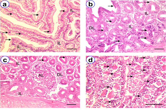
HE staining of duodenum at D30 (a), D90 (b), D180 (c~d) showing villi (V), intraepithelial lymphocytes (arrow), isolatory lymphocytes (IL), diffuse lymphocytes (DL), aggregated lymphocytes (AL); Scale bar = 50 µm (a~c), 100 µm (d).
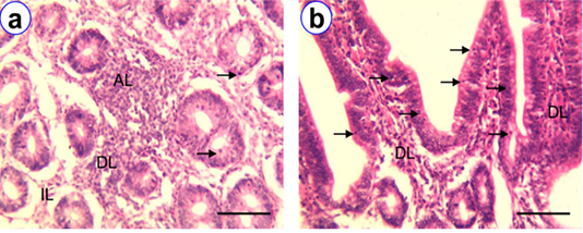
HE staining of jejunum at D30 (a) and D180 (b) showing intraepithelial lymphocytes (arrow), isolatory lymphocytes (IL), diffuse lymphocytes (DL), aggregated lymphocytes (AL); Scale bar = 100 µm (a~b).
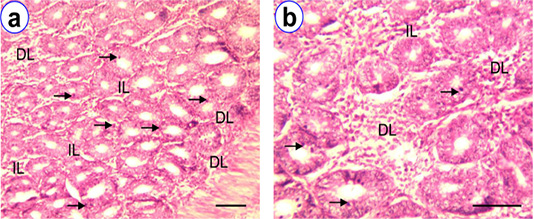
HE staining of ileum at D90 (a~b) showing intraepithelial lymphocytes (arrow), isolatory lymphocytes (IL), diffuse lymphocytes (DL); Scale bar = 50 µm (a), 100 µm (b).
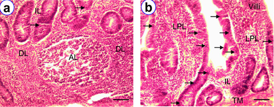
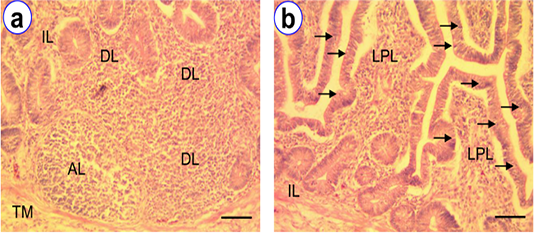
HE staining of cecum at D180 (a~b) showing villi (V), intraepithelial lymphocytes (arrow), isolatory lymphocytes (IL), diffuse lymphocytes (DL), aggregated lymphocytes (AL), lamina proprial lymphocytes (LPL); Scale bar = 50 µm (a~b).
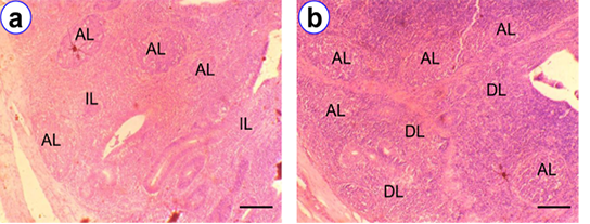
HE staining of cecal tonsil at D90 (a) and D180 (b) showing isolatory lymphocytes (IL), diffuse lymphocytes (DL), aggregated lymphocytes (AL); Scale bar = 50 µm (a~b).
HE staining of colo-rectum at D180 (a~b) showing intraepithelial lymphocytes (arrow), isolatory lymphocytes (IL), diffuse lymphocytes (DL), aggregated lymphocytes (AL); Scale bar = 50 µm (a~b).


