Research Journal for Veterinary Practitioners
Case Report

Description of the Case of a Swim Bladder Torsion in Goldfish (Carassius auratus, Linnaeus 1758)
Andreia Garcês1*, Luís Sousa2, Roberto Sargo2, Filipe Silva2,3, Isabel Pires3
1Inno- Serviços Veterinários, Department of Microbiology, R. Cândido de Sousa 15, 4710-503 Braga, Portugal; 2Wildlife Rehabilitation Centre of University of Trás-os-Montes and Alto Douro (CRAS-UTAD), University of Trás-os-Montes and Alto Douro, Quinta de Prados 5000-801, Vila Real, Portugal; 3CECAV, University of Trás-os-Montes and Alto Douro, Quinta de Prados 5000-801, Vila Real, Portugal.
Abstract | In this paper, the authors report a case of a swim bladder torsion at the ductus communicans in a 15-year-old goldfish (Carassius auratus, Linnaeus 1758). The lesion was observed at post-mortem exam. Its aetiology is not yet well understood, and in the presented case could have resulted of the conjugation of different factors: previously traumas, postural changes, and the corporal condition of the animal. Torsions in swim bladders are not common and still very poorly documented. This report is also a reinforcement of the importance of the post-mortem exam in these animals.
Keywords | Pathology, Fish, Carassius auratus, Torsion, Swim bladder
Received | August 07, 2020; Accepted | August 22, 2020; Published | October 15, 2020
*Correspondence | Andreia Garces, Inno- Serviços Veterinários, Department of Microbiology, R. Cândido de Sousa 15, 4710-503 Braga, Portugal; Email: andreiamvg@gmail.com
Citation | Garces A, Sousa L, Sargo R, Silva F, Pires I (2020). Description of the case of a swim bladder torsion in goldfish (carassius auratus, linnaeus 1758). Res J. Vet. Pract. 8(4): 45-47.
DOI | http://dx.doi.org/10.17582/journal.rjvp/2020/8.4.45.47
ISSN | 2308-2798
Copyright © 2020 Garces et al. This is an open access article distributed under the Creative Commons Attribution License, which permits unrestricted use, distribution, and reproduction in any medium, provided the original work is properly cited.
The swim bladder is a hollow organ filled with a gas, whose main functions are to help the fish to maintain its depth, control its buoyancy, oxygen storage, respiratory functions and communication (hearing and sound production) (Petrick, 1975; Grom, 2013; Smith, 2019). In Carassius auratus (Cyprinidae) is constituted by two chambers – anterior and posterior, linked by a narrow isthmus - ductus communicans. The anterior chamber is bigger than the posterior. The anterior portion of the anterior chamber is connected with the tripus of the Weberian apparatus (physostomous duct) (Grom, 2013). Cyprinidae family members are physostomous species, which means that their swim bladder is connected to the oesophagus by the pneumatic duct (Genten et al., 2009). Due to the connection between the oral cavity and swim bladder in these species, is frequent the occurrence of infections in the lumen of the swim bladder associated to poor water quality by opportunistic pathogenic bacteria (e.g., aeromonads or atypical mycobacteria) (Smith, 2019).
A 15-year-old goldfish (Carassius auratus, Linnaeus 1758) was admitted in the Exotic Animal Clinic from the Veterinary Hospital of Trás-os-Montes and Alto Douro. The owner described changes in the swimming pattern – swimming in circles (circling)- and positive buoyancy. Anamnesis revealed that the animal suffered several traumatic accidents, through its life, such as tank falls. In these episodes, the animal remained on the dry floor for unknown periods. The physical examination confirmed the altered swimming pattern, a curvature of the body to the right side and horizontal nystagmus. The celomic cavity was mildly dilated, and the animal seemed to have a body condition above the normal, nearly obese. A full-body x-ray was obtained with the animal inside of a small container filled with water. Figure 1A shows the radiographic image of the swimming bladder. Insufflation of the swimming bladder was confirmed, with a marked isthmus between the anterior and posterior cameras. Puncture of the posterior camera was performed with a 26G hypodermic needle attached to a 2-millilitre syringe and mild suction was applied to result in retrieving a 2-millilitre air sample. After chamber puncture, the x-ray showed that air was still trapped in the anterior camera of the swimming bladder (Figure 1B) and the animal still had positive buoyancy and right-sided curvature of the body. The quality of the x-ray is not ideal as it was made in a container with water.
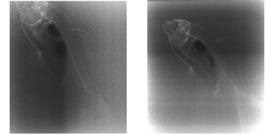
Figure 1: Laterolateral horizontal projection of the goldfish (Carassius auratus, Linnaeus 1758) just before (a) and after (b) puncture of the posterior camera.
Due to the impossibility of correcting this condition, the animal’s advanced age and the lack of welfare, the owner decided for euthanasia. Isoflurane was added to the swimming water and with the animal anaesthetized, 1 mL of pentobarbital sodium solution was administered intravenously. Necropsy was performed immediately after death, according to the techniques described for fish, under appropriate conditions of safety and hygiene (Garcês and Pires, 2017).
On post-mortem examination the fat bodies in the celomic cavity were very enlarged, confirming that this fish was obese (Figure 2).

Figure 2: Morphology and location of the coelomic organs of a goldfish (Carassius auratus, Linnaeus 1758) after incision (SB – swim bladder; FB – fat bodies; St, stomach)
A complete clockwise torsion of the swim bladder (more than 180º) at ductus communicans level was detected. In Figure 3 we can observe the twisting (Figure 3A) and a normal swim bladder without torsion (Figure 3B) to compare. After incision, the lumen of the posterior chamber presents a translucid, limpid mucus in a residual quantity. The wall of the posterior chamber appeared thicker. The bladder had no adhesions to adjacent tissues.
The internal organs didn´t present any visible alterations, except for the swimming bladder and the slightly friable liver.
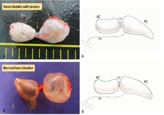
Figure 3: In A swim bladder torsion, the point (arrow) where the torsion happened, and in B a normal swim bladder (anterior chamber -AC, posterior chamber -PC, communicating duct -CD and pneumatic duct -PD). On the left a schematic representation of the torsion (A) and normal swim bladder (B).
Tissue specimens were routinely fixed in 10% neutral buffered formalin and embedded in paraffin, sectioned at 3 m and stained with haematoxylin and eosin (H&E) for histologic examination at the Laboratory of Histology and Anatomic Pathology of UTAD.
The histopathologic exam revealed no alteration in the cellular architecture of the swim bladder. In the anterior chamber, it was observed a single layer of flattened epithelial cells, with a prominent basal lamina below the epithelial cells. The lamina propria of the anterior chamber contained collagen fibrils, elastic fibres, and fibroblasts. The posterior presented of cuboid squamous cells. A thin layer of highly fibrillar elastin adheres closely to the basal lamina of the epithelial cells of the posterior chamber. The tunica externa was composed of two distinct layers of densely packed collagen fibrils. The liver presented a brown pigment in macrophages and hepatic vacuolization consistent with lipidosis (Figure 4). Unfortunately, many structures were degraded due to the rapid decomposition of the corpse.
The swim bladder torsion is a rarely reported condition, with only a few occurrences in ornamental goldfish and rainbow trout (Smith, 2019). References to this pathological condition were found in the textbooks related to pathology, medicine and surgery of fish, but without any proper description of all the case.
Its aetiology is not yet well understood but can be related to buoyancy disorders that can lead to the torsion itself later. The over-insufflation of the swim bladder caused by bacterial diseases, tumours, virus or gas supersaturation; displacement of the posterior chamber of the swim bladder due to tumours, polycystic kidney diseases or trauma; the presence of fluid in the swim bladder; intestinal tympany; rupture of the swim bladder could also be implied in this process (Widgoose, 2001; Woo and Bruno, 2011; Roberts, 2012; Smith, 2019).
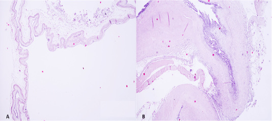
Figure 4: Anterior chamber presented a single layer of flattened epithelial cells, with a prominent basal lamina below the epithelial cells and lamina propria containing collagen fibrils, elastic fibres, and fibroblasts. In B section in the point where the torsion occurred. Many cells were degraded due to the rapid decomposition of the corpse (H&E, 40x).
In Goldfish, this condition could also be explained by its anatomical features. Goldfish, particularly Asian varieties, have short stout bodies can that lead to a unbalance in the swim bladder (Roberts, 2012).
The diagnose of swim bladder torsion was only possible at post mortem examination. In this case, it was not observed any signs of bacterial or viral infections or the presence of neoplastic nodules. We can presume that this torsion could be a consequence of the previously suffered traumas and changes in its posture. Additionally, its anatomical features and the animal body condition (obesity) could contribute to the torsion of the swim bladder.
This report could improve the knowledge on swim bladder pathology in fishes, especially in goldfish (Carassius auratus, Linnaeus 1758) and reinforce the importance of the post-mortem exam in these animals.
Conflict of interests
The authors declare that they have no conflict of interest.
References


