Research Journal for Veterinary Practitioners
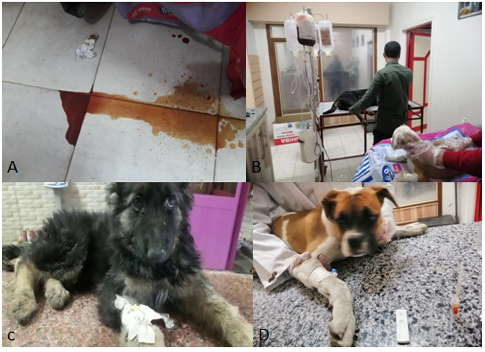
a) Bloody watery diarrhea of CPV infected dog. b) Fluid therapy and blood transfusion for treatment of CPV infected Dogs. c) Husky dog suffering from lethargy and diarrhea due to CPV infection. d) Diagnosis of CPV infection in pit bull dog using Rapid CPV Ag test kit.

a) Bloody watery diarrhea of CPV infected dog. b) Fluid therapy and blood transfusion for treatment of CPV infected Dogs. c) Husky dog suffering from lethargy and diarrhea due to CPV infection. d) Diagnosis of CPV infection in pit bull dog using Rapid CPV Ag test kit.
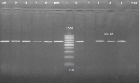
PCR product about 360 bp. Lane 1, 2, 3, 5, 6, 7, 8, 9 and 10 are positive samples while lane 4 is negative sample.
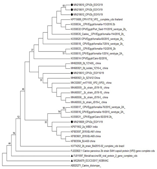
Phylogenetic analysis based on VP2 for the studied isolates against different sequences of canine parvovirus in Gen Bank. Studied samples were marked with dark circle and vaccine strains were marked with dark triangle.
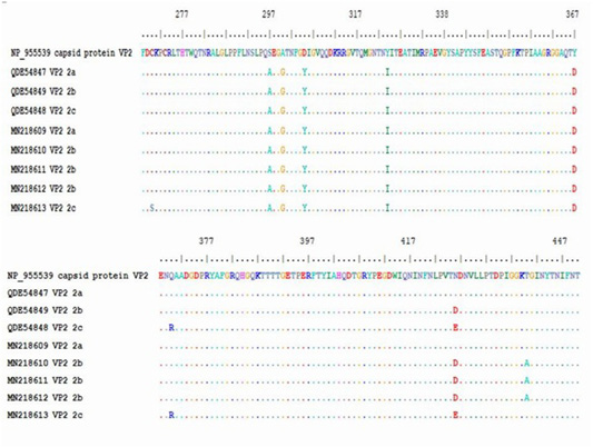
Multiple alignment of translated protein of studied variant (2a,2B,2C) against reference complete (VP2) and partial sequences in Gen-Bank.


