Research Journal for Veterinary Practitioners
Case Report
Research Journal for Veterinary Practitioners 2 (1S): 1 – 3Special Issue – 1 (Epidemiology and Occurrence of PPR in Endemic Situations)
An easy Approach towards Diagnosis of Peste des petits ruminants (PPR) through Clinical Examination
Arslan Tariq1*, Asim Shahzad2, Razia Kausar3, Uzair Ahasan Fiaz1, Syed Sohail Jan1
- Faculty of Veterinary Sciences (FVS), University of Agriculture Faisalabad (UAF), Pakistan
- Department of Veterinary Pathology, Faculty of Veterinary Sciences (FVS), University of Agriculture Faisalabad (UAF), Pakistan
- Department of Anatomy and Histology, Faculty of Veterinary Sciences (FVS), University of Agriculture Faisalabad (UAF), Pakistan
*Corresponding author:dr.arslantariq3418@live.com
ARTICLE CITATION:
Tariq A, Shahzad A, Kausar R, Fiaz UA and Jan SS (2014). An easy Approach towards Diagnosis of Peste Des Petit Ruminant (PPR) through Clinical Examination. Res. j. vet. pract. 2 (1S): 1 – 3.
Received: 2013–10–08, Revised: 2013–10–27, Accepted: 2013–10–28
The electronic version of this article is the complete one and can be found online at
(
http://dx.doi.org/10.14737/journal.rjvp/2014/2.1s.1.3
)
which permits unrestricted use, distribution, and reproduction in any medium, provided the original work is properly cited
ABSTRACT
This case study signifies the diagnosis of Peste des Petits Ruminants (PPR) in a buck of discrete breed on the basis of clinical signs and hematology. Case presented to department of Clinics Medicine and Surgery (CMS), University of Agriculture Faisalabad, Pakistan with chief complains of anorexia, watery diarrhea, ocular discharge and muco–purulent nasal discharge from last 4 days. Clinical examination revealed high fever, increase respiration, pulse rate and capillary refill time. Blood oozing lesions on gums and swollen lymph nodes were seen. Lymphopenia based on complete blood count (CBC) provided another clue to diagnose the disease as PPR on clinical basis. Treatment was done symptomatically for 7 days with amoxicillin, lactated ringer and somogel. Along with, a multivitamin powder was also advised to add in the feed to tone up the animal. Animal recovered completely on 10th day after visit.
A morbillivirus of family Paramyxoviradae produces an important contagious disease in sheep and goat called peste des petits ruminants (PPR). It is also known as “Small ruminant plague” (Zahur et al., 2011; Mahajan et al., 2012). There is very close relationship between PPR virus and Rinderpest virus (Abubakar et al., 2008; Rajak et al., 2005). It results in production losses, abortion in pregnant animals and even death of the animals. This virus causes 100% morbidity and 90% mortality in animals (Intizar et al., 2009). Animals of young age are found to be more susceptible to PPR and cause high mortality in kids and lambs (Zahur et al., 2008).
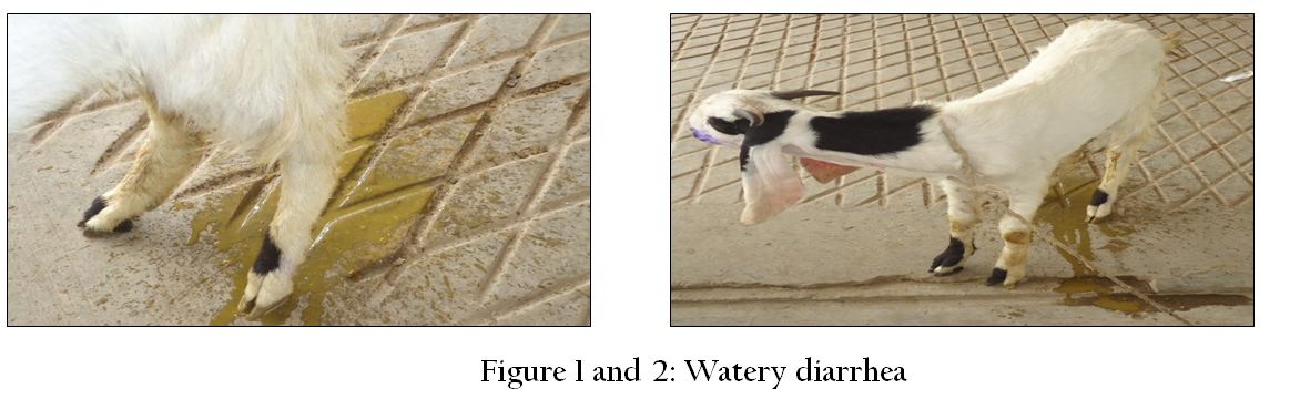
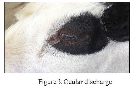
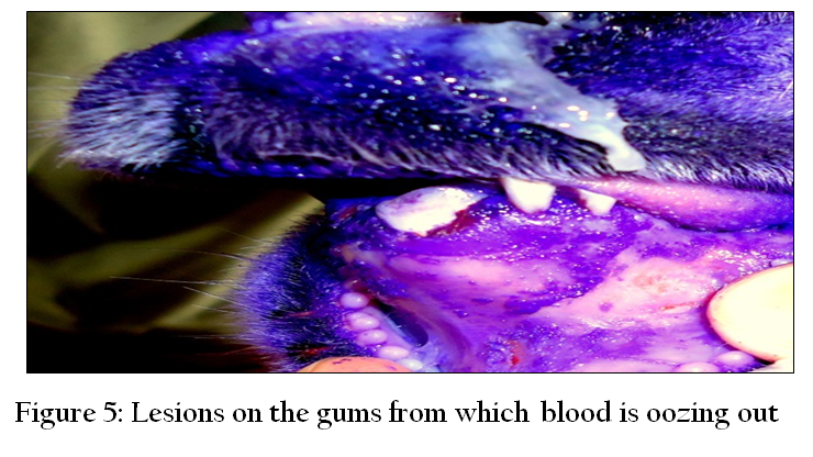
Signs of the disease includes rise in temperature, diarrhea (Figure 1~2), ocular and muco–purulent nasal discharge (Figure 3~4), lesions on the gums from which blood is oozing out (Figure 5), pneumonia, coughing and sneezing and if remain untreated then cause the death of the animal (Mahajan et al., 2012; Intizar et al., 2009).
Lymphopenia is seen in case of PPR virus infection (Rajak et al., 2005). Excluding clinical signs, and history, complete blood count can also be used for diagnosed. Heamagglutination test (HA), compliment fixation test (CFT), immuno–electro–osmo–precipitation test and enzyme linked immuno–sorbent assay (ELISA) (Ezeibe et al., 2004; Intizar et al., 2009) have been used successfully. There is no specific treatment and control is possible only with vaccination. In one report, the sero–prevalence of this disease is found to be 69.64% in Faisalabad and 43.33 % in overall Punjab province. The risk of PPR is 62.5 % in domestic small ruminants around the globe (Khan et al., 2007; Zahur et al., 2008; Zahur et al., 2011).
As the disease is economically very important for developing country like Pakistan, so should be control all over Pakistan by starting free vaccination programs in every part of country and by awareness of farmers about the bad impact of the disease on production and economy.
Approximately 2 year old buck of discrete breed weight about 27 kg visited the outdoor clinic at CMS. The main presenting complaint of animal was that the animal was suffering from diarrhea (Figure 1~2) and anorexia from last 4 days. Ocular and nasal discharge could be seen as in Figure 3 and 4 respectively. Vaccination history against any disease was not found but deworming was done with Oxfendazole about 20 days before. by investigation from owner it was found the recently 3 more animals were bought and introduce into the previous flock of 8 animals and from 2 days later 7 out of 11 were showing the similar signs and symptoms.
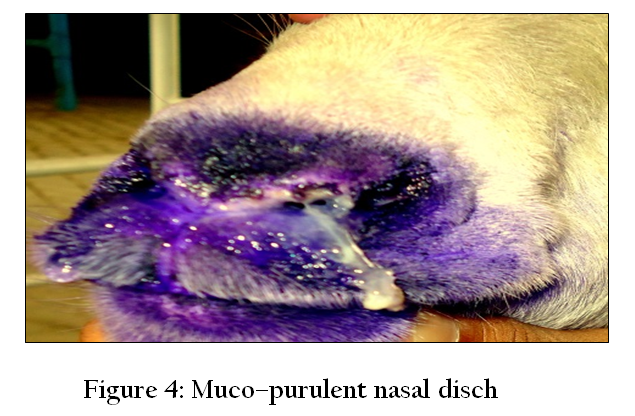
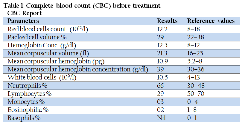
Clinical examination of the animal showed very high fever 104.40F, Respiration rate about 38/min, pulse rate was 112/min and CRT was more than 3 means animals was dehydrated. Lesions from which blood was oozing out could be seen on the gums (Figure 5). Nasal discharge was muco–purulent in nature. Lymph nodes were also palpable. Because of diarrhea the hind quarter was soiled with feces. Feces were check for parasitic ova but result was negative. Complete blood count (CBC) revealed lymphopenia (Table. 1) as an indicator of PPR virus infection (Rajak et al., 2005). On the basis of history, clinical signs and complete blood count (CBC) the disease was diagnosed as PPR.
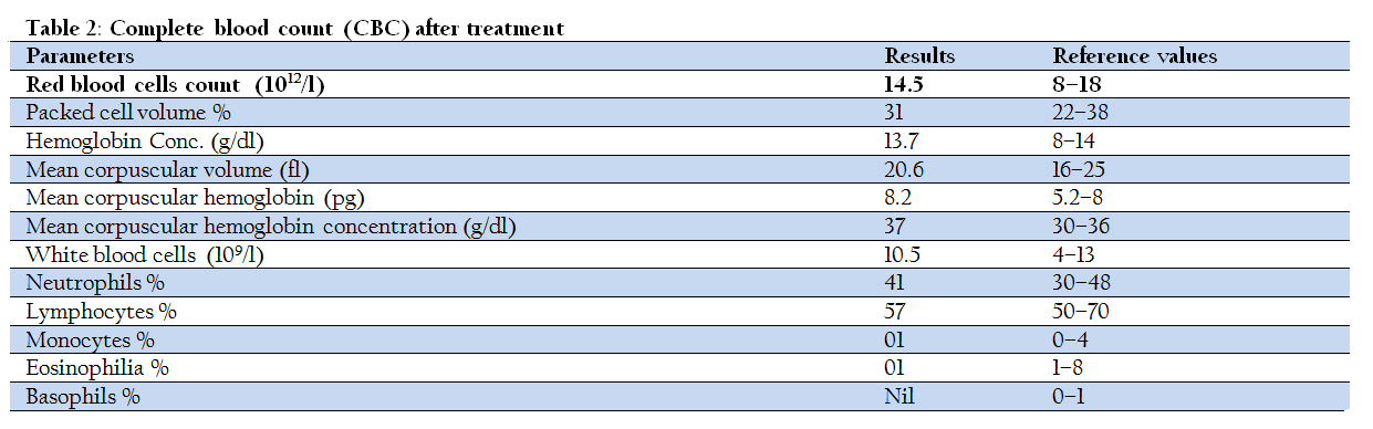
Symptomatic and supportive treatment was done because there is no specific treatment (http:// osp.mans. edu.eg / elsawalhy /Inf–Dis/PPR.htm). Animal was infused with ringer lactate @ 20ml/kg b.w, Metronidazole @ 10mg/kg b.w, Amoxicillin @ 3mg/kg b.w to check the dehydration and diarrhea and secondary infection respectively for 7 days. As take home medicine somogel (Lignocaine INN 0.6%) was recommended 3 times a day for its soothing affect and advice the owner to offer feed to animal just after applying this gel. Oral rehydration solution (ORS) and a multivitamin powder (Bendoz powder) were also recommended for 15 days to rehydrate the animal and to maintain the body condition so that animal could recover as early as possible. Animal started showing recovery signs from 5th day and recovered from disease on 10th day of initial treatment.
PPR is a World Animal Health Organization (OIE) listed disease of sheep and goat caused by morbillivirus. It was first identified in Pakistan in 1991 and remain endemic since then in Central Asia, Pakistan, India, Iran, Iraq and in different parts of Africa. In recent years, this disease has known to cause big economic losses (Zahur et al 2011; Abubakar et al., 2008). As reported by Zahur et al., (2008), the occurrence of disease in different provinces of Pakistan like Punjab, Sindh and Balochistan is 70.93%, 47.98% and 76.74%, respectively. But specifically the prevalence of PPRV is 69.64% in respected area of investigation (Faisalabad).
Close contact is the way of transmission of this virus. The virus come out in every secretion and excretion of infected animal and infects the healthy animals near to it (Abubakar et al., 2008). Transfer of sheep and goats from one place to another and PPR outbreaks are linked closely. Disease stays for 15 days and mostly animal dies in 10–12 days. Morbidity in this disease is 90 % and mortality is 50 – 80 % but mortality may be reach up to 100% (Zahur et al., 2011; Mahajan et al., 2012). In one report by Abubakar et al., (2008) 28–45% abortion rate is seen in pregnant animals in Pakistan.
On clinical examination the animal was found to have high rise in body temperature, diarrhea (Figure 1~2), and ocular discharge (Figure 3), nasal discharge which was muco–purulent (Figure 4), lesion on gums in oral cavity (Figure 5) and dyspnea as seen by the Mahajan in 2012. In complete blood count (CBC), Lymphopenia is seen. Lymphopenia is seen in case of PPR virus infection, which is known to cause immune–suppression and show strong affinity towards the lymphoid tissue and epithelial cell (Rajak et al., 2005).
On clinical basis, we can diagnose the disease as PPR but for further confirmation we can also perform other tests like ELISA, CFT and HA etc. For diagnosis of viral disease, ELISA and CFT are difficult to perform and somewhat expensive. Instead of these a less expensive test is done known as Heamagglutination test (HA). The best thing about HA is that it is effective and easy to perform to diagnose viral diseases in developing countries like Pakistan (Ezeibe et al., 2004).After confirming the disease we treated the disease symptomatically because still there is no specific treatment available for this disease.
As disease is endemic, so the effective vaccination is the best way to control this disease rapidly.
ACKNOWLEDGEMENTS
This case report is especially dedicated to Dr. Asad Manzoor (Lecturer at Department of Clinic Medicine and Surgery, University of Agriculture Faisalabad Pakistan) and Dr. Mumtaz A Khan (Professor at Department of Clinic Medicine and Surgery, University of Agriculture Faisalabad Pakistan) for their inspiration, advice, support and everything they provide me to complete this report.
REFERENCES
Abubakar M, Jamal SM, Hussain M and Ali Q (2008). Incidence of peste des petits ruminants (PPR) virus in sheep and goat as detected by immuno–capture ELISA (Ic ELISA). Small Ruminant Research 75: 256–259
http://dx.doi.org/10.1016/j.smallrumres.2007.12.001
Abubakar M, Ali Q, Khan HA (2008), Prevalence and mortality rate of peste des petitis ruminant (PPR): possible association with abortion in goat. Trop Anim Health Prod. 40(5):317–321.
http://dx.doi.org/10.1007/s11250-007-9105-2
PMid:18509938
Ezeibe MCO, Wosu LO and Erumaka IG, (2004). Standardisation of the haemagglutination test for peste des petits ruminants (PPR). Small Ruminant Research 51: 269–272
http://dx.doi.org/10.1016/S0921-4488(03)00123-8
Intizar M, Ahmad MD, Anjum AA and Hanif A (2009). Comaprative efficacy of peste des petits ruminant (PPR) vaccines available in Pakistan in sheep and goats. Pakistan Veterinary Journal. 29(4): 202–205.
Khan HA, Siddique M, Arshad MJ, Khan QM and Rehman SU (2007). Sero–prevalence of peste des petits ruminants (PPR) virus in sheep and goats in Punjab province of Pakistan. Pakistan Veterinary Journal 27(3): 109–112.
Mahajan S, Agrawal R, Kumar M, Mohan A, and Pande N (2012). Risk of seroconversion to peste des petits ruminants (PPR) and its association with species, sex, age and migration. Small Ruminant Research 104: 195–200.
http://dx.doi.org/10.1016/j.smallrumres.2011.10.009
Rajak KK, Sreenivasa BP, Hosamani M, Singh RP, Singh S.K, Singh R.K and Bandyopadhyay SK (2005). Experimental studies on immunosuppressive effectsof peste des petits ruminants (PPR) virus in goats. Comparative Immunology, Microbiology & Infectious Diseases 28: 287–296.
http://dx.doi.org/10.1016/j.cimid.2005.08.002
PMid:16188317
Saravanan P, Sen A, Balamurugan V, Bandyopadhyay SK and Singh RK (2008). Rapid quality control of a live attenuated Peste des petits ruminants (PPR) vaccine by monoclonal antibody based sandwich ELISA. Bioologicals 36: 1–6.
http://dx.doi.org/10.1016/j.biologicals.2007.03.005
PMid:17644410
Zahur AB, Ullah A, Hussain M, Irshad H, Hameed A, Jahangir M and Farooq MS (2011). Sero–epidemiology of peste des petits ruminants (PPR) in Pakistan. Preventive Veterinary Medicine 102: 87–92.
http://dx.doi.org/10.1016/j.prevetmed.2011.06.011
PMid:21788090
Zahur AB, Irshad H, Hussain M, Ullah A,Jahangir M, Khan MQ and Farooq MS (2008). The epidemiology of peste des petits ruminant in Pakistan. Rev. sci. tech. Off. int. Epiz. 27 (3): 877–884.
http://www.merckmanuals.com/vet/appendixes/reference_guides/hematologic_reference_ranges.html


