Research Journal for Veterinary Practitioners
Case Report

Dystocia in a Non-Descript cow due to Pelvic Fracture and Left Hock Flexion
Alok Kumar Rathore, Amit Kumar, Jitendra Kumar Agrawal*, Atul Saxena
Department of Veterinary Gynaecology and Obstetrics, College of Veterinary Science & Animal Husbandry, U.P.Pandit Deen Dayal Upadhyay Pashu Chiktsa Vigyan Vishwavidyalaya Evam Go Anusandhan Sansthan (DUVASU), Mathura (U.P) India.
Abstract | The present case reports successful per vaginal delivery of dead male fetus in posterior presentation which was impacted in the birth canal due to narrow pelvis and hock flexion,thereafter it’s therapeutic management.
Keywords | Dystocia, Fetotomy, Cow, Pelvic fracture, Hock flexion
Editor | Muhammad Abubakar, National Veterinary Laboratories, Islamabad, Pakistan.
Received | February 01, 2018; Accepted | February 23, 2018; Published | March 30, 2018
*Correspondence | Jitendra Kumar Department of Veterinary Gynaecology and Obstetrics, College of Veterinary Science & Animal Husbandry, U.P.Pandit Deen Dayal Upadhyay Pashu Chiktsa Vigyan Vishwavidyalaya Evam Go Anusandhan Sansthan (DUVASU), Mathura (U.P) India; Email: jituvet11@gmail.com
Citation | Rathore AK, Kumar A, Agrawal JK, Saxena A (2018). Dystocia in a non-descript cow due to pelvic fracture and left hock flexion. Res. J. Vet. Pract. 6(1): 7-9.
DOI | http://dx.doi.org/10.17582/journal.rjvp/2018/6.1.7.9
ISSN (Online) | 2308-2798
Copyright © 2018 Rathore et al. This is an open access article distributed under the Creative Commons Attribution License, which permits unrestricted use, distribution, and reproduction in any medium, provided the original work is properly cited.
INTRODUCTION
Pelvic fracture is oneof the main causes of narrowing of the pelvis (Dubay, 1987) due to altered shape and volume of the birth canal (Sloss and Dufty, 1980). Pelvic fracture may occur due to accidental injuries by vehicle mishap or falling on a hard object, rolling of dam to relive torsion, falling on iron stake (Noakes et al., 2009; Singh et al., 2015). In animals with narrow pelvis, there is a lack of progress in the second stage of labor, as the chances of fetus being stuck in the pelvic inlet are more (Samantha, 2011). If the fetus enters the pelvis partially, there is severe non-progressive straining (Purohit et al., 2011). The incidence of narrow pelvis in cattle and buffalo has been earlier reported between 7.79% (Phogat et al., 1992) to 9.2% (Sharma et al., 1992).
The present case report communicates dystocia due to pelvic fracture in cattle and it’s successful per vaginal delivery through fetotomy.
CASE HISTORY AND CLINICAL OBSERVATIONS
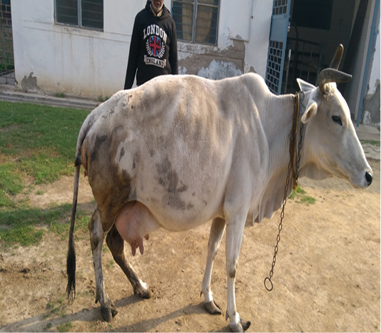
A full term pregnant non-descript cow in its seventh parity (aged about 10 years) was brought to TVCC, COVAS, Mathura with the history of acute straining since last 20-25 hours, ruptured water bags with one limb protruding out of vulva. Inclined shape of hind portion of the cow was indicating the possibility of the pelvic fracture (Figure 1). Vaginal examination revealed the dilated cervix with mild lubrication of the genital tract and reduced pelvic diameter. Dead male fetus was palpable in posterior longitudinal presentation, lumbo sacral position and left hock flexion.
TREATMENT
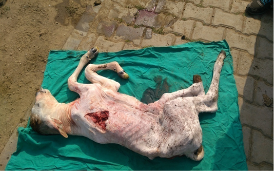
Animal was properly restrained in it’s lateral recumbency. On per vaginal examination, left hind limb was impacted in the birth canal due to which manual correction was not possible, hence, it was decided to amputate the left hind limb just below the hock joint using percutaneous fetotomy. Lubrication of the birth canal was done using heavy liquid paraffin (approximately 3 litres). After fetotomy, traction was applied on both the limbs using obstetrical chains. Controlled and systemic traction resulted in delivery of entire dead male fetus (Figure 2). Animal was given intravenous fluid therapy (Inj. DNS @ 3 litre and Inj. RL @ 3 litre) along with Ceftriazone 4.5 gm intravenously,Flunixin meglumine @ 2.2 mg/kg b.wt intramuscularly, Chlorpheneramine maleate 0.5 mg/kg b.wt intramuscularly and Inj. Oxytocin 70 IU intramuscularly. Intrauterine therapy (4 bolus of Cleanex®, Dosch Pharmaceuticals Ltd.) was also carried out simultaneously. Animal was discharged, advising the follow-up care and management.
DISCUSSION
Congenital and acquired deformities of the pelvis, cervix, vagina or vulva lead to dystocia (Sloss and Dufty, 1980). The constriction/obstruction of the birth canal may be due to pelvic abnormalities, vulvar or vaginal stenosis, neoplasms of the vagina and vulva, vaginal cystocele, incomplete cervical dilation, uterine torsion and ventral displacement of the uterus (Purohit et al., 2011). Pelvic abnormalities of the mother which results in dystocia include small size of the pelvis (Jackson 1995), pelvic deformities or exostoses (Roberts 1986), osteomalacia and hypoplasia of vagina and vulva (Kodagali, 2003).
In the present case, narrowing of the birth canal was due to pelvic fracture as a result of falling on the hard floor a year before. Although the fetus was in posterior presentation but due to hock flexion and less pelvic space, normal delivery was not possible. Sharma et al., (1992) also reported similar case of dystocia due to narrow pelvis resultant of an accident. Generally, in case of the pelvic fracture, caesarean operation is recommended but in the present case, proper lubrication, controlled and systemic traction made the per-vaginal delivery possible. Even though the animal recovered uneventfully, but its future fertility was questionable, hence, owner was advised not to breed the animal further.
ACKNOWLEDGEMENTS
Authors are highly thankful to Dr. Atul Saxena,Professor and Head, Department of Veterinary Gynaecology and Obstetrics, College of veterinary Science and AnimalHusbandry, DUVASU, Mathura for encouragementand providing necessary facilities to carry out this work.
Conflict of interest
There is no conflict of interest.
Authors Contribution
All authors contributed equally.
REFERENCES


