Advances in Animal and Veterinary Sciences
Short Communication
Advances in Animal and Veterinary Sciences. 1 (1S): 18 – 19Special issue-1 (Veterinarians approaches for safeguarding animal health and production)
Detection of Group A Rotavirus in Faeces of Diarrhoeic Bovine Porcine and Human Population from Eastern India by Reverse Transcriptase–Polymerase Chain Reaction
Anjan Mondal1, Kuldeep Sharma2, Yashpal Singh Malik2, Siddhartha Narayan Joardar1*
- Department of Veterinary Microbiology; West Bengal University of Animal and Fishery Sciences; P.O.–Belgachia, Kolkata–700037, West Bengal, India
- Division of Biological Standardization, Indian Veterinary Research Institute (IVRI) Izatnagar 243 122, Bareilly, Uttar Pradesh India
*Corresponding author:joardar69@gamil.com
ARTICLE CITATION:
Mondal A, Sharma K, Malik YS, Joardar SN (2013). Detection of group a rotavirus in faeces of diarrhoeic bovine porcine and human population from eastern India by reverse transcriptase–polymerase chain reaction. Adv. Anim. Vet. Sci. 1 (1S): 18 – 19.
Received: 2013–09–04, Revised: 2013–09–16, Accepted: 2013–09–16
The electronic version of this article is the complete one and can be found online at
(
http://nexusacademicpublishers.com/table_contents_detail/4/99/html
)
which permits unrestricted use, distribution, and reproduction in any medium, provided the original work is properly cited
ABSTRACT
The present study describes the distribution of group A rotavirus in animals and human population of eastern state of India– West Bengal. During the study, a total of 211 samples were collected from diarrhoeic bovine, porcine and human population (below 6 months of age). Of which , 26 (12.32%) samples were found positive for group A rotavirus by VP6 gene based reverse transcriptase– polymerase chain reaction (RT–PCR) assay. A total of 89 bovine, 82 porcine faecal and 40 human stool samples were screened by RT–PCR assay for the detection of group A rotavirus, of which 10 bovine (11.23%), 9 porcine (10.97%) and 7 human (17.5%) samples were detected positive. The study revealed baseline information to understand rotavirus epizootology and future prophylactic strategies against rotavirus in eastern India.
Rotaviruses are recognized as the major viral etiological agents causing diarrhoea in neonates of all species including animals, human and birds. Rotavirus accounts for more than 6 million deaths in infants worldwide (Estes and Kapikian, 2007), which are more in developing countries including India (Parasar et al., 2006). Rotavirus have accounted for 25% mortality in young animals and causes severe economic losses in livestock sector (Fagiolo et al., 2005). The virus belongs to the genus Rotavirus under the family Reoviridae and group A rotaviruses are major pathogens causing acute gastroenteritis in children and animals (Ciarlet et al., 2002).
Combined studies on the prevalence of rotavirus associated diarrhoea in bovine and porcine species of animals and human have not been documented in eastern India. This study was aimed to understand the burden and epidemiology of rotavirus infection in West Bengal, one of the eastern states of India. The results of this study are expected to be considered as baseline information for future studies to determine genotypes and subsequent vaccine against rotavirus.
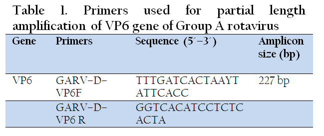
A total of 211 samples were collected from bovine (n=89), porcine (n=82) and human (n=40) (below the age of 6 months) affected with acute diarrhoea from different districts of West Bengal viz. Purulia (n=35), Burdwan (n=34), Nadia (n=56) and Kolkata (n=86). The samples were transported to laboratory for processing using ice cold container (Thermoflask, Milton, India). The samples were stored at –20oC for extraction of viral RNA. A 10% (w/v) faecal suspension of the faecal material was prepared in phosphate buffer saline (PBS; pH 7.2). Centrifugation was carried out, after thorough vortexing, at 7500 rpm for 20 min to remove the coarse debris. Total RNA was extracted using RiboZol RNA extraction reagent (Amresco, USA) as per manufacturer instruction. The dsRNA was subjected to reverse transcription as per the method described by Iturriza–Gomara et al. (2004). The synthesized cDNA was stored at –20oC till further use. Detection of group A rotavirus, amplification of partial length VP6 gene was carried out using primers and conditions as optimized by Kattoor et al. (2013). The primer sequences and nucleotide position of oligonucleotide primers are shown in Table 1.
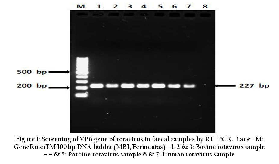
All the 211 samples including 89 bovine, 82 porcine faecal and 40 human stool samples were screened by the VP6 gene based RT–PCR assay. PCR was carried out by using primers for the group specific VP6 gene which generated expected amplicon size 227 bp (Figure 1). In total 12.32% (26/211) diarrhoeic faecal samples were positive for group A rotavirus. On RT–PCR analysis, 10 bovine (11.23%), 9 porcine (10.97%) and 7 human (17.5%) samples were found positive for the presence of group A rotavirus. The RT–PCR offers many advantages besides high sensitivity and specificity in detection of rotavirus in faecal samples (Fedorova et al., 2005; Kang et al., 2004). It helps in the detection of viral nucleic acid during initial stages of infection without waiting for higher virus titre and development of immune response in the affected host species (Niture et al., 2010). Detection of rotavirus infection in reservoir animals and carriers is another advantage of RT–PCR (Niture et al., 2010). The PCR techniques are used throughout the world for the group A rotavirus typing in strains obtained directly from faecal extracts and/or cell culture (Basera et al., 2010; Mondal et al., 2011).
In India, Gulati et al. (1996) was the first to determine the genetic diversity of bovine group A rotavirus using RT–PCR. In a study by Basera and co–workers (2010), group A rotavirus was found in 7.81% (10/128) samples from bovine calves. Chinsangaram et al. (1995) detected group A rotavirus from 94% (44/47) calves, directly by RT–PCR in faecal samples. In the present study, we detected high incidence of rotavirus among human (17.5%), followed by bovine (11.23%) and porcine (10.97%) samples. The findings are in agreement with the report from Western India that revealed 20.25% of human rotaviruses followed by 12.5% of bovine and 7.84% of avian rotaviruses (Niture et al., 2011). Earlier, 9.73% incidence of bovine group A rotavirus was reported from Mumbai region (Western part of India) (Mondal et al., 2012). The differences in the prevalence of rotavirus could be some factors like hygienic measures, proper access to maternal colostrum, nutrition, season and climatic factors such as rainfall, temperature, relative humidity etc.
In conclusion, the group A rotavirus is prevalent in bovine and porcine species of animals and human in eastern Indian state, West Bengal. RT–PCR assay was evaluated as sensitive and specific assay for the rapid detection of group A rotavirus in faecal samples. In Indian subcontinent, human live in close proximity to their livestock, often in poor sanitary conditions, contamination of water and food is possible. It may be responsible for possible zoonotic transmission of rotaviruses. So, in order to obtain a much better perspective in terms of the circulating genotypes in different species and regions of the country, molecular epidemiological surveillance of the circulation of group A rotaviruses in varied host species needs to be carried out.
ACKNOWLEDGEMENT
The work was supported by Rajib Gandhi Fellowship, offered by Govt. of India, to the first author. Authors wish to thank the Vice Chancellor, West Bengal University of Animal and Fishery Sciences, for providing necessary infrastructure facility to carry out the work.
REFERENCES
Basera SS, Singh R, Vaid N, Sharma K, Chakravarti S and Malik YPS (2010). Detection of Rotavirus Infection in Bovine Calves by RNA–PAGE and RT–PCR. Indian J. Virol. 21(2):144 – 147.
http://dx.doi.org/10.1007/s13337-010-0017-9
PMid:23637494 PMCid:PMC3550708
Ciarlet M, Conner ME, Finegold MJ and Estes MK (2002). Group A rotavirus infection and age–dependent diarrheal disease in rats: a new animal model to study the pathophysiology of rotavirus infection. J. Virol. 76(1): 41– 57.
http://dx.doi.org/10.1128/JVI.76.1.41-57.2002
PMid:11739670 PMCid:PMC135688
Chinsangaram J, Schore CE, Guterbock W, Weaver LD and Osburn BI (1995). Prevalence of group A and group B rotaviruses in the feces of neonatal dairy calves from California. Comp. Immun. Microbiol. Infect. Dis. 18(2): 93 – 103.
http://dx.doi.org/10.1016/0147-9571(95)98850-H
Estes MK and Kapikian AZ (2007). Rotaviruses. In: Knipe DM, Howley PM, Griffin DE, Lamb RA, Martin MA, Roizman B and Straus SE (ed.), Fields virology, 5th ed. Lippincott Williams & Wilkins, Philadelphia, PA pp. 1917 – 1974.
Fagiolo A, Cristina R, Ogla L and Antonio B. (2005). In: Antonio, B. (Ed.), Buffalo Pathologies. Buffalo Production and Research, FAO, Rome. pp. 249 – 296.
Fedorova OF, Novikova NA, Epifanova NV, Lukovnikova LB, Kniagina ON, Gracheva EV and Bogacheva EM. (2005).Optimization of RT–PCR for the identification of VP4 gene of Group A rotaviruses and evaluation of its diagnostic efficiency. Vopr. Virusol. 50: 39 – 41.
PMid:15747872
Gentsch J, Gray J, Itturiza–Gomara M, Klena J, Kirkwood C, Armah G and Page NA. (2009). Manual of rotavirus detection and characterization methods. Department of immunization, vaccines and Biologicals, World Health Organization pp. 53.
Gulati BR, Nakagomi O, Koshimura Y, Nakagomi T and Pandey R (1996). Relative frequencies of G and P types among rotaviruses from Indian diarrheic cow and buffalo Calves. J. Clin. Microbiol. 37(6): 2074 – 2076.
Iturriza–Gomara M, Kang G and Gray J. (2004). Rotavirus genotyping: keeping up with an evolving population of human rotaviruses. J. Clin. Virol. 31:259 – 265.
http://dx.doi.org/10.1016/j.jcv.2004.04.009
PMid:15494266
Kang G, Iturriza–Gomara M, Wheeler JG, Crystal P, Monica B, Ramani S, Primrose B, Moses PD, Gallimore CI, Brown DW and Gray J. (2004). Quantitation of group A rotavirus by real–time reverse–transcription polymerase chain reaction: correlation with clinical severity in children in South India. J. Med. Virol. 73:118 – 122.
http://dx.doi.org/10.1002/jmv.20053
PMid:15042658 PMCid:PMC2459214
Kattoor JJ, Sharma K, Kumar N, Batra M, Jindal N, Yadav AS and Malik YPS. (2013). Distribution Frequency of Avian Rotaviruses in India, 2011-2013. Advan. Anim. Vet. Sci. (in press).
Mondal A, Majee SB and Bannalikar AS. (2011). Molecular characterization of VP7 gene of bovine group A rotavirus. Anim. Sci. Rep. 5(1): 27 – 33.
Mondal A, Aich R, Majee S and Bannalikar, AS. (2012). Determination of bovine rotavirus G serotype by polymerase chain reaction. Trop. Anim. Health. Prod. 44(4):763 – 767.
http://dx.doi.org/10.1007/s11250-011-9962-6
PMid:21892766
Niture GS, Karpe AG, Prasad M, Bhonsle AV and Patil SV. (2010). Detection of Group D Avian Rotaviruses among Layer Poultry from Western India. Int. J. Poult. Sci. 9 (1): 72 – 76.
http://dx.doi.org/10.3923/ijps.2010.72.76
Niture GS, Karpe AG and Prasad M. (2011). Characterization of buffalo, poultry and human rotaviruses in Western India. Vet. Archiv. 81(3): 307 – 319.
Parashar UD, Gibson C, Bresse JS and Glass RI. (2006). Rotavirus and severe childhood diarrhea. Emerg. Infect. Dis. 9:565 – 572
http://dx.doi.org/10.3201/eid0905.020562
PMid:15207040 PMCid:PMC2972763


