Advances in Animal and Veterinary Sciences
Research Article
Adv. Anim. Vet. Sci. 6(7): 281-285
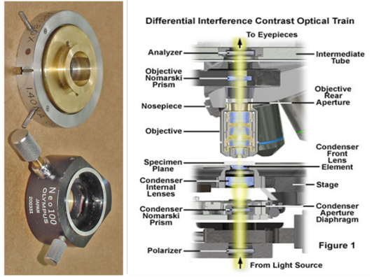
Figure 1
Shows the DIC optical path
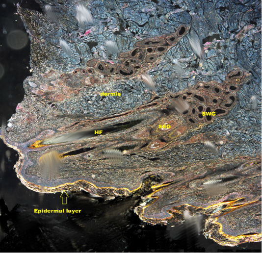
Figure 2
Shows DICM image of the skin of the camel revealing all layers of the skin (X10). HF: Hair follicle; SWG:Sweat glands; SEG: Sebaceous gland
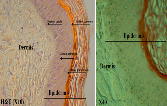
Figure 3
Shows the epidermis layers under DICM
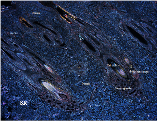
Figure 4
Shows DICM image of the dermis layer , SR: Stratum reticular (X10)
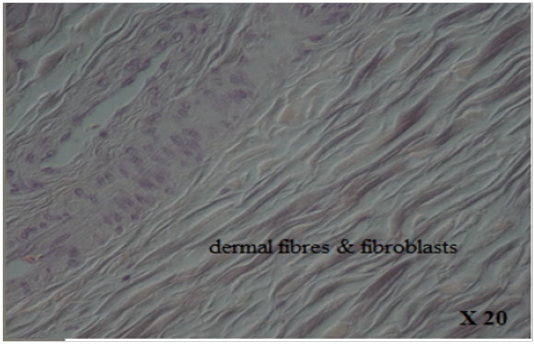
Figure 5
Shows DICM image of the dermal fibres & fibroblasts
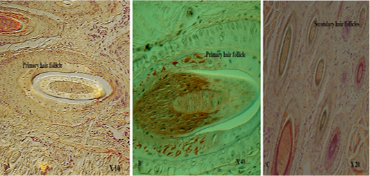
Figure 6
Shows DICM image of primary hair follicle (A. X10; B. X 40; C. X20)
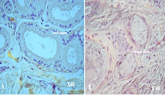
Figure 7
Shows DICM image of A. sweat glands ( X 40); B. Sebaceous glands (X 40)


