Advances in Animal and Veterinary Sciences
Research Article
Adv. Anim. Vet. Sci. 6(2): 81-87
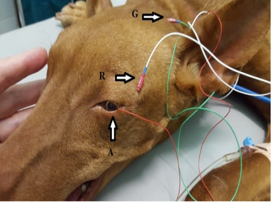
Figure 1
Position of the electrodes for ERG examination. A-active electrode in lens shape put on the cornea, R-reference needle electrode put on 1 cm of lateral eye border, G-ground needle electrode put on the external occipital protuberance.
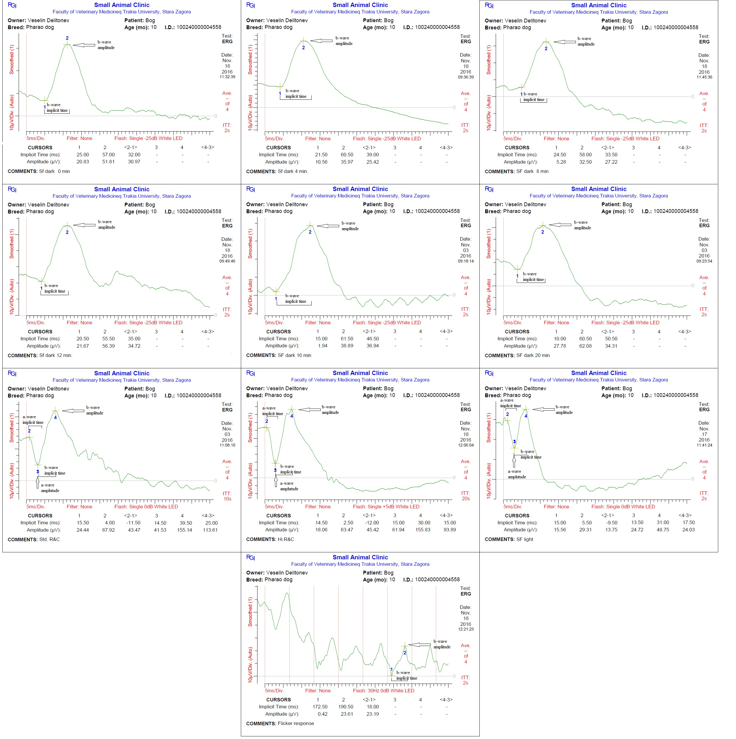
Figure 2
Representative waveforms from a dog in the study during individual ERG tests.


