Advances in Animal and Veterinary Sciences
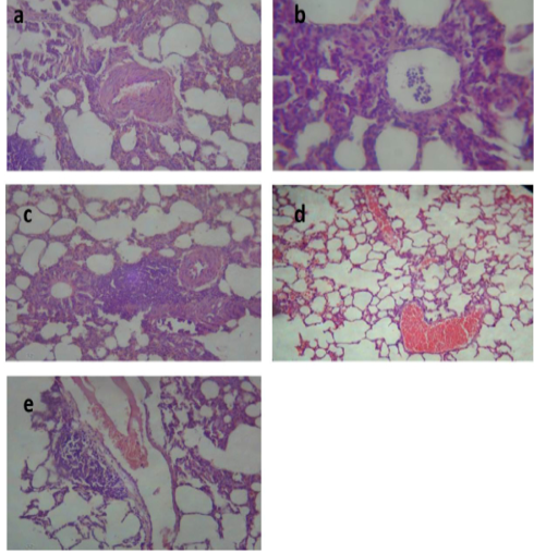
Lung of group (II) showing slight congestion of thickened pulmonary blood vessels (a) (H&E ×520). Lung of group (III) showing pneumonic area represented by thickening of the interalveolar septa by extensive leukocytic infiltration mostly polymorphonuclears (b) (H&E ×400), peribronchial lymphoid hyperplasia (c) and severe vascular congestion (d) (H&E ×100). Lung of group (IV) showing vascular congestion with perivascular leukocytic aggregation (e) (H&E ×100).
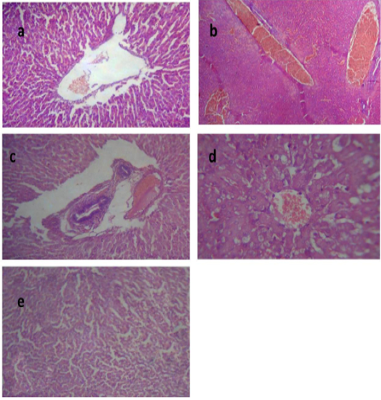
Liver of group (II) showing congested central vein and dilated hepatic sinusoids (a) (H&E ×400). Liver of group (III) showing severe congestion of the central veins (b). Cholangitis represented thickening of the bile duct wall by vascular congestion, edema and leukocytic infiltration besides cholestasis (c) (H&E ×100).Vacuolation of the hepatocytes (d) (H&E ×400).Liver of group (IV) showing interstitial hemorrhage and normal tissue architecture (e) (H&E ×520).
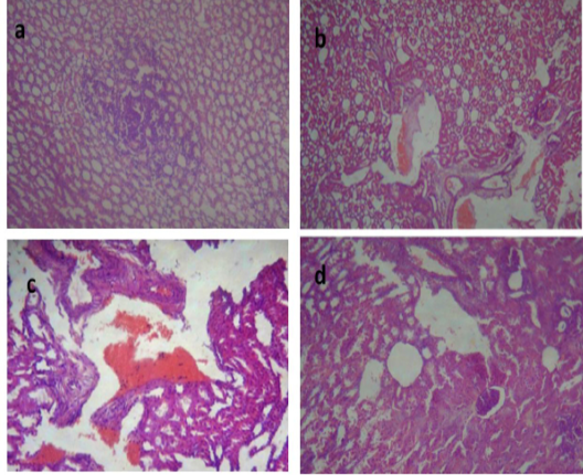
Kidney of group (II) showing focal leukocytic aggregation in the renal medulla (a) (H&E ×400). kidney of group (III) showing vascular congestion and hemorrhage (b) (H&E ×400) with fibrous connective tissue proliferation (c) (H&E ×100). Kidney of group (IV) showing cystic dilatation of few renal tubules, renal casts, hydropic degeneration of the epithelial lining of some renal tubules and vascular congestion (d) (H&E ×400).
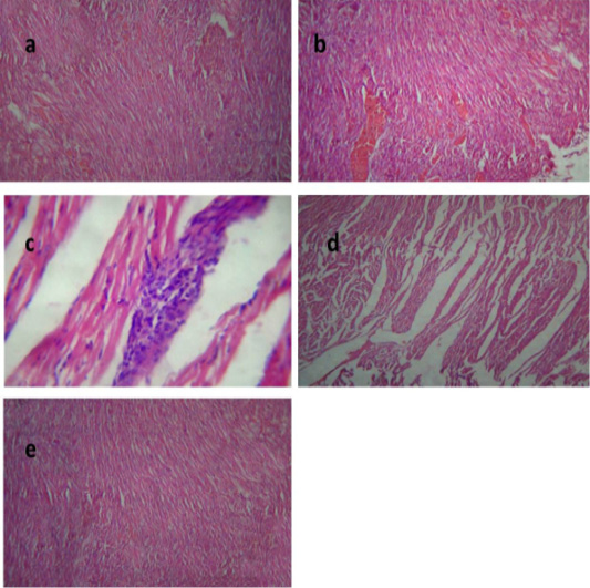
Heart of group (II) showing mild congestion of cardiac blood vessels (a) (H&E ×400). Heart of group (III) showing sever vascular congestion (b), leukocytic infiltration between the myocardiocytes (c) and intermuscular edema (d) (H&E ×100). Heart of group (IV) showing mild congestion of cardiac blood vessels (e) (H&E ×400).


