Advances in Animal and Veterinary Sciences
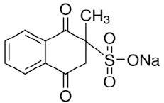
Chemical structure of menadione (Castro et al., 2008)
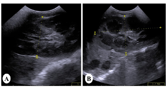
Ultrasound images of the kidney of vitamin K3-exposed horses showing (A); enlargement of the right kidney with loss of differentiation between cortex and medulla whereas, the left kidney showing increasein the echogenicity of the renal cortex making the corticomedullary junction more prominent (B)
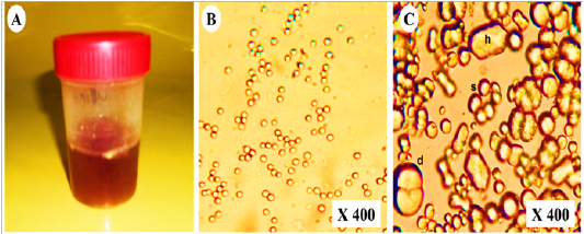
Representative photo showing (A); dark red urine contained (B); non- organized dumble (d), spherical-radiated(s) and hexagonal(h) shaped calcium carbonate crystals with (C); organized erythrocytes in the urine of vitamin K3-exposed horses
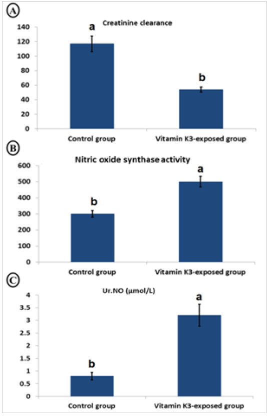
Char bars showing changes in creatinine clearance (A), serum nitric oxide synthase activity (B) and urinary nitric oxide (Ur.NO) (C) in vitamin K3-exposed horses comparing with the control horses
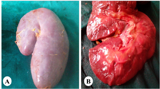
Representative photo showing enlargement of the kidney (A) with cortical and medullary congestion (B) of horses exposed orally to vitamin K3.
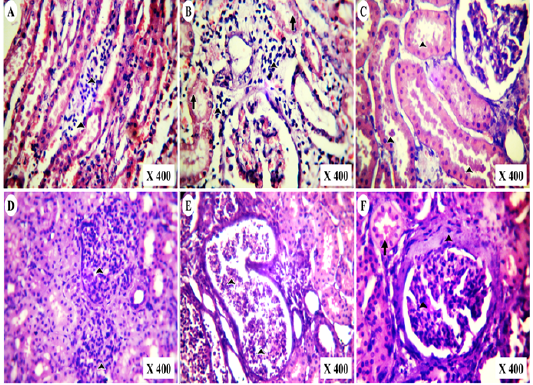
Photomicrographs of HE-stained kidney section of vitamin K3-exposed group showing (A); medullary lymphocytic aggregations (arrowheads),(B); cortical lymphocytic aggregations (arrowheads) with marked tubular necrosis and (C); globular eosinophilic proteinisuos casts (arrowheads) Also, the experimental cases showing (D); proliferative glomerulonephritis (arrowheads) with protein casts inside the renal tubules, (E); tubular casts of polymorph nuclear cells (arrowheads) and tubular cystic dilation besides (F); periglomerular fibrosis with mesangial cells proliferation (Arrowheads) and globular protein casts (arrow).


