Advances in Animal and Veterinary Sciences
Case Report
Adv. Anim. Vet. Sci. 5(11): 460-462
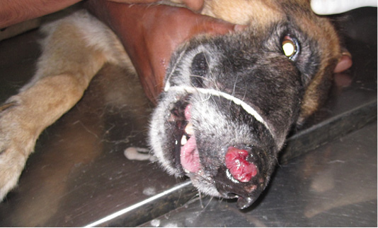
Figure 1
Reddish pedunculated mass completely obliterating the right nasal passage and protruding through right nostril
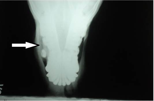
Figure 2
Radiography of rostral view revealing a radio opaque mass (arrow) in the right nostril
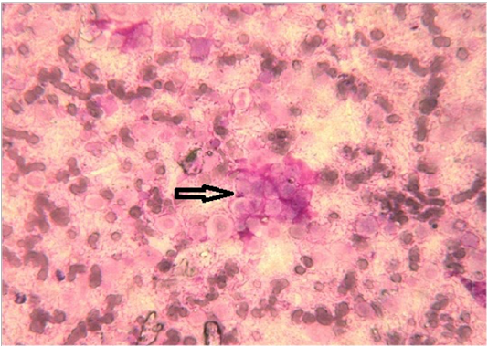
Figure 3
Cytology picture showing clusters (arrow) of chondrocytes (Leishman’s stain×1000)
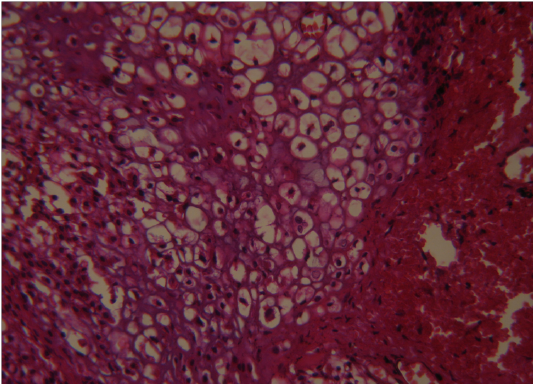
Figure 4
Tumour mass showing pleomorphic chondrocytes with mitotic (arrows) figures (H&E×400)
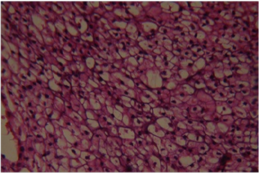
Figure 5
Sublingual lymphnodealso revealingpleomorphic chondrocytes (H&E×400)


