Advances in Animal and Veterinary Sciences
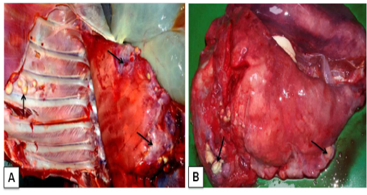
Corynebacterium pseudotuberculosis in goat: A) Caseo-calcified nodules of variable sizes in different lobes of pneumonic lungs and even in the chest wall (arrows); B)Small pea to the walnut size of abscesses with greenish pus scattered throughout the lung parenchyma (arrows)
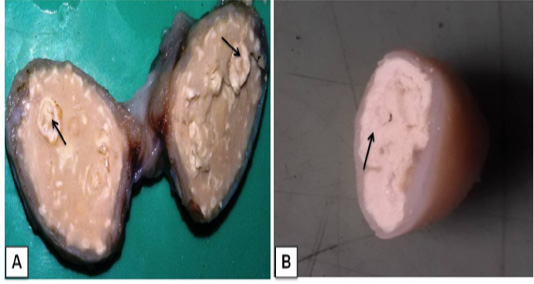
Lymphadenitis in goat. A) A cross-sectional view of affected bronchial lymph node hard calcium granules that deposited with abscesses (arrows); B) The mediastinal lymphnode showedcharacteristic lamellate (onion skin or onion ring), pathognomonic signs of CLA (arrow)
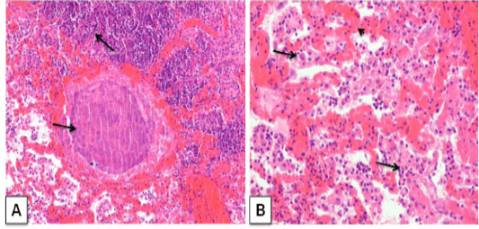
Corynebacterium pseudotuberculosis abscess in lung section of a goat. ; A) caseative necrotic mass with bacterial colonies within the lung parenchyma (arrows); H&E. (X 100) B) Lung section showing vascular congestion (arrow head), infiltration of neutrophils with macrophage in the alveoli (arrows). H&E. (X 200)
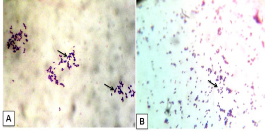
Corynebacterium pseudotuberculosis in goat: A) Bacterial culture gram’ staining showed gram positive cocco-bacilli arranged in Chinese pattern; B) Impression smear from heart also given gram positive cocco-bacilli arranged in Chinese pattern. (x1000)
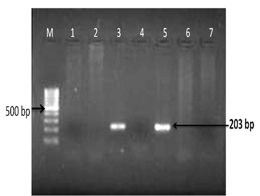
Detection of Corynebacterium pseudotuberculosis DNA in affected lung and lymph node tissue samples: PCR for pld gene regions; Lanes- 3 and 5 are positive amplification (203 bp); Lanes- 1, 2, 4 and 6 are negative; Lane 7- negative control and M-100 bp ladder


