Advances in Animal and Veterinary Sciences
Case Report

Clinical Management of Parasitic Gastroenteritis (PGE) Concurrent with Mycoplasmosis and Orf in Sheep
Faez Firdaus Abdullah Jesse1,3*, Yusuf Abba2, Innocent Damudu Peter1, 4, Asinamai Athliamai Bitrus1, Idris Umar Hambali1, 4, Nurul Liyana Jamaluddin1, Abdul Wahid Haron1
1Department of Veterinary Clinical Studies, Faculty of Veterinary Medicine, Universiti Putra Malaysia, 43400 Serdang, Selangor, Malaysia; 2Department of Veterinary Pathology, Faculty of Veterinary Medicine, University of Maiduguri, P.M.B, 1069, Borno State, Nigeria; 3Institute of Tropical Agriculture and Food security, Universiti Putra Malaysia, 43400 Serdang, Selangor, Malaysia; 4Faculty of Veterinary Medicine, University of Maiduguri, P.M.B 1069 Maiduguri, Nigeria.
Abstract | A 1-year-old male ovine, Black Dorper, was presented to the Large Animal Ward of University Veterinary Hospital, University Putra Malaysia with a complaint of pasty diarrhoea. The farm appeared to be endemic for parasitic gastroenteritis (PGE) and animals have developed resistance to albendazole. Physical examination revealed a pale mucous membrane with absence of capillary refill time and a FAMACHA score of 4. The animal had pyrexia, tachycardia and tachypnia. Additionally, scabby lesions were present on the mouth region and pasty faeces on the anal region. Complete blood count revealed a PCV of 14%, normocytic hypochromic anaemia, neutrophilia with left shift, lymphopenia and thrombocytosis. Serum biochemistry result revealed uraemia and low creatinine, hyperbilirubinemia and hypoalbuminemia. McMaster faecal egg count showed 3,000 eggs per gram of Strongyles. Mycoplasma ovis organism was observed on Giemsa-stained thin blood smear. Blood transfusion was done on the second day of hospitalization and the PCV value gradually improved. Levamisole was given as anthelmintic and oxytetracycline for the Mycoplasmosis. The condition of the animal improved and it was discharged on the 9th day of hospitalization.
Keywords | Parasitic gastroenteritis, Mycoplasma ovis, Orf, Anaemia, Parasitic gastroenteritis, Blood transfusion
Editor | Kuldeep Dhama, Indian Veterinary Research Institute, Uttar Pradesh, India.
Received | July 07, 2017; Accepted | August 28, 2017; Published | September 01, 2017
*Correspondence | Faez Firdaus Abdullah Jesse, Immunology Section, Department of Veterinary Clinical Studies, Faculty of Veterinary Medicine, University Putra Malaysia, 43400 Serdang, Selangor, Malaysia; Email: jesseariasamy@gmail.com
Citation | Jesse FFA, Abba Y, Peter ID, Bitrus AA, Hambali IU, Jamaluddin NL, Haron AW (2017). Clinical management of parasitic gastroenteritis (pge) concurrent with mycoplasmosis and orf in sheep. Adv. Anim. Vet. Sci. 5(9): 358-361.
DOI | http://dx.doi.org/10.17582/journal.aavs/2017/5.9.358.361
ISSN (Online) | 2307-8316; ISSN (Print) | 2309-3331
Copyright © 2017 Jesse et al. This is an open access article distributed under the Creative Commons Attribution License, which permits unrestricted use, distribution, and reproduction in any medium, provided the original work is properly cited.
INTRODUCTION
Parasitic gastroenteritis is caused by nematode species either singly or in combination. In Malaysia, the important species causing PGE are Haemonchus spp., Trichostrongylus spp., and Oesophagostomum spp (Abdullah et al., 2016). Haemonchosis in sheep can be divided into three stages which are hyper acute, acute and chronic. Hyper acute stage is characterized by death within one week; acute is characterized by the presence of severe anaemia and generalized oedema, while chronic stage is established when there is presence of anaemia accompanied by progressive weight loss (Mohammed et al., 2016). The most effective diagnostic procedure for haemonchosis is through detection of larval stage (L3) in fecal samples (Nasai et al., 2016).
Mycoplasmosis is an infection caused by Mycoplasma ovis organism in sheep. The infection is often asymptomatic but in young animals with a heavy intestinal burden, it can cause severe haemolytic anaemia. Chronic infection usually occurs in adults leading to poor weight gain, exercise intolerance, decreased wool production and mild anaemia (Jesse et al., 2015). Mycoplasmosis is endemic in most ruminant farms in Malaysia. In 2015, an incidence rate of 93.6% was observed in a goat farm in Ladang Angkat, Selangor, Malaysia (Jesse et al., 2015).
Contagious ecthyma or orf, is a highly contagious zoonotic viral disease caused by Parapoxvirus. Then virus produces scab like lesions in the mouth and muzzle of affected animals (Abdullah et al., 2015). In Malaysia, the virus is endemic in small ruminant populations with documented reports (Abdullah et al., 2015; Sadiq et al., 2017). Once a farm is infected with the virus, it is advisable to isolate sick animals and cull in order to prevent spread to other animals. This report documents the clinical management of parasitic gastro-enteritis with concurrent mycoplasmosis and orf in a sheep.
CASE REPORT
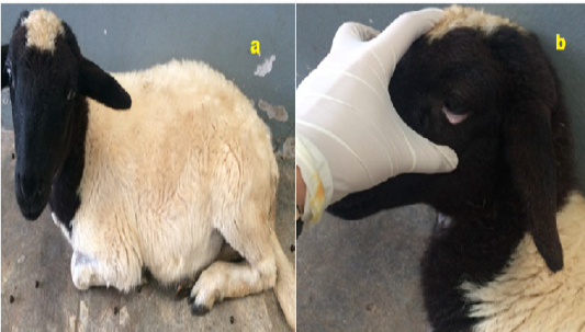
A 1-year-old male, Black Dorper sheep was presented to the University Veterinary Hospital, UPM with a complaint of diarrhoea. Based on the history, the farm is endemic with PGE and the animals have developed resistance to albendazole. There was no record of vaccination in the farm and the deworming status is not up to date. The animal was managed semi-intensively. Upon physical examination; the sheep was pyretic with a temperature of 40.7°C, having tachycardia and tachypnea. The animal weighed 12kg with a body condition score of 2.5, was dull and depressed, had a pale mucous membrane with capillary refill time that was inaccessible (Figure 1). According to the FAMACHA chart, a score of 4 out of 5 was observed. There was presence of scabby lesion at the mouth region with a faeces pasted anal region.
Sample Collection and Analysis
Blood sample was collected for complete blood count and serum biochemistry, and hemo-parasite screening. Faecal sample was collected for parasitology. The hemogram revealed normocytic hypochromic anaemia, neutrophilia with left shift, lymphopenia and thrombocytosis. The serum biochemistry results showed evidence of uraemia, hypocreatininemia, hyperbilirubinemia, hypoproteinemia characterized by hypoalbuminemia and hypoglobulinemia (Table 1). Faecal egg count was performed using McMaster technique and an egg count of 3,000 e.p.g was determined. Giemsa-stained thin blood smear revealed coccoid shaped Mycoplasma ovis organism on the surface of red blood cells. This high burden of parasitemia indicates a clinical mycoplasmosis infection. Thus, the final diagnosis for this case was Parasitic Gastroenteritis concurrent with Mycoplasmosis and Contagious Ecthyma (Orf).
Table 1: Hematological and biochemical parameters observed in the sheep.
|
Hematology |
Results |
Reference |
|
Erythrocytes (RBC) x1012/L |
4.24 |
9 – 15 |
|
Haemoglobin g/L |
37.3 |
90 – 150 |
|
PCV L/L |
0.14 |
0.27 – 0.45 |
|
MCV fL |
33 |
28 – 40 |
|
MCHC g/L |
266 |
310 – 340 |
|
Leukocytes (WBC) x109/L |
9.58 |
4 – 12 |
|
Band Neutrophils x109/L |
0.19 |
0 |
|
Seg. Neutrophils x109/L |
6.80 |
0.7 – 6.0 |
|
Lymphocytes x109/L |
1.92 |
2.0 – 9.0 |
|
Monocytes x109/L |
0.57 |
< 0.75 |
|
Eosinophils x109/L |
0.10 |
< 0.75 |
|
Basophils x109/L |
0 |
< 0.3 |
|
Thrombocytes x109/L |
1047 |
250 – 750 |
|
Plasma protein g/L |
38 |
60 – 75 |
|
Icterus Index Unit |
5 |
< 15 |
|
Biochemical Parameters |
Results |
Reference |
|
Sodium mmol/L |
147 |
139-152 |
|
Potassium mmol/L |
5.5 |
3.9-5.4 |
|
Chloride mmol/L |
108 |
95-103 |
|
Urea mmol/L |
9.1 |
2.8-7.1 |
|
Creatinine µmol/L |
67 |
106-168 |
|
Bilirubin, Total µmol/L |
7.3 |
1.7-6.8 |
|
Bilirubin, Conjugated µmol/L |
2.4 |
<4.6 |
|
γ-GT U/L |
37 |
30-50 |
|
AST U/L |
118 |
50-100 |
|
Total protein (Serum) g/L |
40.4 |
55-70 |
|
Albumin g/L |
19.0 |
25-35 |
|
Globulin g/L |
21.4 |
25-45 |
|
A:G Unit |
0.9 |
0.5-1.2 |
Management Plan
Fercobasang, 2mL, IM was given for 7 days as an iron supplement in order to correct the anaemia. Kaolin Pectin, (30mL/25kg) was given Per Os for 6 days to correct the diarrhoea. Levamisole suspension, 5mL, Per Os was given as an anthelmintic agent. Flunixin Meglumine, (2.2mg/kg), I.M was given for 5 days. The Orf lesion at the mouth region was dabbed with iodine and Woundsarex® as a local antiseptic. Oxytetracycline, 1mL/10kg, I.M was given every 48 hours for the Mycoplasmosis.
During the second day of hospitalization, blood transfusion was done because of the low PCV value and the deteriorating condition of the animal. Donor blood was collected in a CPDA-1 blood bag. The donor and recipient blood were compatible by cross-match and the donor blood tested negative for blood parasites screening. An intravenous port was established via the jugular vein and an initial 3mL of blood was transfused in the first 15 minutes and the animal was observed for signs of any adverse reaction. After 15 minutes, the rate of transfusion was increased to 3 drops/second. A total of 102 mL of blood was successfully transfused in about 4 hours. After transfusion, the animal was flushed with Lactated Ringer’s solution and Duphalyte. The PCV of the animal slowly increased to 19% at day 9 of hospitalization. The animal was discharged on the third week of hospitalization. The prognosis of the animal was good since the PCV increased and the mucous membrane colour was restored. The appetite of the animal also improved.
DISCUSSION
Haemonchus contortus is a common cause of PGE in sheep. It feeds on blood, resulting in hypoalbuminaemia which in turn leads to decreased osmotic pressure that eventually results in oedema. PGE may also cause immunosuppression which leads to secondary infections (Abdullah et al., 2016). Each worm is estimated to suck about 0.05mL of blood per day by ingestion or seepage form the lesions (Qamar et al., 2009). This results in normocytic hypochromic anemia as was observed in this case. Diarrhoea associated with hemonchosis also results in dehydration and hypovolemia (Mohammed et al., 2016). In this case PGE was treated with levamisole; a cholinergic agonist belonging to the imidazothiazole group. The drug induces an outward influx of sodium ion currents, which depolarises neuromuscular transmission resulting in paralysis of the worm (Ap, 1994). PGE co-infection with other parasitic diseases such as eperythrozoonosis and mycoplasmosis has been reported previously. In this instance, the con-infecting pathogen resulted in aggravation of the clinical presentation of the disease (Jesse et al., 2015; Abdullah et al., 2016).
Mycoplasmosis was also observed as a concurrent infection in this case. Mycoplasmosis is often asymptomatic in the subclinical stage and can later progress to severe anaemia or even death in young animals, thus resulting in huge economic losses. In Malaysia, the first case of mycoplasmosis in sheep was described in 1994, which was concurrent with copper toxicity (Fatimah et al., 1994). A study conducted in 2015 revealed a prevalence of 93.6% in goats (Jesse et al., 2015). In other countries, North Central Nigeria reported a prevalence of 25.8% (Akwuobu et al., 2014), North Africa reported a prevalence of 6.28% (Rjeibi et al., 2015) and West Iran reported a prevalence of 76.2% (Khezri et al., 2014). The information on the prevalence of mycoplasmosis is important in order to sensitize farmers to the realities of the condition in the field. In this case, Mycoplasma ovis also contributed to the anemia observed since the organism has been shown to induce moderate to severe anemia in farm animals. As a therapeutic remedy, Oxytetracycline was administered and it proved effective. Oxytetracycline works by binding to the 30S ribosomal subunit and inhibiting protein synthesis. The other effective choice of antibiotic against Mycoplasma ovis is Enrofloxacin. It has a bactericidal activity that works by inhibiting the DNA and RNA synthesis (Mazaheri et al., 2014).
In this case, mild Orf lesions were observed in the mouth and feet. The lesions were routinely dressed in order to prevent secondary bacterial infection. Orf is an endemic disease in Malaysia (Abdullah et al., 2015). Previous outbreaks with severe lesion distribution have been recently reported (Sadiq et al., 2017). In severe outbreaks, the lesions on the lips, gums and tongue affect the feeding and can result in death especially when secondary bacterial infection sets in (Zamri-Saad et al., 1992). Since Orf is a viral disease and self-limiting, there is no specific treatment and the infection usually clears up within a few weeks. In order to prevent the spread of the infection, the affected animals in a herd should be identified and isolated, and the pen should be thoroughly disinfected.
CONCLUSIONS
In this case, PGE with concurrent Mycoplasmosis and Orf was reported. Normocytic hypochromic anemia observed was severe but effectively managed with chemotherapy and blood transfusion. Serum biochemical alterations observed was also associated with the blood loss due to PGE. Prompt institution of therapy with Fecobsang, Koalin pectin, levamisole and oxytetracycline was effective in curtailing the menace of PGE and Mycoplasmosis in the sheep.
ACknowledgements
The authors wish to appreciate the technical assistance of Staff of Parasitology and Microbiology Laboratories of the Faculty of Veterinary Medicine, UPM.
Conflict of interest
There is no conflict of interest regarding the publication of this work.
authors contribution
All authors contributed equally.
REFERENCES


