Advances in Animal and Veterinary Sciences
Research Article

A Comparative Study in some Morphological and Histological Features of the Live in Gull (Laruscanus) and Mallard duck (Anas platyrhynchos)
Iman M. Khaleel1*, Khalid Ibrahim Al-Khazraji2, Muna Hussein AL-Aameli3
1Department of Anatomy and Histology, College of Veterinary Medicine, University of Baghdad, Iraq; 2Department of Anatomy and Histology, College of Veterinary Medicine, University of Diyala, Iraq; 3Department of Anatomy and Histology, Faculty of Veterinary Medicine, University of Kerbala, Iraq.
Abstract | This study was conducted to evaluate the morphological and histological features of the liver in two species of birds that vary in their food habitats; gull and mallard. Histomorphologically, the liver in these species is composed of the left (small) and right (big) lobes and the right lobe in gull was subdivided into two parts. Histologically, fibrous capsule covered the liver parenchyma without any clearly defined lobules in both species. The hepatic architecture comprised of hepatic cells organized as irregular plates or cords, which radiated around the central vein and the blood sinusoids distributed among those plates. The portal triads, enclosed by fibrous connective tissue, contains branch of hepatic artery, bile duct andportal vein. Several histological structures shown variations in both species including; capsule appeared thinner in mallard than in gull with mean thickness of 80.52±6.38µm and 86.23± 7.14µm respectively, and the diameters of central veins differed where mallard has 461.15 ± 11.25 µm and gull carried 387.42 ± 9.06 µm size. These morphological and histological features highlight fundamental differences in these species can be used as species-identification markers.
Keywords | Liver, Hepatic artery, Laruscanus, Anas platyrhynchos
Editor | Kuldeep Dhama, Indian Veterinary Research Institute, Uttar Pradesh, India.
Received | June 03, 2017; Accepted | July 10, 2017; Published | July 28, 2017
*Correspondence | Iman M. Khaleel, Department of Anatomy and Histology, College of Veterinary Medicine, University of Baghdad, Iraq; Email: talibhussein36@yahoo.com
Citation | Khaleel IM, Al-Khazraji KI, Al-Aameli MH (2017). A comparative evaluation of morphological and histological features of gull (laruscanus) and mallard duck (anas platyrhynchos) lungs. Adv. Anim. Vet. Sci. 5(7): 307-311.
DOI | http://dx.doi.org/10.17582/journal.aavs/2017/5.7.307.311
ISSN (Online) | 2307-8316; ISSN (Print) | 2309-3331
Copyright © 2017 Khaleel et al. This is an open access article distributed under the Creative Commons Attribution License, which permits unrestricted use, distribution, and reproduction in any medium, provided the original work is properly cited.
Introduction
The liver is regarded as an essential and biggest gland of the body, and it plays a crucial role in numerous physiological processes such as synthesis of blood proteins, production and secretion of bile, detoxification, nutrients absorption, metabolism of several substances and storing metabolites (Saez et al., 2012; Odokuma and Omokara, 2015). The bird liver is divided into two portions forming the right and left lobe (Al-Abedi, 2015). The size or subdivision of these two lobes can vary among different species. The right lobe appeared in more instance greater than the left one as in ostrich and bustard (Stomelli et al., 2006; Baily et al.,1997), however, some species of birds have an analogous sizes of the hepatic lobes as in galliformes (Schmidt et al., 2003). Hepatic parenchyma in birds showed similarity with that observed in most mammals except the variation in some histological characteristics (El-Zoghby, 2005). In coot birds, the hepatic parenchyma is subdivided into defined lobules and the hepatocytes organized around the central vein in radiation manner (Selman, 2013). The present study was carried out to investigate the morphological and histological features of livers in two species of birds that vary in their food habitat including carnivorous and omnivorous represented by gull (Laruscanus) and mallard duck (Anas platyrhynchos), respectively.
Materials and Methods
Liver of forty birds, comprised a twenty gull (Laruscanus) and twenty mallard duck (Anas platyrhynchos), were used in this comparative study. To represent diversification, birds were obtained from different areas in Baqubah, Diyala Province, Iraq. After measuring the body weight, all birds were eusthanized using a combination of Diazepam and Ketamine at dose rates of 5 mg/kg and 25 mg/kg of body weight (intramuscular), respectively, (Schindala, 1999). Half of the liver from each sampled species was examined grossly, whereas the other half underwent to the histological study. A sensitive balance was used to estimate the liver weight. The specimens from different lobes were taken and immediately fixed in 10% neutral buffered formalin for histological analysis and staining with Hematoxline & Eosin stain (Bancroft and Stevens, 2013). The histological structures were measured using ocular micrometer. Statistical analysis of the experimental results was conducted using t-test in the Statistical Package for the Social Sciences (SPSS) version 13.00 (Joda, 2008).
Results and Discussion
The morphological examination of the liver in both species of birds shown liver as a big gland situated in mid-coelomic cavity of the body. Anatomically, it was located in closed correlation with the spleen and gizzard, and caudal to the heart (Figure 1 and 2). It has dark brown color in gull whereas the color of the liver in mallard was reddish brown. This variation in the color of the liver between the two species may be related to the difference in their nutrition as nutritional state of the birds may affect the liver color (Clark, 2005).
The liver in those two species was composed of asymmetrical lobes (n=2), which were connected together cranially at the midline. It appeared that the left lobe in two species was smaller than right lobe (Figure 1 and 2). Consistently, Hussein and Hussein,2016 shown similar findings in fowl and common moorhen, however, (Ibrahim et al., 2016) noticed the left lobe of the liver was greater than the right one in local moorhen. In our study, we observed that the left lobe was undivided in two species of birds in disagreement with (Stomell et al., 2006; Nickel et al.,1977). This study revealed that the right lobe in mallard species was undivided, as that observed by (Al-AAaraji, 2015) in turkey, whereas this lobe in gull species showed two divisions at the caudal end (Figure 2).
It was observed that the relative liver weight to total body weight was 2.08% in the gull, compared with 1.95% in mallard. This significantly (p ˂ 0.05) higher relative liver weight in gull than those reported in mallard, could be reflected by the negative correlation between liver weight and body weight in birds. This explanation was coinciding with (Else et al., 2004), who recorded relative weight of the
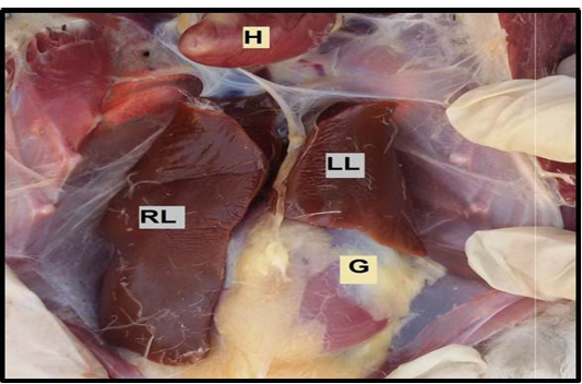
Figure 1: The liver in mallard duck. Showing the left lobe (LL), right lobe (RL) of the liver, heart (H), gizzard (G).
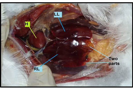
Figure 2: The liver in gull. Showing the left lobe (LL), right lobe (RL) which contains two parts, a presence Heart (H).
liver was 2.71% of total body weight in zebra finches. The change in the weight of the liver may be associated with the type of food reception and higher metabolism rate in the body (Ankney, 1977). Histologically, the current study showed that the liver parenchyma in gull and mallard species covered by a capsule, which consisted of irregular dense connective tissue, comprised by the collagen fibers and elastic fibers (Figure 3 and 4) as shown previously (Bacha and Wood, 1990; Subhan, 2009) in other avian species such as chicken and geese. The capsule was noticed thinner in mallard than in gull with mean thickness of 80.52 ± 6.38 µm and 86.23± 7.14µm, respectively.
In this study the liver lobules were not clearly identified in the parenchyma of both species due to absence of indiscernible hepatic connective tissue septa. This finding was not compatible with observations previously made by Selman, (2013) in coot bird, but agreed with reports of El-Zoghby, (2005) in quail, Bacha and Wood (1990) in chicken, and Al-AAaraji (2015) in turkey. The hepatic parenchyma in both species of birds was consisted of hepatocytes that were organized as irregular plates or cords of thi-
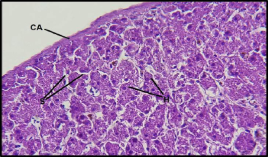
Figure 3: Histological section of the liver in gull: Showing the thick capsule (CA), hepatocyte (H), sinusoids (S). (H&E 40X).
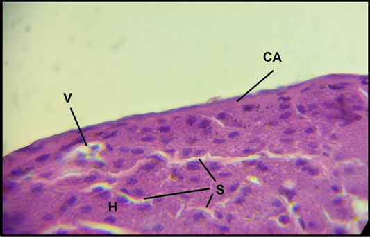
Figure 4: Histologicalsection of the liver in mallard duck s: Showing the capsule (CA), hepatocyte (H), sinusoids (S), vein (V). (H&E 100X).
ck cells, which radiated around the central vein, forming small acini or lobules, and the blood sinusoids were distributed among those plates (Figure 5 and 6). This results resembled description in the common moorhen (Hussein and Hussein, 2016), and differs with report of Hickey and Elias (1954) who reported that the hepatocytes arranged in cords mostly with one cell thick in birds, in contrast to just two thick cells formed hepatocyte plates in turkey and chicken (Bacha and Wood, 1990; Bhatanagar and Singh, 1982).
The diameters of central veins differed between mallard and gull with 461.15 ± 11.25 µm and 387.42 ± 9.06 µm, respectively. The hepatocytes appeared large in both species and their nuclei shown a large, rounded and centrally located dark distinct nucleoli (Figure 7 and 8).The shape of these cells varied in the two species; they were polyhedral in mallard, but irregular to oval in gull. This variation in the shape of these cells could be related to the species differences. The blood sinusoids were distributed between hepatic plates throughout the hepatic parenchyma, and were narrower in gull compared with those showed in mallard.
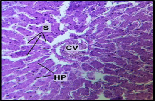
Figure 5: Histological section of the liver in mallard duck: Showing Central vein (CV), Hepatocyte Plates (HP), Sinusoids (S). (H&E 40X).
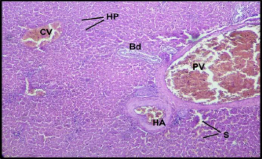
Figure 6: Histological section of the liver in gull : Showing Central vein (CV), Hepatocyte plates (HP), Sinusoids (S) Portal vein (PV), Hepatic artery (HA), Bile duct(Bd). (H&E 20X).
They were irregular in shape and lined by flattened endothelial cells with existence of large Kupffer’s cells. These observations were agreed with Attia and Soliman (2005) in liver of ostrich.
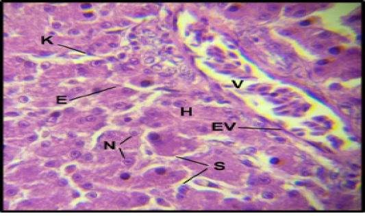
Figure 7: Histological section of the liver in mallard duck: Showing Vein (V), Hepatocyte (H), Sinusoids (S). Nucleus of hepatocyte (N), kupffer cell (K), Endothelial cell of sinusoid (E), Endothelial cells of Vein (EV). (H&E 100X).
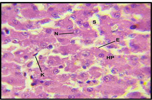
Figure 8: Histological section of the liver in gull: Showing Hepatocyte Plates (HP), sinusoids (S), Nucleus of hepatocyte (N), kupffer cell (K), Endothelial cell of sinusoid (E). (H&E 100X).
This study showed that the hepatic tissue in both species encompassed several regions, which were enclosed by connective tissue and dispersed throughout the parenchyma without a defined arrangement. These regions represented the portal triad, and each region contains the terminal branches of portal vein, hepatic artery and bile duct (Figure 9 and 10), as that proved in many other domestic birds and vertebrates (Elizabeth and Frye, 2001).
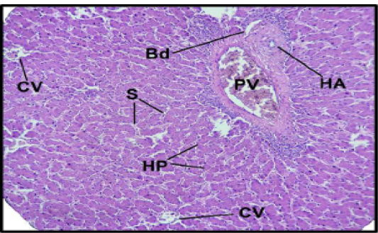
Figure 9: Histological section of the liver in mallard duck : Showing the portal triad. Portal vein (PV), Hepatic artery (HA), Bile duct (Bd), Central vein (CV) Sinusoids (S), Hepatic plate (HP)( H&E 20X).
The bile duct was lined with epithelium of simple cuboidal, while the portal vein had a large lumen with thin wall and lined by endothelial cells. The hallmark of hepatic artery was thick wall, small and winding lumen lined by endothelial cells as shown by Hussein and Hussein (2016) in the liver of moorhen and domestic fowl. These vessels and ducts varied in diameters between the two species, the mean diameter of bile duct, hepatic artery and portal vein were 135.5± 4.85 µm, 231. 64 ± 7.08 µm and 786.15± 13.20 µmin mallard, respectively, whereas in gull they were147.21± 3.16 µm, 201.75± 6.17µm and 625. 5 ± 12.23 µm, respectively.
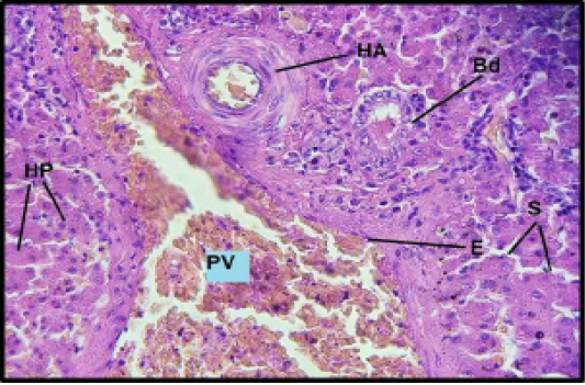
Figure 10: Histological section of the liver in gull: Showing the portal triad. Portal vein (PV), Hepatic artery (HA), Bile duct (Bd) Sinusoids (S), Hepatic plate (HP), Endothelial cells of portal vein (E). ( H&E 40X).
In conclusion, findings of this study highlight some of the key morphological and histological differences in the major organ of the body in two avian species. These differences would be used as marker of species identification and to be applied in comparative pathological investigations in health and disease.
COnflict of interest
There is no conflict of interest.
Authors Contribution
Iman: planning research and writing, Khalid: collecting samples and writing and Muna: preparing the histological slides.
References


