Advances in Animal and Veterinary Sciences
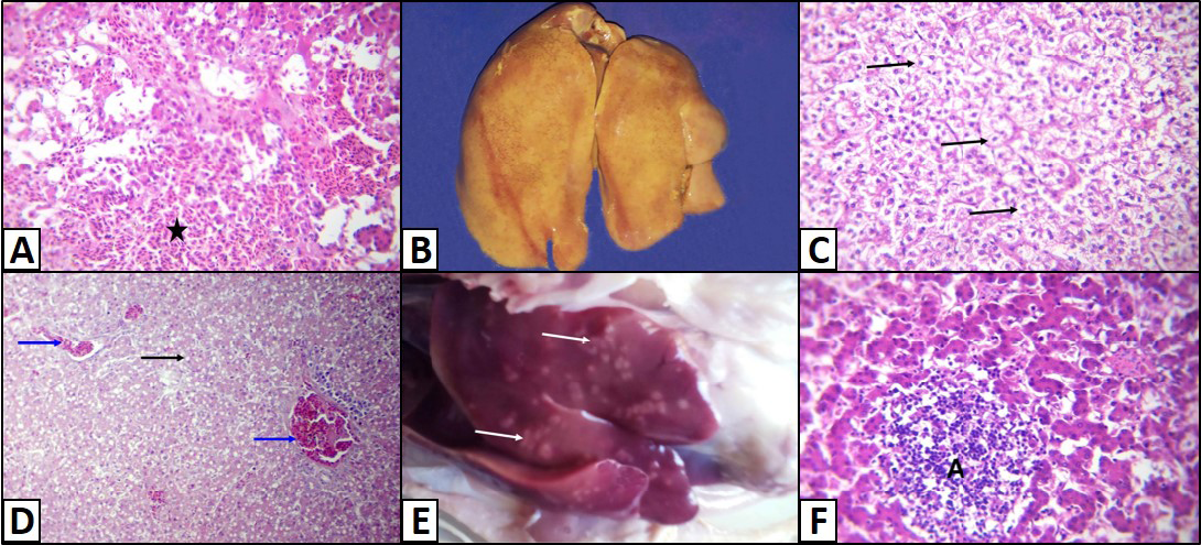
Liver of a White broiler chicken A) Infected by E.coli showing extravasations of numerous nucleated erythrocytes (asterisk); B) Infected by E.Coli showing enlargement, yellow in colour and soft in texture; C) Hydropic degeneration of the hepatocytes (arrows); D) infected by E. Coli showing fatty change of the hepatocytes (arrow) and congestion of the blood vessels (blue arrows); E) Infected by Salmonella showing multifocal to diffuse whitish patches (arrows); infected by E. Coli showing Hepatocytic necrosis with inflammatory cells infiltrations (A), H&E. (X250)
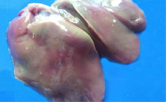
Liver of a Saso-breed chicken infected by salmonella showing Bronze discoloration
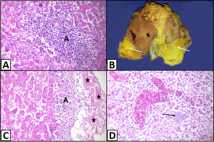
Liver of a White broilers chicken A) Infected by E. coli showing inflammatory cells infiltrations and fibrosis in the portal area (A); B) Infected by E. coli showing grayish whitish fibrin layers (arrows); C) Infected by E. coli showing eosinophilic fibrillar masses (stars) with presence of heterophils infiltration within or under these masses (A); D) Infected by E. coli showing hepatitis that represented by infiltration of inflammatory cells associated with bacterial colonies in the hepatic parenchyma (arrow), H&E. (X250)
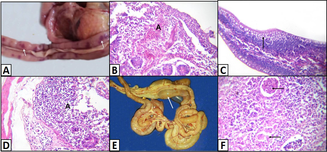
Intestine of A) Saso-breed chicken with coccidiosis showing congestion with multifocal dark red hemorrhagic patches (arrows); B) Balady-breed chicken infected by E. coli and Coccidian spp. showing numerous nucleated erythrocytes extravasated from blood vessels (A); C) White broilers chicken with mixed infection (E. coli and Salmonella) showing hyperplasia of goblet cells (arrow); D) Saso-breed chicken infected by E. coli showing intense inflammatory cells infiltrations in deep layer of the mucosa (A); E) Balady-breed chicken with mixed infection (E. coli, Clostridia, Coccidian spp.) showing ballooning of intestine with gases (arrow); F) White broilers chicken infected by E. coli showing different stages of coccidian parasite (arrows), H&E. (X250)
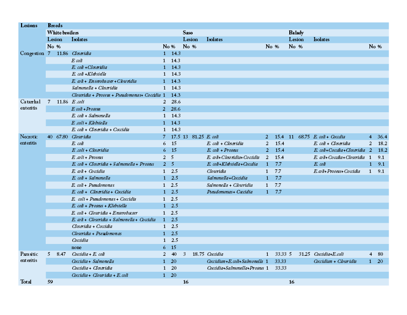
Number and percentage of histopathological lesions of the intestine of chicken breeds in relation to isolated bacteria and parasites


