Advances in Animal and Veterinary Sciences
Research Article
Adv. Anim. Vet. Sci. 4(11): 580-583
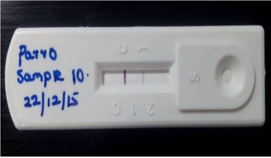
Figure 1
Detection of Canine parvoviral antigen in faecal sample by Lateral flow assay: T) Test line represents presence of CPV in test sample, C) band indicates Validity of the result
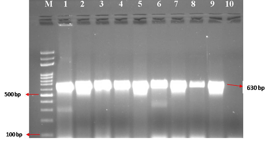
Figure 2
CPV specific amplicons (630 bp) by PCR using H specific primers: Lane M) 100 bp ladder; Lane 1) Positive control (CPV DNA); Lane 2-9) DNA samples isolated from faeces and blood; Lane 10) Negative control


