Advances in Animal and Veterinary Sciences
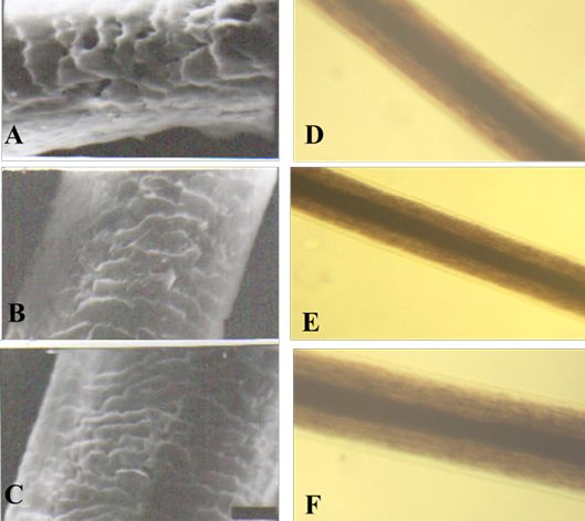
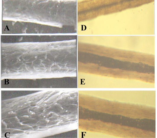
SEM of the hair shaft of Urus americanus
A: proximal part, B: middle part and C: distal part; note the thickness of hair shaft, note also the overlapped short and wide imbricated scales of equal hastate with saw margin and without long free blade (X1000). The other side shows the three morphological regions of hair (the cuticle, the medulla and the cortex) along the hair shaft of Urus americanus (D: proximal part, E: middle part and F: basal part) under light microscope (X400)
SEM of the hair shaft of (Ceropithecus mitis)
A: proximal part, B: middle part and C: distal part; note the thickness of hair shaft, note also the overlapped moderate length and wide imbricated scales of equal hastate with saw margin and without long free blade (X1000). The other side shows the three morphological regions of hair (the cuticle, the medulla and the cortex) along the hair shaft (D: proximal part, E: middle part and F: basal part) of Ceropithecus mitis under light microscope (X400)
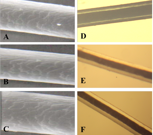
SEM of the hair shaft of Amotracus lervia
A: proximal part, B: middle part and C: distal part; note the thickness of hair shaft, note also the overlapped short and wide imbricated scales of equal hastate with smooth margin and without long free blade (X1000). The other side shows the three morphological regions of hair (the cuticle, medulla and the cortex) along the hair shaft of Amotracus lervia (D: proximal part, E: middle part and F: basal part) under light microscope (X400)
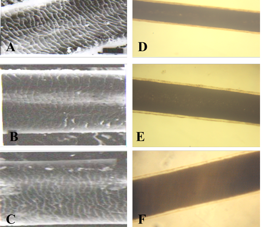
SEM of the hair shaft of Lama
A: proximal part, B: middle part and C: distal part; note the thickness of hair shaft, note also the overlapped moderate length and moderate width imbricated scales of equal hastate with saw margin and without long free blade (X1000). The other side shows the three morphological regions of hair (the cuticle, the medulla and the cortex) along the hair shaft of Lama glama (D: proximal part, E: middle part and F: basal part) under light microscope (X400)
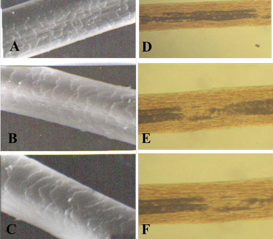
SEM of the hair shaft of Camelus bactrianus
A: proximal part, B: middle part and C: distal part; note the thickness of hair shaft, note also the overlapped moderate length and moderate width imbricated scales of equal hastate with saw margin and without long free blade (X1000). The other side shows the three morphological regions of hair (the cuticle, the medulla and the cortex) along the hair shaft of Camelus bactrianus (D: proximal part, E: middle part and F: basal part) under light microscope (X400)


