Advances in Animal and Veterinary Sciences
Research Article

Ultrasonography and Ultrasonographic Guided Biopsy of the Liver in Pregnant Toxemic Does
Amr Gamal Kotb, Wafaa Mahmoud Abd El-Razik, El-Abas Mohamed El-Nagar, Shaimaa Mohamed Gouda*
Department of Animal Medicine, Faculty of Veterinary Medicine, Zagazig University, Egypt.
Abstract | Clinical and biochemical examinations are still the most common tools for diagnosis of pregnancy toxemia (PT) in does. In the present study, ultrasonography and ultrasonographic guided biopsy of the liver were used as additional tools in evaluation of such disease. Fifteen pregnant toxemic does as well as fifteen apparently healthy does (control) were used. The disease was tentatively diagnosed by the history and the clinical assessment and confirmed diagnosis was achieved by positive ketone-urea, hypoglycemia and elevation of beta hydroxy butyric acid level (βHBA). Ultrasonographic examination of the liver was performed at the area extending from the 7th to the 12th right intercostal space using a 3.5 to 5 MHz convex transducer. Liver biopsy samples were obtained under ultrasonographic guidance and submitted for histopathological examination. Necropsy findings were recorded in 3 does with PT. Does with hyperechogenic hepatic parenchyma and visible blood vessels had a good prognosis and respond to treatment while does with hyperechogenic hepatic parenchyma and invisible blood vessels had a bad prognosis. The result of histopathology and necropsy revealed presence of different degrees of fatty liver which was mild in good prognostic group (n=9) and severe in bad prognostic group (n=6). In conclusion, ultrasonography can be used as a useful and practical tool to evaluate the state of the liver in does with PT.
Keywords | Biopsy, Does, Fatty liver, Pregnancy toxemia, Ultrasonography
Editor | Kuldeep Dhama, Indian Veterinary Research Institute, Uttar Pradesh, India.
Received | June 23, 2015; Revised | July 02, 2015; Accepted | July 03, 2015; Published | July 06, 2015
*Correspondence | Shaimaa Mohamed Gouda, Zagazig University, Egypt; Email: dr.shaimaagouda@yahoo.com
Citation | Kotb AG, El-Razik WMA, El-Nagar EM, Gouda SM (2015). Ultrasonography and ultrasonographic guided biopsy of the liver in pregnant toxemic does. Adv. Anim. Vet. Sci. 3(8): 466-472.
DOI | http://dx.doi.org/10.14737/journal.aavs/2015/3.8.466.472
ISSN (Online) | 2307-8316; ISSN (Print) | 2309-3331
Copyright © 2015 Kotb et al. This is an open access article distributed under the Creative Commons Attribution License, which permits unrestricted use, distribution, and reproduction in any medium, provided the original work is properly cited.
INTRODUCTION
In Egypt, goats represent one of most important animals among livestock, as they possess great capacity to convert the poor feedstuffs to very useful products for human as meat, milk and wool.
In late pregnancy, energy deficits often results in pregnancy toxaemia in ewes and does (Pugh and Baird, 2012). Pregnancy toxemia follows a period of negative energy balance and impaired gluconeogenesis resulting in hypoglycemia, fat mobilization, ketonemia, and ketonuria (Rook, 2000). When the hepatic uptake of lipids exceeds its oxidation and secretion excess lipids are stored as tricycle glycerol and fatty liver syndrome is developed. This condition is associated with decreased metabolic functions of the liver. Liver can be categorized into normal or mild, moderate or severe fatty liver. The latter can be subdivided into non-encephalopathic severe fatty liver and hepatic encephalopathy (Bobe et al., 2004). Diagnosis of pregnancy toxemia is based on history, clinical signs and serum biochemical analyses. Assessment of some prognostic tools as such as liver function test, renal function test and cardiac biomarker has been established (Abdelaal et al., 2013).
Ultrasonography was established as one of the principle imaging techniques in veterinary practice. It allows the clinician to obtain instant information about a wide range of body systems and in some cases, the dynamic function of organs can be assessed. It had been used for examination of the liver in healthy condition (Braun and Steininger, 2011) and pathological conditions as fatty liver in goats (Gonenci et al., 2003), hepatic abscesses in cattle (Abdelaal et al., 2014) and hydatid cyst in ovine (Guarnera et al., 2001; Hussein and Elrashidy, 2014).
In the current study, ultrasonographic assessment of the liver and the ultrasonographic guided biopsy of the liver were suggested as an additional prognostic tool for pregnancy toxemia in does.
MATERIALS AND METHODS
This work was conducted in Faculty of Veterinary Medicine, Zagazig University, Egypt from February 2013 to September 2014. This study was carried out on 30 late pregnant Balady does. Their ages were (3 ± 0.5 years) and their weights were (28.5 ± 2.5 kg). Control group animals (n=15) were reared indoor and obtained from the Faculty farm. Each animal was fed on commercial concentrate mixture consisting of 35% wheat bran, 32% Cotton seed cake, 30% crushed maize and supplemented with 2% limestone and 1% common salts. Rice straw was available throughout the pregnancy period, green fodder as Egyptian clover (Trifolium alaxandrinum), darawa, and maize fodder was offered when available, and the ration was offered into two equal portions twice daily. Water was accessible at all times. Rational, natural pasture (Green herbage, grass and remnant of plant, barseem and darawa) was fed when available.
The diseased group (n=15) were admitted to the hospital of Veterinary Medicine, Zagazig University with the signs of anorexia, dullness and scanty feces at late pregnancy.
Clinical Examination
All animals were subjected to physical examination which included evaluation of general condition, visible mucous membrane, heart rate, respiratory rate and rectal temperature as described previously by Pugh and Baird (2012).
Biochemical Profile
Two blood samples were collected by the jugular venepuncture, the first with sodium fluoride and the second without anticoagulant. Blood glucose level was estimated in the first sample. After centrifugation of the second blood sample, serum samples were collected and then frozen at –20°C for 1 week and subsequently analysed for biochemical parameters. The commercial test kits (Biomerieux, Egypt) were used to determine the concentrations of total proteins, albumin, beta hydroxy butyric acid, blood urea nitrogen, creatinine, triglyceride and total cholesterol. The activities of alanine aminotransferase (ALT), aspartate aminotransferase (AST) and γ-glutamyl transpeptidase (GGT) were also measured in serum samples. The biochemical analyses of the selected parameters were conducted using a spectrophotometer (RIELE GmbH & Co, Berlin, Germany) according to the standard protocol of the suppliers.
Urinalysis
About 5 ml of fresh and well-mixed urine samples were collected in a clean and dry plastic container from each animal. Urine reagent strips were used according to the directions of the manufacturer (Roche combur urine strips®, Boehringer Monnheim, Germany) to estimate ketone bodies.
Ultrasonographic Examination
After diagnosing the case as pregnancy toxemia using the physical examination and urine analysis, transcutaneous ultrasonographic examination was performed using ultrasound unit (Sunbright Sun-806F, CHINA) supplied with convex probe with frequency range 3.5 to 5 MHz. After clipping the hair and application of coupling gel, the liver was examined on the right intercostal spaces (ICS) from dorsal to ventral according to the technique used in goat by earlier workers (Braun and Steininger, 2011). The position of the liver, the texture of the parenchyma and visibility of hepatic vessels were assessed.
Liver Biopsy and Histopathology
Biopsies were obtained under ultrasonographic guidance on all diseased does and 3 healthy does as described previously by Nesspoli et al. (2010). The site was shaved and disinfected by alcohol 70% and Povidone Iodine 10% (Betadine® solution). The place was desensitized using a local anesthetic agent (Lidocaine hydrochloride spray). The liver biopsy needle (disposable, semi-automatic needle 16G X 20cm - USA) was inserted into the abdominal cavity until it appears inside the liver parenchyma. The piece of the liver obtained by this procedure was usually weighting approximately 100-200 mg. After the procedure, the liver was ultrasonography scanned to assess the presence of hematoma or active bleeding. All does subjected to biopsy were kept under close observation for a period of 6 hours post-procedure and received vitamin K as a prophylactic measurement. All collected liver specimens were examined macroscopically for the colour, appearance and consistency, and then put in 15% formaldehyde until processed and stained by hematoxylin and eosin (H&E) as described by Johnson et al. (2004).
Therapeutic Trials
Medical Treatment of Diseased Does
All diseased does received medical treatment as described by Pugh and Baird (2012). Five hundred ml of Dextrose 25% (Al-Ameria Pharmaceutical Company, Egypt) at dose of 500 ml was administrated by intravenous route. One to two hundred ml of propylene glycol (Al Gomhoria Company, Egypt) was given orally for 5-10 days and 20 ml of calcium (Cal-Bor- Mag, Adwia, Egypt) was administrated intramuscularly for 4 days. Termination of pregnancy was completed by 1 ml dexamethasone intramuscular (dexacortyl200 mg/100 ml Coophavet. France).
Caesarean Section
Out of 15 PT does, 9 not responding to medical treatment and submitted to caesarian section (CS). The operation was done routinely using xylazine (Rombun 2%, Bayer, Germany) and local infiltration anesthesia of the left flank (lidocaine 2%, Vetoquinol, France).
Necropsy Findings
Out of 9 cases which submitted to CS, 6 cases not responding and die during or immediately after operation. Necropsy findings were recorded in 3 cases out 6 does which not respond to either medical or surgical trials.
Statistical Analysis
Data biochemical profile of both healthy and diseased groups is presented as mean ± SD and the analysis was conducted using SPSS 16.0., Student’s t-test. Differences between parameters considered significant at a level of P ≤ 0.05.
Table 1: Clinical findings of does suffered from pregnancy toxemia
|
Parameters |
PT does |
|
|
Number |
Percent |
|
|
Rectal temperature °C Normal (38.8-39.2oC) Hypothermic Hyperthermic |
3 6 6 |
20 40 40 |
|
Heart rate (beats/minute) Normal (75-90 per minute) Brady cardic Tachy cardic |
3 6 6 |
20 40 40 |
|
Respiratory rate (breaths/minute) Normal (20-30 per minute) Decreased Increased |
3 6 6 |
20 40 40 |
|
Rumen activity (contractions/minute) Normal (3-4 per 2 minutes) Decreased or absent |
3 12 |
20 80 |
|
Dullness appearance |
15 |
100 |
|
Anorexia |
15 |
100 |
|
Tympany |
15 |
100 |
|
Scanty feces |
15 |
100 |
|
Recumbancy |
6 |
40 |
|
Aceteone smelling |
6 |
40 |
|
Nervous signs |
6 |
40 |
|
Ictrus |
2 |
13.3 |
RESULTS
Clinical Findings
As shown in Table 1, all diseased does had dullness and dummy appearance, did not respond to external stimuli and showed absence of menace reflex, anorexia, mild tympany and scanty feces. Additional signs included recumbency, acetone odor from breathing and nervous signs like blindness, circling, grinding on their teeth, incoordination, star-gazing, tremors, and convulsions in 6 cases (Figure 1). Icterus was visible in conjunctival and vulval mucosa in 2 cases. The vital parameters included temperature; ruminal motility; pulse and respiratory rate were normal in control group, but in PT does, these parameters were increased (n=6), decreased (n=6) or were within normal range (n=3).
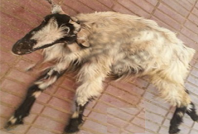
Figure 1: A doe with PT and exhibit nervous signs include incoordination, star-gazing, tremors, and convulsions
Biochemical Profile
The PT does had hypoglycemia, hypoproteinemia, hypoalbuminaemia and significant increase in Beta hydroxyl butyric acid (βHBA). There were significant increases in Alanine Amino Transferase, Aspartate Amino Transferase and Gamma Gultamic Transferase. Non-significant changes in blood urea nitrogen and creatinine levels were detected. Serum cholesterol showed significant decrease in PT does, while triglyceride showed significant increase (Table 2).
Table 2: The biochemical and urinalysis of healthy and PT does
|
Parameters |
Control group |
PT does |
|
Biochemistry |
||
|
Glucose mg/dl |
64.33±4.18 |
26.85±1.26* |
|
Total protein g/dl |
6.21±0.13 |
4.15±0.10* |
|
Albumin g/dl |
3.54±0.06 |
2.44±0.08 |
|
βHBA mmol/L |
0.45 ± 0.16 |
4.46±1.6* |
|
AST u/l |
49.87±0.77 |
68.73 ± 0.82* |
|
ALT u/l |
26.73±0.62 |
43.87±1.2* |
|
GGT u/l |
31.07±0.73 |
45.40 ± 0.88* |
|
BUN mmol/l |
2.1±0.1 |
2.7±0.2 |
|
Creatinine µmol/L |
72.0±4.0 |
75.5±3.5 |
|
Cholesterol mg/dl |
78.47±1.15 |
43.87±1.40* |
|
Triglycerides mg/dl |
65.07±1.70 |
109.63±1.34* |
|
Urinalysis |
||
|
Keton-urea |
-ve |
++ ve |
|
Glucose-urea |
-ve |
- ve |
Values are represented by means ± SD; Different superscripts in the same row indicate a significant difference at (P ≤ 0.05)
Urinalysis
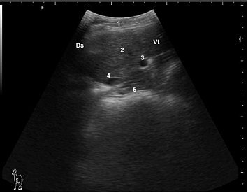
Figure 2: Ultrasonogram of hepatic parenchyma and hepatic blood vessels in a healthy late pregnant doe viewed from the 10th ICS on the right side
1: Lateral abdominal wall; 2: Liver parenchyma; 3: Portalvein; 4: Caudal vena cave; 5: Omasum; Ds: Dorsal; Vt: Ventral
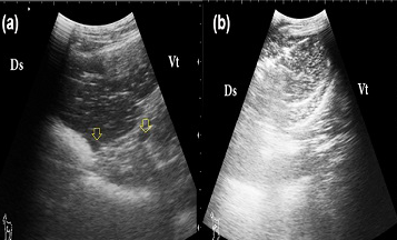
Figure 3: Ultrasonogram of liver in PT does
A: a good prognosis case; the liver appears more echogenic than the normal with visible hepatic vessels (arrows); B: a bad prognosis case: the liver appears more echogenic than the normal with invisible hepatic vessels; Ds: Dorsal; Vt: Ventral
Ketone bodies were negative in the urine of all healthy pregnant does, while it was positive in the urine of PT does (+/++).
Ultrasonographic Examnation
The normal parenchymal pattern of the liver consisted of numerous fine echoes homogenously distributed over the entire area of the liver (Figure 2). The caudal vena cava has a triangular shape in cross section because it is embedded in the sulcus of the vena cava in the liver. The vein was seen in all examined Does, usually limited to the 7th to 12th ICS. The diameter of the caudal vena cava noticed greater than that of the portal vein and they decreased from the 9thto the 12th ICS. The portal vein appeared rounded in cross section and biforcated in longitudinal section surrounded by echogenic wall. It was seen in all ICSs in which liver parenchyma is visible except for the 5th, 6thand 7th ICS. In PT does, 9 does had more echogenic liver parenchyma than the normal while the visibility hepatic vessels still clear, these does were respond to therapeutic trials. Increase hepatic echogenicity with complete invisibility of hepatic vessels was detected in 6 does. These cases were not responding to therapeutic trials (Figure 3).
Liver Biopsy, Histopathological and Necropsy Findings
Biopsy needle appeared as hyperechogenic structure among the hepatic parenchyma (Figure 4). The specimen of PT liver was greasy in consistency and floated on the surface of a test tube filled with water while it sink in the bottom at normal condition.
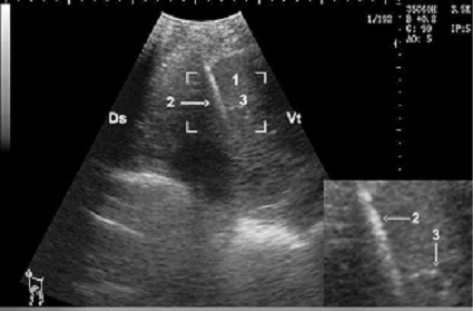
Figure 4: Hepatic biopsy in a doe, notice the needle is clearly visible as hyper-echoic structure (zoom area) within the hepatic parenchyma
1: Liver parenchyma; 2: Biopsy needle; 3: Portal vein; Ds: Dorsal; Vt: Ventral
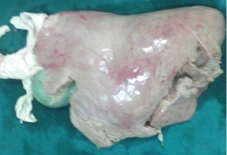
The histopathological examination of obtained samples showed changes in the liver cells, the hepatocytes throughout the liver had abnormally extensive cytoplasmic vacuoles represented fatty liver and increased by the progress of the disease (Figure 5). Necropsy findings of the liver obtained from 3 dead PT does characterized by yellow colour appearance, friable and having a greasy texture (Figure 6).
Therapeutic Trials
Nine PT does were respond to treatment (medical= 6 and caesarian section= 3) while other 6 cases not responding to treatment and 3 of them were submitted to necropsy.
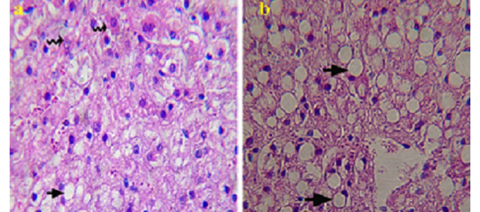
A: showing mild macro vesicular fatty change (arrow) and numerous normal hepatocytes (zigzag arrow) (good prognosis); B: showing severe vesicular fatty change (arrow) and less normal hepatocytes (bad prognosis). H & E Χ 1200
DISCUSSION
Presence of anorexia, depression and scanty feces in all pregnancy toxemic Does was in agreement with the results recorded by Rook (2000). Acetone odour was smelled at breathing in 6 cases of severe PT as reported by Abdelaal et al. (2013). Development of icterus in 2 cases with PT can be attributed to an increase in total bilirubin in the blood in case of fatty liver as suggested by West (1990), Staufenbiel et al. (1991), Rehage et al. (1996) and Radostits et al. (2007). In the current study, nervous manifestations including muscular tremors, deviated head and neck, apparent blindness and grinding on teeth were recorded in 6 does with advanced PT. These findings were similar to those previously observed by Rook (2000), Witsch et al. (2012) and Abdelaal et al. (2013). Nervous manifestations may be developed due to impairment of glucose utilization or inability of the nerve cells to utilize sugar as a result of high cortisol levels. The temperature, ruminal motility, heart and respiratory rates in the control group were similar to the values in previous studies that established for healthy Does in the same race by Balikci et al. (2007). Where, these parameters were significantly affected in PT does when compared with control group. This result was similar to Van Saun (2000), Rook (2000) and Abdelaal et al. (2013) who reported increase of the heart and respiratory rates in does with early PT while decreasing in does with advanced PT. In 3 cases these parameters were within normal range. These results were in accordance to Kabakci et al. (2003) and Barakat et al. (2007) as they found that temperature, heart and respiratory rates values in Does with sub clinic and clinic groups of pregnant toxemia were usually normal.
The mean value of serum glucose was significantly decreased in all does with PT as previously reported by Hefnawy et al. (2010) and Abdelaal et al. (2013). The importance of glucose in the pregnant does as the major source of energy to the fetus is well known. Therefore, pregnant does are at high risk of developing pregnancy toxemia due to the rapid fetal growth. The decrease of the serum total protein associated with PT may be due to the impairment of liver function, which agreed with Ulvund (1990). The obtained results for serum protein in diseased animals showed a significant drop in the albumin concentration. The mean value of serum beta hydroxyl butyric acid in PT goats showed a marked elevation compared with clinically healthy pregnant ones. This significant increase could be attributed to the lipolysis of tissue and the release of long chain fatty acids which were converted by the liver into ketones (Roubies et al., 2003) in goat. Moreover, this increase could be attributed to disturbance in carbohydrate and fat metabolism leading to hypoglycemia and mobilization of fat stores which lead to hepatic ketogenesis. Many authors prefer to measure enzyme activities and considered them sensitive and specific to assess the liver function (Rehage et al., 1996; Pechova et al., 1997; Komatsu et al., 2002). In this study, it was observed that serum hepatic enzymes (ALT and AST) increased significantly in PT does. The altered liver function could be associated with pregnancy toxemia related hepatic insufficiency. In the present study, kidney function parameters (BUN and creatinine) in PT goats, showed non-significant changes. Concerning serum lipid pattern, the low serum cholesterol level can be thought to be caused by a fat infiltration in the liver and a low output of lipoprotein which agreed with Sevinc et al. (2003). The observed increase in serum triglycerides in diseased animals was in agreement with Barakat et al. (2007) while Drackley et al. (2001) recorded that, triglyceride (TG) level in cows with moderate to severe fatty liver was not different from healthy cows. The elevation of triglyceride may be attributed to lipolysis of tissue and the release of long chain fatty acids which were stored as TG in liver or converted to ketone bodies.
Ketonuria has been detected in PT does which was agreed with Barakat et al. (2007), Gonzalez (2011) and Abdelaal et al. (2013). The urine diagnostic strip is a rapid and useful approach for securing field cases. Presence of ketone bodies in urine suggested that when fetal needs for glucose exceeds the dietary supply there would be increased lipolysis and incomplete oxidation of the 2-carbon radical due to a relative lack of oxaloacetate in the tricarboxylic acid cycle, which lead to build up aceto acetyl CoA. Hydrogenation of acetyl CoA results in the formation of acetone by decarboxylation, which in return, descend in urine and milk.
Ultrasonography, the liver can be imaged on the right side, and in Balady doe was always visible from the 7th to the 12th ICS in all examined animals. Braun and Steininger (2011), Braun et al. (2013) reported that, Saanen does liver was always visible from the 7th to the 9th ICS while the 5th and 6th and the 10th to the 12th ICS the liver could be seen only in some of the does. The normal parenchymal pattern of the liver consists of numerous fine echoes homogenously distributed over the entire area of the liver that appeared more echogenic than the cortex of the kidney and a comparatively less echogenic than the spleen. The results confirmed to that reported by Braun (1990), Braun and Hausammann (1992), Braun and Steininger (2011), Braun et al. (2013) and Alsafy et al. (2013). In the current study, the liver of PT does appeared more echogenic than the normal and hepatic vessels appeared either visible or not. The visibility of blood vessels give an idea about the severity of the diseases, it considered severe and bad prognosis when the blood vessels were invisible. The hyperechoic features of fatty liver may be attributed to changes in the nature of the liver tissues that increase the attenuation of the ultrasound beam (Cebra et al., 1997; Szebeni et al., 2006).
Ultrasound-guided liver biopsy, histopathology, necropsy findings and the result of therapeutic trials confirmed the ultrasonographic findings and provided positive results. The results coincided with that previously reported in humans (Caturelli et al., 1992). However, in humans, ultrasonographic diagnosis of fatty liver was found sufficient and biopsy was not recommended, where no other indications for biopsy existed (Szebeni et al., 2006).
CONCLUSION
Ultrasonography and ultrasonographic guided biopsy may be considered as rapid tools for evaluation the state of the liver in PT does and it may enable the veterinarian to evaluate such cases.
AKNOWLDGEMENT
The authors thank all staff members in Animal Medicine and Dr. Hany Lotfy, (department of Theriogenology, Faculty of Veterinary Medicine, Zagazig University, Egypt) for their continuous help and support during conduction of this work.
CONFLICT OF INTERESTS
The authors declare that there is no conflict of interests
AUTHORS CONTRIBUTION
Amr Kotb and Shaimaa Gouda performed clinical, laboratory and ultrasonographical work as well as writing the paper. Wafaa Abdel-Razik and Abbas El-Nagar supervised and performed the plan of the work.
REFERENCES


