Advances in Animal and Veterinary Sciences
Research Article
Advances in Animal and Veterinary Sciences 1 (2): 59–64Prevalence and Antimicrobial Susceptibility Patterns of Bovine and Ovine Staphylococcus aureus Isolates in Maiduguri, Nigeria
Pwaveno Huladeino Bamaiyi1*, Augustine Tochukwu Aniesona2,3
- Department of Veterinary Medicine, University of Maiduguri. Nigeria
- Department of Veterinary Microbiology and Parasitology, University of Maiduguri. Nigeria
- Department of Microbiology, Faculty of Science, University of Maiduguri. Nigeria
*Corresponding author: phbamaiyi@gmail.com
ARTICLE CITATION:
Bamaiyi PH, Aniesona AT (2013). Prevalence and antimicrobial susceptibility patterns of bovine and ovine Staphylococcus aureus isolates in Maiduguri, Nigeria. Adv. Anim. Vet. Sci. 1(2): 59–64
Received: 2013–05–15, Revised: 2013–06–06, Accepted: 2013–06–10
The electronic version of this article is the complete one and can be found online at
(
http://www.nexusacademicpublishers.com/table_contents_detail/4/49/html
)
which permits unrestricted use, distribution, and reproduction in any medium, provided the original work is properly cited
ABSTRACT
A study was conducted to determine the prevalence and antibiotic susceptibility patterns of Staphylococcus aureus from Bovine and Ovine species in Maiduguri metropolis. Two hundred and eighty one (n =281) clinical and non–clinical swab samples from Bovine (n=133) and Ovine (n=148) species were collected; the samples were screened for incidence of Staphylococcus aureus and susceptibility of the isolates to various antimicrobial agents using standard procedures. Eighty two (61.7%) of the Bovine and 91 (61.5%) of the Ovine samples were S. aureus positive. The isolates were further tested for haemolysin production and subjected to antibiotic sensitivity tests against 16 antibiotics representing various classes of antimicrobial agents, 10 antibiogroups (Groups A – K) were obtained and the antimicrobial resistance index (MARI) showed that as high as 86.1% (149/173) of the screened isolated pathogens had MARI above 0.2 (> 0.2). The haemolysin tests showed 34.1% of the isolates to be alpha–haemolytic. The MARI value is an indication of misuse or abuse and the haemolysin test results suggests a possibility of cross–species infections or acquisition of mobile genetic elements from other sources by the pathogens. There is widespread misuse of antimicrobials and signs of resistance development by the pathogen in Maiduguri, Nigeria. The use of antibiotics should be regulated to arrest this development and such regulation should be enforced by appropriate authority.
INTRODUCTION
Staphylococcus aureus (S. aureus) is an important pathogen of domestic ruminants and man (Bukharie et al., 2001; Grundmann et al., 2006; Fitzgerald, 2012), and is regarded as one of the most versatile and devastating zoonotic pathogen responsible for causing widespread outbreaks of serious infection, food poisoning, colonization, animal mastitis and pneumonia affecting man and animals (Fthenakis, 1998; Simoons–Smit et al., 2000; Kesah et al., 2003; Pengov and Ceru, 2003; Eftekhar and Dadaei, 2011; Ateba Ngoa et al., 2012; Dhama et al., 2013). S. aureus can also cause a number of infections in animals, such as abortions in Ewes (Edwards et al., 2008), staphylococcosis in rabbits (Hermans et al., 1999, 2003), pneumonia and osteomyelitis complex in poultry (Akbar and Anal, 2013; Monecke et al., 2013). In the various farms where such infections have been diagnosed, cumbersome preventive and control measures had to be undertaken on S. aureus, and despite all these measures, new infections continued to occur, treatment is often associated with poor success (Zecconi and Scali, 2013a) and attempts at producing a viable vaccine have failed and more research is needed in this aspect (Proctor, 2012; Sadeghi, 2012). One of the reasons for these failures are not only because infections originate from both exogenous and endogenous sources to the host, but more importantly due to the ability of the pathogen to rapidly develop resistance to antimicrobial agents commonly employed for intervention by the clinicians (Grundmann et al., 2006; Hasman et al., 2010; Gharsa et al., 2012; Porrero et al., 2012). Besides their role as commensals on the skin and mucosal surfaces, their occurrence on the respiratory and gastrointestinal tracts of worm blooded animals; they have also been involved in a wide variety of animal diseases (Werckenthin et al., 2001). These pathogens are responsible for disease when the host defense or immunity is compromised or lacking. Several manifestations of disease such as pyemic abscesses, suppurative arthritis and urinary tract infections in domestic and wild animals could be attributed to numerous virulent factors, such as the production of toxins and enzymes (Pengov and Ceru, 2003; Gharsa et al., 2012). S. aureus has several factors that contribute to the pathogenesis of infection (Gharsa et al., 2012), Several reports of drug resistant S. aureus isolated from animals have been documented; few reports of S. aureus species are from horses (Werckenthin et al., 2001; Alves et al., 2009; van Duijkeren et al., 2010), dairy herds (Fthenakis, 1998; Normanno et al., 2007; Alves et al., 2009), pet dog (Fitzgerald, 2012; Yamamoto et al., 2013), avian species, cats, rabbits, pigs, goats and sheep (Van Duijkeren et al., 2010; de la Fuente et al., 2011; Zecconi and Scali, 2013b). However, some studies (Seguin et al., 1999; Zecconi and Scali, 2013b) emphasized the possibility of exchange of the organism between species of animals.
Previous workers have reported studies on S. aureus in different parts of Nigeria in both human and animal subjects (Chigbu and Ezeronye, 2004; Isara et al., 2010; Olajubu et al., 2012). Maiduguri, the capital of Borno State, Nigeria is located in the extreme north of the Sahel region and accounts for about one quarter of the total ruminant population in Nigeria (Majiyagbe and Lamorde, 1997). Despite the large population of these animals in the region, the prevalence and the antibiotic susceptibility pattern of S. aureus among Bovine and Ovine species in Maiduguri metropolis have not been adequately studied and documented. The ability to isolate S. aureus and the determination of the antibiograms is very crucial for clinicians to select empiric antimicrobial therapy, rational formulation of public health policies and providing useful information on the global surveillance of this pathogen (Shittu et al., 2006).
Present study was designed to comparatively evaluate trends in colonization or infections and determined the extent of S. aureus prevalence as a widespread problem among Bovine and Ovine species in Maiduguri. The study also accessed the antibiograms of the isolates to determine their sensitivity to commonly used antibiotics.
MATERIALS AND METHODS
Sample Collection
The Bovine and Ovine clinical and non–clinical samples for analysis in this study were collected from septic wounds, abscess, nasal and skin swabs from animals in Maiduguri. The study lasted between February 2006 to November, 2007. Two hundred and eighty one (281) specimens; Bovine (n =133) and Ovine (n = 148) clinical and non–clinical samples were collected from different animal species using sterile swab sticks (Evepon sterile, Anambra State. Nigeria). University of Maiduguri Veterinary Teaching Hospital (UMVTH) and Modu Sheriff (State) Veterinary Clinic both located in Maiduguri, Northeastern Nigeria served as the sources of clinical specimen while various commercial farms and house hold farm animals served as non–clinical subjects. Each specimen was accompanied by details of the species from which the samples was collected as described by Cook and Pezzlo (Cook and Pezzlo, 1992). Verbal consent was sort prior to sample collection and farm/herds owner’s permission or consents were obtained before sampling of non–clinical subjects. The labelled samples were transported to the laboratory as soon as possible for processing or stored at refrigeration temperature (4oC) where processing was not possible.
Sample Analysis
Collected swabs samples were streaked over blood and nutrient agar plates (Oxoid) and were incubated at 37oC for 24–48 hours. After incubation, colony types were visualized for characteristic morphology of S. aureus subcultured on mannitol salt agar (LAB M, Landashire, UK) and identified phenotypically by conventional methods as Gram’s staining, catalase test, coagulase test as described by Cowen and Steel (Cowan and Steel, 1993) and Bello (Bello, 2002).
Antibiotic Susceptibility Test
A light suspension for each of the isolates was made in peptone water and was incubated, the inoculum was standardized to appropriate density of recommended 0.5 McFarland, which is equivalent to the turbidity of a Barium chloride standard (Brown et al., 2005; CLSI, 2008). The suspension was poured over 4mm thick Mueller–Hinton agar plates. The agar surface was allowed for few minutes and antibiotic impregnated discs (Mast Diagnostics, UK) containing: Ofloxacin (5µg), Erythromycin (15µg), Gentamycin (10µg), Ciprofloxacin (5µg), Clindamycin (2µg), Penicillin (10Units), Fusidic acid (10µg), Chloramphenicol (30µg), Nalidixic acid (30µg), Streptomycin (30µg), Tetracycline (30µg), Rifampicin (5µg), Mupirocin (5µg), Oxacillin disc (1µg) and Neomycin (30µg) were placed on the agar (about 10mm apart), over–turned and incubated at 37oC for 24 hours, then the zones of inhibition pattern were read following the CLSI guidelines (CLSI, 2008). Interpretative zone diameters or susceptibility breakpoints for resistance to Fusidic acid, Neomycin and Streptomycin which were not stated in the CLSI guidelines were considered as follows; ≤ 14 mm– Fusidic acid (Daxboeck et al., 2004), ≤ 16 mm– Neomycin and ≤ 14 mm– Streptomycin (Kim et al., 2004). Growth within a 14mm zone of inhibition with the 5µg Mupirocin disc indicated low–level resistance (Udo et al., 1999).
Multiple Antibiotic Resistance Index (MARI)
Determination of multiple antibiotic resistance index (MARI) was determined for each isolate by dividing the number of antibiotic to which the organism was resistant by the total number of antibiotics tested (Krumperman, 1983; Paul et al., 1997; Olayinka et al., 2006).
RESULTS
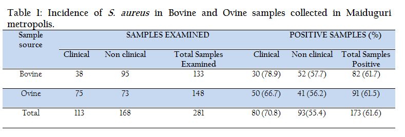
A total of two hundred and eighty one (281) specimens; Bovine (n =133) and Ovine (n = 148) clinical and non–clinical samples were collected from the animal species, Table 1 shows the details of prevalence of S. aureus according to different sources. Bovine clinical samples accounted for 30/38 (78.9%) isolates while 52/95 (57.7%) S. aureus isolates were obtained from the non–clinical samples. Ovine clinical samples showed 50/75 (66.7%) positive and 41/73 (56.2%) were positive from non–clinical samples. Hence, of the total 281 samples examined for S. aureus, 173 (61.6%) were positive.
Antimicrobial Susceptibility Testing (AST)
Antimicrobial Susceptibility Testing (AST) on the 173 isolates was done by disc diffusion method only, using various concentrations of antimicrobial agents representing various antimicrobial classes. AST results detailed in Table 2, showed that the highest level of resistance (100%) by all the isolates were found against Nalidixic acid. Test against Penicillin showed that 67/82 (81.7%) of Bovine isolates were resistant and 88/91 (96.7%) of Ovine isolates were resistant.
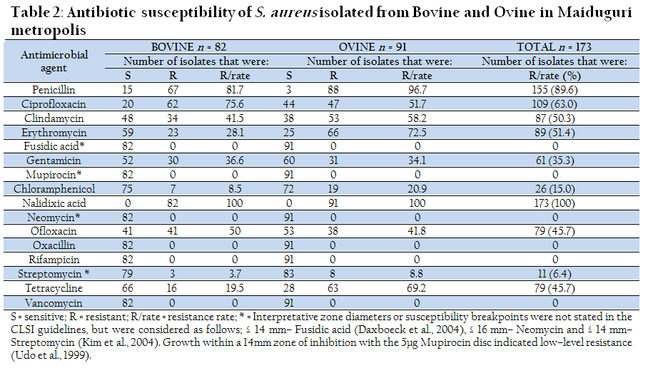
Table 2: Antibiotic susceptibility of S. aureus isolated from Bovine and Ovine in Maiduguri metropolis
Tetracycline tests against the isolates indicated very high resistance among Ovine 63 (69.3%) isolates, while Bovine isolates showed remarkably higher susceptible population of 66 (80.5%). All the isolates were 100% susceptible to Fusidic acid, Neomycin, Oxacillin, Rifampicin and Vancomycin. Comparatively, Ovine isolates had lower 47 (51.7%) Ciprofloxacin resistant population than Bovine isolates which showed 62 (75.6%) Higher sensitivity to Clindamycin was observed among Bovine isolates as they showed sensitivity in above 50% of the population while Ovine isolates test showed that <50% were sensitive to the drug. 72.5% isolates from Ovine showed high resistance to Erythromycin, but Bovine isolates had lower resistant population of 28.1%. Less than 50% of the Bovine, and Ovine isolates were resistant to Gentamicin. Only 7 (8.5%) of Bovine isolates showed resistance to Chloramphenicol, although Ovine isolates showed 29.9%. All the isolates showed high sensitivity to Streptomycin. Generally, S. aureus isolates from Ovine samples indicated higher antimicrobial resistance than the Bovine isolates.
Multi antimicrobial resistance index (MARI)
The multi antimicrobial resistance index (MARI) gives a direct suggestion of the probable sources of an microorganism. According to previous workers, MARI greater than 0.2 indicates that an organism must have originated from an environment where antibiotics are often used or abused (Krumperman, 1983; Paul et al. 1997; Olayinka, et al. 2006). A careful look at the results (Table 3) shows that all of the isolates in group A, were resistant to only three of the sixteen antimicrobial agents tested. The calculation (not shown) proved that all the isolates in the group had MARI equal to 0.2. Hence, only 5.2% (9/173) fall within this group. 86.1% (149/173) of the isolates are within Group B – J, and all had MARI above 0.2 (MARI > 0.2), while 8.7% (15/173) of the isolates (Group K) were susceptible to all the antibiotics tested, hence their MARI = 0.
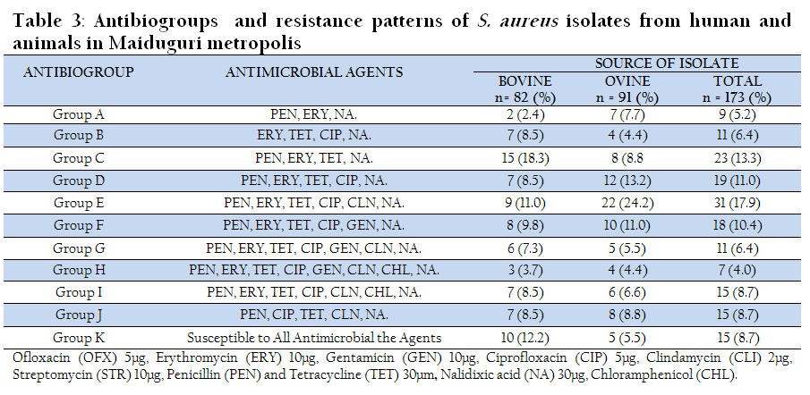
Table 3: Antibiogroups and resistance patterns of S. aureus isolates from human and animals in Maiduguri metropolis
Antibiogroups
Ten antibiogroups (Groups A – K) were detected in this study based on the resistance pattern of isolates. Bovine and Ovine isolates showed their highest level of resistance in Groups C– 18.3%, and E–24.2% Lowest resistance population in bovine isolate was 2.4% (Group A), while Ovine had a population of 4.4% in both Group B and H as the lowest resistant population. In Group L, 15 (8.7%) of the total isolates were susceptible to all the eight classes of antimicrobial agents tested. Comparatively, Bovine isolates presented more isolates 10 (12.2%) that were susceptible to all the antimicrobial agents tested, Ovine isolates were 5 (5.5%) (Table 3).
Hemolysin Production Test
Twenty nine (35.4%) of the Bovine isolates were α–hemolytic, 32/82 (39.0%) were β–haemolytic 9/82 (11.0%) produced γ–hemolysins and only twelve (14.6%) were non–hemolytic. Among the Ovine isolates, the α–haemolytic strains were 30/91 (33.0%), β–haemolysin positive isolates were 41/91 (45.1%) and 13/91 (14.3%) of γ–haemolytic strains were found among ovine isolates, 7/91 (7.7%) were negative for haemolysin production. (Table 4). Overall haemolysin production test result showed that Ovine isolates had the higher prevalence rate of 92.3% (84/91) of haemolytic strains compared to Bovine isolates with 85.4% (70/82) prevalence rate.
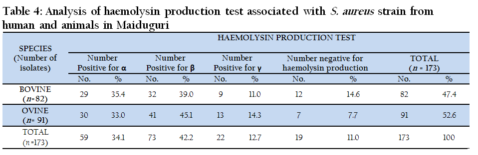
Table 4: Analysis of haemolysin production test associated with S. aureus strain from human and animals in Maiduguri
The β–haemolytic strains were 42.2% (73/173) which were the most prevalent among all the isolates tested, followed by α–haemolytic strains that accounted for 34.1% (59/173). The γ–haemolytic strains were 12.7% (22/173) while the non–haemolytic group gave 11.0% (19/173). Of the 173 isolates from Bovine and Ovine tested, 89.0% (154/173) were either α, β or γ–haemolytic.
DISCUSSION
The results of this study demonstrated a high prevalence of S. aureus among the animals investigated, the clinical and non–clinical samples of both animals revealed >50% positive for S. aureus. These results are similar to those reported from Umudike, Nigeria (Achi and Ugbogu, 2006), where S. aureus incidence among sheep, goat, cattle, pigs and rabbits were reported to be high in healthy animals. Thus both clinically healthy and unhealthy animals can carry S. aureus with zoonotic and pathogenic potentials. The result is also an indication that S. aureus is one of the most common pathogen in most infections which is in agreement with the report of earlier workers (Werckenthin et al., 2001; Oranusi et al., 2003; Olaoye and Onilude, 2009; de la Fuente et al., 2011) were they explained that the pathogen begins with colonization of the host, which usually culminate into clinical conditions in the animals and is suspected to be the source of most resistant strains infecting both man and animals (Seguin et al., 1999; Simoons–Smit et al., 2000; Lee, 2003). AST of the isolates tested against different classes of antibiotics showed that great populations of resistant strains were found among both animal species. All of the isolates were resistant to Nalidixic acid, this could be due to the fact that Nalidixic acid is an antibiotic used against gram negative bacteria which explains why there seems to be high level of resistance on the isolates.
This study indicated that a greater percentage (86.1%) of the isolates had MARI above 0.2, this underscores the need for effective antibiotic policy in both veterinary and human consumption in Maiduguri. Susceptibility testing serves as a guide to the physician in selecting appropriate drug to which a particular microorganism is susceptible, thereby producing effective therapy. The observation of high population of resistant strains among these animals poses a great public concern for humans and a risk to other animal population in and around Maiduguri owing to the fact that people rely on the animals for food, labour, transport and have constant and prolonged contact with the animals in the area of study.
Many of the Bovine isolates (35.4%) were α–haemolytic, this also applies to the Ovine isolates where 33.0% (30/91) were recorded as α–haemolysin positive. Alpha–haemolytic (α–haemolysin positive) strains of S. aureus are known to be more of human biotypes (Dinges et al., 2000), the isolation of these strains from animals in such a high number is an indication that a cross species infection or contamination had occurred between human and animals. Equally, as many as 39.0% and 45.2% Bovine and Ovine isolates were discovered to be β–haemolytic, these β–haemolytic biotypes are known to predominantly be of animal origins (Dinges et al., 2000; Achi and Ugbogu, 2006; Hasman et al., 2010; de la Fuente et al., 2011; Espinosa–Gongora et al., 2012). These results suggest that the close association of human and animals in Maiduguri may have led to the transfer or exchange of different specie specific biotypes. This is in agreement with previous reports that constant contact with animals could result into exchange or just jumping of these pathogens (Umoh et al., 1991; Oranusi et al., 2006).
Ten resistance patterns (antibiogroups) were found (table 3). The predominant pattern was in group E where resistance to six antibiotics (penicillin, erythromycin, tetracycline, ciprofloxacin, Clindamycin and Nalidixic acid) was observed. This is evidence that S. aureus isolates of animal origin are relatively resistant to many commonly used antibiotics which is similar but slightly higher than results reported from Umudike, Nigeria (Achi and Ugbogu, 2006). This high resistance could be attributed to extensive use of antibiotics in Veterinary establishments to combat infections or in subtherapeutic doses in feeds. Most of the antibiotics tested for are commonly used in both human and veterinary interventions in clinical cases or as prophylaxis, the high resistance patterns are also not entirely strange since there is the possibility of resistant strains originating from other sources and spread to the animals through close contact. The presence of multidrug–resistant isolates in these animals constitutes a reservoir of antibiotic resistant genes, which can spread to other pathogenic species. These strains might have acquired the genes from other species living in the surrounding environment through conjugational genetic transfer (Forbes and Schaberg, 1983) and through mutation of an already existing bacterial gene or horizontal transfer of a resistance gene from another bacterium (Grundmann et al., 2006).
It has been significantly proven that the use of antimicrobial agents for enhancing growth, chemoprophylaxis and treatment of animals meant for food increases the possibility of resistance in human pathogens (Singer et al., 2003; Ternak, 2005; Martinez, 2009). Therefore, the reduction or strict regulation and judicious use of antibiotics use in veterinary medicine is important to minimize the emergence of resistant strains. In this study, we recommend that the choice of antibiotics used for intervention by clinicians (both human and veterinary) and feed formulators must take into account the emergence of drug resistant strains. Constant evaluation of the antibiotic susceptibility patterns for commonly used antimicrobial agents in the study environment is carried out to be informed and take rational decisions on the most efficacious agents to employ during infections.
ACKNOWLEDGEMENT
We sincerely acknowledge the assistance of the staff of University of Maiduguri Veterinary Teaching Hospital and Modu Sheriff Veterinary Hospital in Maiduguri during our sample collection. The efforts of the laboratory staff of University of Maiduguri teaching hospital in media preparation are also appreciated.
REFERENCES
Achi OK and Ugbogu GA (2006). Characterization of drug resistant Staphylococcus aureus of animal origin. J. Biol. Sci. 6: 1088–1092.
http://dx.doi.org/10.3923/jbs.2006.1088.1092
Akbar A and Anal AK (2013). Prevalence and antibiogram study of Salmonella and Staphylococcus aureus in poultry meat. Asian Pac. J. Trop. Biomed. 3: 163–168.
http://dx.doi.org/10.1016/S2221-1691(13)60043-X
Alves PDD, McCulloch JA, Even S, Le Maréchal C, Thierry A, Grosset N, Azevedo V, Rosa CA, Vautor E and Le Loir Y (2009). Molecular characterisation of Staphylococcus aureus strains isolated from small and large ruminants reveals a host rather than tissue specificity. Vet. Microbiol. 137: 190–195.
http://dx.doi.org/10.1016/j.vetmic.2008.12.014
PMid:19157725
Ateba Ngoa U, Schaumburg F, Adegnika AA, Kösters K, Möller T, Fernandes JF, Alabi A, Issifou S, Becker K, Grobusch MP, Kremsner PG and Lell B (2012). Epidemiology and population structure of Staphylococcus aureus in various population groups from a rural and semi urban area in Gabon, Central Africa. Acta Trop. 124: 42–47.
http://dx.doi.org/10.1016/j.actatropica.2012.06.005
PMid:22750045
Bello CSS (2002). Laboratory manual for students of medical microbiology, 2nd ed. Satohgraphics press, Jos, Nigeria.
Brown DFJ, Edwards DI, Hawkey PM, Morrison D, Ridgway GL, Towner KJ and Wren MWD (2005). Guidelines for the laboratory diagnosis and susceptibility testing of methicillin-resistant Staphylococcus aureus (MRSA). J. Antimicrob. Chem. 56: 1000–1018.
http://dx.doi.org/10.1093/jac/dki372
PMid:16293678
Bukharie HA, Abdelhadi MS, Saeed IA, Rubaish AM and Larbi EB (2001). Emergence of methicillin-resistant Staphylococcus aureus as a community pathogen. Diag. Microbiol Infect. Dis. 40: 1–4.
http://dx.doi.org/10.1016/S0732-8893(01)00242-5
Chigbu C and Ezeronye O (2004). Antibiotic resistant Staphylococcus aureus in Abia state of Nigeria. Afr. J. Biotech. 2: 374–378.
CLSI (2008). Performance Standards for Antimicrobial Susceptibility Testing; Eighteenth Informational Supplement. CLSI document 28, M100–S18.
Cook J and Pezzlo M (1992). Specimen receipt and accessioning, in: Isenberg, H. (Ed.), Clinical Microbiology Procedures Handbook. pp. 1–4.
Cowan SF and Steel KJ (1993). Manual for identification of medical bacteria, 3rd ed. Cambridge University Press, Cambridge.
Daxboeck F, Assadian O, Apfalter P and Koller W (2004). Resistance rates of Staphylococcus aureus in relation to patient status and type of specimen. J. Antimicrob. Chem. 54: 163–167.
http://dx.doi.org/10.1093/jac/dkh277
PMid:15175269
De la Fuente R, Ballesteros C, Bautista V, Medina A, Orden JA, Domínguez-Bernal G and Vindel A (2011). Staphylococcus aureus subsp. anaerobius isolates from different countries are clonal in nature. Vet. Microbiol. 150: 198–202.
http://dx.doi.org/10.1016/j.vetmic.2010.12.022
PMid:21236606
Dhama K, Rajagunalan S and Chakraborty S (2013). Food-borne pathogens of Animal origin-Diagnosis, prevention and control and their zoonotic significance-A review. Pakistan J. Biol. Sc. 16: 1076–1085.
http://dx.doi.org/10.3923/pjbs.2013.1076.1085
PMid:24506006
Dinges MM, Orwin PM and Schlievert PM (2000). Exotoxins of Staphylococcus aureus. Clin. Microbiol. Rev. 13: 16–34.
http://dx.doi.org/10.1128/CMR.13.1.16-34.2000
PMid:10627489 PMCid:PMC88931
Edwards JF, Lassala AL and Spencer TE (2008). Staphylococcus-associated abortions in ewes with long-term central venous catheterization. Vet. Path. 45: 881–888.
http://dx.doi.org/10.1354/vp.45-6-881
PMid:18984790
Eftekhar F and Dadaei T (2011). Biofilm Formation and Detection of IcaAB Genes in Clinical Isolates of Methicillin Resistant Staphylococcus aureus. Iranian J. Basic Med. Sci. 14: 132–136.
Espinosa-Gongora C, Chrobak D, Moodley A, Bertelsen MF and Guardabassi L (2012). Occurrence and distribution of Staphylococcus aureus lineages among zoo animals. Vet. Microbiol. 158: 228–231.
http://dx.doi.org/10.1016/j.vetmic.2012.01.027
PMid:22369899
Fitzgerald JR (2012). Livestock-associated Staphylococcus aureus: origin, evolution and public health threat. Trends Microbiol. 20: 192–198.
http://dx.doi.org/10.1016/j.tim.2012.01.006
PMid:22386364
Forbes BA and Schaberg DR (1983). Transfer of resistance plasmids from Staphylococcus epidermidis to Staphylococcus aureus: evidence for conjugative exchange of resistance. J. Bacteriol. 153: 627–634.
PMid:6822476 PMCid:PMC221678
Fthenakis G (1998). Susceptibility to antibiotics of staphylococcal isolates from cases of ovine or bovine mastitis in Greece. Small Ruminant Res. 28: 9–13.
http://dx.doi.org/10.1016/S0921-4488(97)00057-6
Gharsa H, Ben Slama K, Lozano C, Gómez-Sanz E, Klibi N, Ben Sallem R, Gómez P, Zarazaga M, Boudabous A and Torres C (2012). Prevalence, antibiotic resistance, virulence traits and genetic lineages of Staphylococcus aureus in healthy sheep in Tunisia. Vet. Microbiol. 156: 367–373.
http://dx.doi.org/10.1016/j.vetmic.2011.11.009
PMid:22176760
Grundmann H, Aires-de-Sousa M, Boyce J and Tiemersma E (2006). Emergence and resurgence of meticillin-resistant Staphylococcus aureus as a public-health threat. Lancet 368: 874–885.
http://dx.doi.org/10.1016/S0140-6736(06)68853-3
Hasman H, Moodley A, Guardabassi L, Stegger M, Skov RL and Aarestrup FM (2010). Spa type distribution in Staphylococcus aureus originating from pigs, cattle and poultry. Vet. Microbiol. 141: 326–331.
http://dx.doi.org/10.1016/j.vetmic.2009.09.025
PMid:19833458
Hermans K, De Herdt P, Devriese LA, Hendrickx W, Godard C and Haesebrouck F (1999). Colonization of rabbits with Staphylococcus aureus in flocks with and without chronic staphylococcosis. Vet. Microbiol. 67: 37–46.
http://dx.doi.org/10.1016/S0378-1135(99)00028-0
Hermans K, Devriese LA and Haesebrouck F (2003). Rabbit staphylococcosis: difficult solutions for serious problems. Vet. Microbiol. 91: 57–64.
http://dx.doi.org/10.1016/S0378-1135(02)00260-2
Isara A R, Isah EC, Lofor PVO and Ojide CK (2010). Food contamination in fast food restaurants in Benin City, Edo State, Nigeria: Implications for food hygiene and safety. Public Health 124: 467–471.
http://dx.doi.org/10.1016/j.puhe.2010.03.028
PMid:20630547
Kesah C, Ben Redjeb S, Odugbemi TO, Boye CSB, Dosso M, Ndinya Achola JO, Koulla-Shiro S, Benbachir M, Rahal K and Borg M (2003). Prevalence of methicillin-resistant Staphylococcus aureus in eight African hospitals and Malta. Clin. Microbiol. Inf. 9: 153–156.
http://dx.doi.org/10.1046/j.1469-0691.2003.00531.x
PMid:12588338
Kim H. Bin, Jang H, Nam HJ, Lee S, Kim BS, Park WB, Lee KD, Choi YJ, Park SW, Oh M, Kim E, Choe KW and Lee YS (2004). In Vitro Activities of 28 Antimicrobial Agents against Staphylococcus aureus Isolates from Tertiary-Care Hospitals in Korea : a Nationwide Survey In Vitro Activities of 28 Antimicrobial Agents against Staphylococcus aureus Isolates from Tertiary-Care Hosp. Antimicrob. Agents Chem. 48: 1124–1127.
Krumperman PH (1983). Multiple antibiotic resistance indexing of Escherichia coli to identify high-risk sources of fecal contamination of foods . Multiple Antibiotic Resistance Indexing of Escherichia coli to Identify High-Risk Sources of Fecal Contamination of Foodst. Appl. Environ. Microb. 46: 165–170.
PMid:6351743 PMCid:PMC239283
Lee JH (2003). Staphylococcus aureus Strains Isolated from Major Food Animals and Their Potential Transmission to Humans Methicillin (Oxacillin) -Resistant Staphylococcus aureus Strains Isolated from Major Food Animals and Their Potential Transmission to Humans. Appl. Environ. Microb. 69: 6489–6494.
http://dx.doi.org/10.1128/AEM.69.11.6489-6494.2003
PMid:14602604 PMCid:PMC262320
Majiyagbe KA and Lamorde AG (1997). Nationally Coordinated Research Programme on Livestock Diseases: Subsectorial goals, Performance and Medium-term research plans. Trop. Vet. 15: 75–83.
Martinez JL (2009). Environmental pollution by antibiotics and by antibiotic resistance determinants. Environmental pollution (Barking, Essex : 1987) 157: 2893–2902.
Monecke S, Ruppelt A, Wendlandt S, Schwarz S, Slickers P, Ehricht R and Jäckel SC De (2013). Genotyping of Staphylococcus aureus isolates from diseased poultry. Vet. Microbiol. 162: 806–812.
http://dx.doi.org/10.1016/j.vetmic.2012.10.018
PMid:23140941
Normanno G, Corrente M, La Salandra G, Dambrosio A, Quaglia NC, Parisi A, Greco G, Bellacicco AL, Virgilio S and Celano G V (2007). Methicillin-resistant Staphylococcus aureus (MRSA) in foods of animal origin product in Italy. Int. J. Food Microbiol. 117: 219–222.
http://dx.doi.org/10.1016/j.ijfoodmicro.2007.04.006
PMid:17533002
Olajubu AF, Osinupebi OA, Ismail L, Bosede O and Deji-Agboola AM (2012). Pattern of hospital associated infections in a teaching hospital in Nigeria. Asian Pac. J. Trop. Dis. 2: S869–S873.
http://dx.doi.org/10.1016/S2222-1808(12)60282-5
Olaoye OA and Onilude AA (2009). Assessment of microbiological quality of sachet–packaged drinking water in Western Nigeria and its public health significance. Public Health 123: 729–734.
http://dx.doi.org/10.1016/j.puhe.2009.09.015
PMid:19880150
Olayinka BO, Olonitola OS, Olayinka AT, Nwogu CC (2006). Effects of Sodium chloride and urea on antibiotic susceptibility patterns of S. aureus strains isolated from urine of suspected UTI patients. Nigerian J. Microbiol. 20: 676–680.
Oranusi S, Galadima M and Umoh V (2006). Toxicity test and bacteriophage typing of Staphylococcus aureus isolates from food contact surfaces and foods prepared by families in Zaria, Nigeria. Afr. J. of Biotech. 5: 362–365.
Oranusi S, Umoh V and Kwaga JK (2003). Hazards and critical control points of kunun–zaki, a non–alcoholic beverage in Northern Nigeria. Food Microbiol. 20: 127–132.
http://dx.doi.org/10.1016/S0740-0020(02)00072-2
Paul S, Bezbaruah RL, Roy MK and Ghosh AC (1997). Multiple antibiotic resistance (MAR) index and its reversion in Pseudomonas aeruginosa. Letters in applied microbiology 24: 169–171.
http://dx.doi.org/10.1046/j.1472-765X.1997.00364.x
PMid:9080693
Pengov A and Ceru S (2003). Antimicrobial drug susceptibility of Staphylococcus aureus strains isolated from bovine and ovine mammary glands. J. Dairy Sci. 86: 3157–3163.
http://dx.doi.org/10.3168/jds.S0022-0302(03)73917-4
Porrero MC, Hasman H, Vela AI, Fernández–Garayzábal JF, Domínguez L and Aarestrup FM (2012). Clonal diversity of Staphylococcus aureus originating from the small ruminants goats and sheep. Vet. Microbiol. 156: 157–161.
http://dx.doi.org/10.1016/j.vetmic.2011.10.015
PMid:22112857
Proctor RA (2012). Is there a future for a Staphylococcus aureus vaccine? Vaccine 30: 2921–2927.
http://dx.doi.org/10.1016/j.vaccine.2011.11.006
PMid:22115633
Sadeghi M (2012). Two Dimensional Structural Analysis and Expression of a New Staphylococcus aureus Adhesin Based Fusion Protein. Iran J. Basic Med. Sci 15: 725–738.
Seguin JC, Walker RD, Caron JP, Kloos W, George CG, Hollis RJ, Jones N, Pfaller MA and Jones RN (1999). Methicillin–Resistant Staphylococcus aureus Outbreak in a Veterinary Teaching Hospital : Potential Human–to–Animal Transmission Methicillin–Resistant Staphylococcus aureus Outbreak in a Veterinary Teaching Hospital : Potential Human–to–Animal Transmission. J. Clin. Microbiol. 37: 1459–1463.
PMid:10203505 PMCid:PMC84801
Shittu A, Lin J and Kolawole D (2006). Antimicrobial Susceptibility Patterns of Staphylococcus aureus and Characterization of MRSA in Southwestern Nigeria. Wounds 18: 77–84.
Simoons–Smit AM, Savelkoul PH, Stoof J, Starink TM and Vandenbroucke–Grauls CM (2000). Transmission of Staphylococcus aureus between humans and domestic animals in a household. European J. Clin. Microbiol. Inf. Dis. 19: 150–152.
http://dx.doi.org/10.1007/s100960050450
PMid:10746507
Singer RS, Finch R, Wegener HC, Bywater R, Walters J and Lipsitch M (2003). Antibiotic resistance—the interplay between antibiotic use in animals and human beings. Lancet Inf. Dis. 3: 47–51.
http://dx.doi.org/10.1016/S1473-3099(03)00490-0
Ternak G (2005). Antibiotics may act as growth/obesity promoters in humans as an inadvertent result of antibiotic pollution? Med. hypotheses 64: 14–16.
http://dx.doi.org/10.1016/j.mehy.2004.08.003
PMid:15533603
Udo EE, Farook VS, Mokadas EM, Jacob LE and Sanyal SC (1999). Molecular Fingerprinting of Mupirocin–Resistant Methicillin–Resistant Staphylococcus aureus from a Burn Unit. Int. J. Inf. Dis. 3: 82–87.
http://dx.doi.org/10.1016/S1201-9712(99)90014-0
Umoh VJ, Adesiyun AA and Gomwalk NE (1991). Bacteriophage typing of Staphylococci aureus strains from fermented milk products and raw milk. Trop. Vet. 9: 131–138.
Van Duijkeren E, Moleman M, Sloet van Oldruitenborgh–Oosterbaan MM, Multem J, Troelstra A, Fluit AC, Van Wamel WJB, Houwers DJ, De Neeling AJ, Wagenaar JA (2010). Methicillin–resistant Staphylococcus aureus in horses and horse personnel: an investigation of several outbreaks. Vet. Microbiol. 141: 96–102.
http://dx.doi.org/10.1016/j.vetmic.2009.08.009
PMid:19740613
Werckenthin C, Cardoso M, Martel JL, Schwarz S, Erckenthina CW, Ardosob MC, Artelc JM and Chwarzd SS (2001). Antimicrobial resistance in staphylococci from animals with particular reference to bovine Staphylococcus aureus, porcine Staphylococcus hyicus, and canine Staphylococcus intermedius. Vet. Res. 32: 341–362.
http://dx.doi.org/10.1051/vetres:2001129
PMid:11432424
Yamamoto T, Hung WC, Takano T and Nishiyama A (2013). Genetic nature and virulence of community–associated methicillin–resistant Staphylococcus aureus. BioMedicine 3: 2–18.
http://dx.doi.org/10.1016/j.biomed.2012.12.001
Zecconi A and Scali F (2013a). Staphylococcus aureus virulence factors in evasion from innate immune defenses in human and animal diseases. Immunol. lett. 150: 12–22.
http://dx.doi.org/10.1016/j.imlet.2013.01.004
PMid:23376548
Zecconi A and Scali F (2013b). Staphylococcus aureus virulence factors in evasion from innate immune defenses in human and animal diseases. Immunol. Lett. 150: 12–22.
http://dx.doi.org/10.1016/j.imlet.2013.01.004
PMid:23376548


