Advances in Animal and Veterinary Sciences
Case Report
Successful Management of Upper Eyelid Laceration in a Dromedary Camel (Camelus dromidarius)
Biswadeep Jena1*, Abrar Ahmed2, Nilesh Kumar Pagrut3
1Department of Veterinary Surgery & Radiology; 2Department of Veterinary Gynaecology & Obstetrics; 3Department of Veterinary Pathology, M. J. F. college of Veterinary and Animal Sciences, Rajasthan University of Veterinary and Animal Sciences, Rajasthan, India.
Abstract | A female dromedary camel was presented with clinical signs of lacerated left upper eyelid, profuse lacrimation due to continuous irritation of avulsed large, edematous tarsal flap. In order to correct the thwarted vision, continuous irritation and profuse lacrimation, a decision was made to repair the avulsed part of the lacerated eyelid under deep sedation and local infiltration. Obligation of proper surgical techniques and maintenance of adequate postoperative measures rewarded with uneventful recovery.
Keywords | Dromedary Camel, Eyelid, Laceration, Two-layer closure technique, Sedation
Editor | Kuldeep Dhama, Indian Veterinary Research Institute, Uttar Pradesh, India.
Received | January 06, 2015; Revised | January 22, 2015; Accepted | January 23, 2015; Published | January 26, 2015
*Correspondence | Biswadeep Jena, Rajasthan University of Veterinary and Animal Sciences, Rajasthan, India; Email: biswadeep44@gmail.com
Citation | Jena B, Ahmed A, Pagrut NK (2015). Successful management of upper eyelid laceration in a dromedary camel (Camelus dromidarius). Adv. Anim. Vet. Sci. 3(2): 133-135.
DOI | http://dx.doi.org/10.14737/journal.aavs/2015/3.2.133.135
ISSN (Online) | 2307-8316; ISSN (Print) | 2309-3331
Copyright © 2015 Jena et al. This is an open access article distributed under the Creative Commons Attribution License, which permits unrestricted use, distribution, and reproduction in any medium, provided the original work is properly cited.
The eyelids are musculo-fibrous plica of which the upper eyelid is more substantial and mobile. During blinking, these are intermittently swept over the eyeball to evenly dispense the lacrimal fluid to maintain the uncovered parts moist and to protect it from external stimuli (Maggs, 2008; Siddiqui and Telfah, 2010). Full thickness tearing of either of the eyelids often occurs as a result of browsing on thorny bushes and trees, accidental rubbing or, invasion by a barbed wire is very common (Bishnoi and Gahlot, 2004; Gahlot et al., 2007; Siddiqui and Telfah, 2010; Gharu et al., 2013; Ramdan, 2014).
About 10 years old female dromedary camel used for draught purposes in Chomu, Jaipur (Rajasthan, India), was presented in the month of November 2014 with a lacerated wound in the left upper eyelid. Because of the tear, lid margin avulsed in the shape of a large, edematous flap hanging down over the cornea causing continuous irritation, profuse lacrimation and subsequent soiling around the eye (Figure 1). Hence a decision was made to repair the avulsed part of the lacerated eyelid in order to relieve the obstructed of vision and profuse lacrimation.
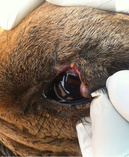
Figure 1: Showing avulsed large, edematous tarsal flap with lacrimation and soiling around the left eye
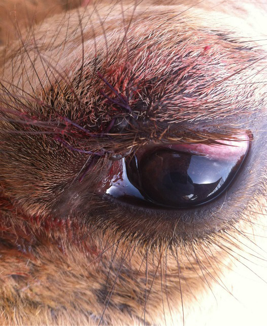
Figure 2: Showing reapposed eyelid margins with a standard two-layer closure technique using 2-0 Vicryl without penetrating the skin, margin or, conjunctiva
As the procedure had to be conducted under field conditions without any application of general anaesthesia, the animal was physically restrained in right lateral recumbency and deeply sedated by intravenous administration of 10 ml Xylazine (Xylaxin®; Indian Immunologicals Ltd). After attainment of sedation, the external jugular vein was catheterised and approximately 5 litres of normal saline was administered intra-operatively. In order to achieve complete anaesthesia and akinesia of upper eyelid, 10 ml of 2% Lidocaine hydrochloride (Xylocaine®; Astra Zeneca Ltd) local anaesthetic solution was deposited subcutaneously at multiple sites about 0.5 cm from the margin of dorsal eyelid and selectively desensitizing the auriculopalpebral branch of the facial nerve at the notch of zygomatic arch, using a 2.5 cm 20 gauge needle. The long hairs around the site were closely trimmed and the area was cleaned with 1% boric acid solution. The eyeball was protected with a cotton pad soaked in normal saline to prevent falling of hairs into the eye. Because of the irregular and soiled edges of the torn eye lid, it was subjected to “freshening” of wound margins by gentle scraping with a scalpel blade until they were devoid of noticeable debris and exudates. After freshening, the eye lid margins re-apposed with a standard two-layer closure technique using soft, braided, buried, absorbable suture 2-0 Vicryl® (polyglactin 910, Ethicon Inc.) on a reverse cutting needle without penetrating the skin, the margin or, the conjunctiva (Figure 2). Systemic (60 ml Oxytetracycline®; Zydus AHL intravenously) and topical antibiotics (Chloromycetin Apli Caps®; Pfizer India Ltd.) used perioperatively. The animal was recovered within 60 minutes after the onset of sedation. Post-operatively owner was instructed to apply topical antibiotic ointment (Neosporin® Ophthalmic Ointment; GlaxoSmithKline Pharmaceuticals Ltd.) twice daily for 2 weeks and to tie the animal in isolation away from any field objects or poles to avoid self-mutilation. During re-examination after 3 weeks, there was little evidence of the trauma to the eyelid. The camel recovered uneventfully and was permitted to return to its normal draught duties.
Being a highly vascular structure, the eyelids are susceptible to severe edema and distortion after even relatively minor injury which leads to thwarted vision, continuous irritation and profuse lacrimation. Complete resection of the indignant eyelid should be avoided because of its aesthetic value and permanent exposure of the eyeball to the external environmental stimuli alluring to problems like conjunctivitis. Hence an earnest effort should be made to refurbish the injured eyelid unless it is beyond the realm of repair. Sutures in the eyelids must be soft, pliable and should go through partial thickness of the skin; otherwise they will irritate the sclera and cornea. Bacterial flora in the conjunctival sac and surroundings may readily invade this area leading to severe postoperative septic blepharitis. Hence a course of systemic antibiotics with good spectrum of activity against gram-positive organisms should be instituted. Postoperatively, an ophthalmic ointment is preferred as it provides lubrication and protects the cornea during wound healing (Maggs, 2008; Siddiqui and Telfah, 2010).
ACKNOWLEDGEMENT
The authors are thankful to the Dean, M.J.F. college of Veterinary and Animal Sciences, Rajasthan University of Veterinary and Animal Sciences, Rajasthan, India for his support and cooperation in carrying out the study.
COMPETING INTERESTS
The authors declare that they have no competing interests.
REFERENCES


