Advances in Animal and Veterinary Sciences
Research Article
Adv. Anim. Vet. Sci. 3 (1): 71 - 78
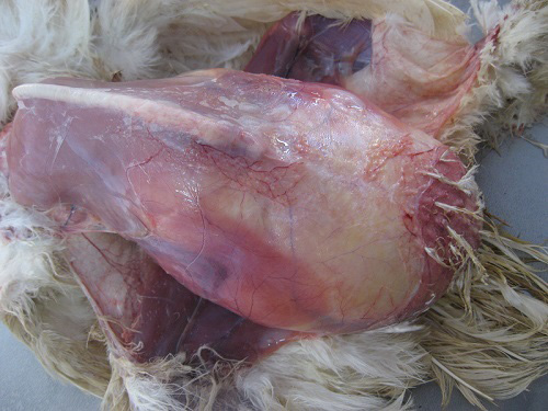
Figure 1
Affected bird showing marked distension of the abdomen.
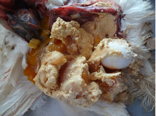
Figure 2
Abdominal cavity showing coagulated yolk deposits and eggs.
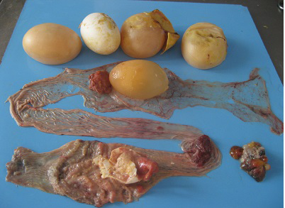
Figure 3
Oviduct lumen showing the partially formed egg, blood tinged albumin and yolk material and shell membrane
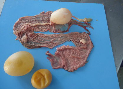
Figure 4
Oviduct lumen containing inflammatory exudate coated egg and wall showing inflammatory changes in salpingitis
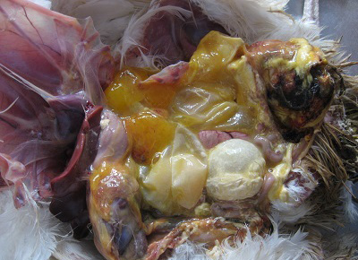
Figure 5
Cloacal region showing blackish discoloration and thin shelled eggs in peritoneal cavity in vent trauma
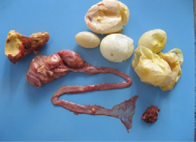
Figure 6
Marked thickening of uterine and vaginal wall in oviduct neoplasm


