Advances in Animal and Veterinary Sciences
Research Article
Adv. Anim. Vet. Sci. 3 (1): 27 - 33
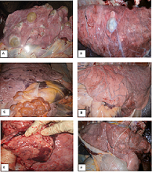
Figure 1
A. Polycystic lung containing large yellow fluid filled viable cyst and abscess; B. Old degenerated cyst and fibrosis in lung; C. Viable cyst in the pleura and lung in clustered appearance; D. Severe interstitial emphysema and marked distention of the veins (blue color) in middle and caudal lobe; E. Red Hepatization (consolidation) indicating pneumonia; F. Abscess in all over the lung parenchyma.
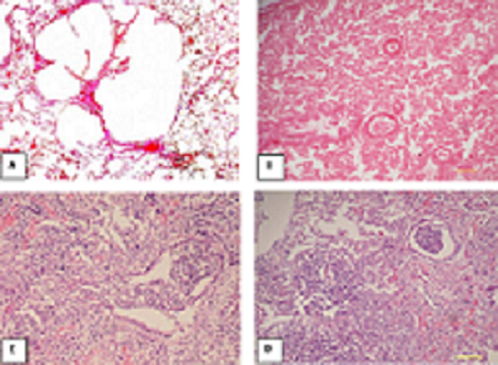
Figure 2
![]() A. Pulmonary congestion and alveolar emphysema; B. Pulmonary atelectasis; C. Purulent bronchopneumonia in lung, accumulation of neutrophils in bronchiolar airway; D. Pulmonary hepatization (pneumonic lung) where alveolar space is filled with copious exudates.
A. Pulmonary congestion and alveolar emphysema; B. Pulmonary atelectasis; C. Purulent bronchopneumonia in lung, accumulation of neutrophils in bronchiolar airway; D. Pulmonary hepatization (pneumonic lung) where alveolar space is filled with copious exudates.


