Advances in Animal and Veterinary Sciences
Research Article
Seroprevalence of Brucellosis in Sheep and Humans in District Kohat, Pakistan
Muhammad Altaf Hussain1, Rahmatullah Rind2, Muhammad Adil3, Momen Khan4, Farmanullah1, Arbab Sikandar3*, Usman Waheed3, Mohammad Salim5
1Lasbela University of Agriculture, Water & Marine Sciences, Uthal, Pakistan; 2Sind Agriculture University, Tandojam, Pakistan; 3University of Veterinary & Animal Sciences, Lahore, (Jhang campus), Pakistan; 4Department of Livestock & Dairy Development, Khyber Pakhtunkhwa, Pakistan; 5University of Veterinary and Animal Sciences, Lahore, Pakistan
Abstract | Brucellosis is a bacterial disease and is caused by genus Brucella. It is highly prevalent zoonotic disease in developing countries including Pakistan. This study was conducted to determine the prevalence of brucellosis in sheep and humans of district Kohat, Khyber Pakhtunkhwa. For this purpose, one hundred blood samples from each humans and sheep (50 each from both sexes) were randomly collected at three different tehsils (Lachi, Seni Gumbat and Kohat) of district Kohat. The serum samples were tested for presence of anti-Brucella antibodies by Rose Bengal Plate Test (RBPT) and Serum Agglutination Test (SAT). Moreover, a total of 50 milk samples were also collected from various sheep herds for subsequent conduction of milk ring test (MRT). The estimated prevalence of ovine brucellosis was 12.12%, 09% and 08.82% in Lachi, Seni Gumbat and Kohat, respectively, with an overall prevalence of 10% in the district. Whereas, SAT and RBPT demonstrated the prevalence as 7% and 6%, respectively, in humans. The results of current study validated that brucellosis was widespread in district Kohat with a relatively higher prevalence being recorded in ewes and occupationally exposed women than their male counterparts. Furthermore, the prevalence of brucellosis was significantly higher in sheep and humans of Lachi and Seni Gumbat tehsils as compared to Kohat. Results of the current study may support the health care organizers to formulate suitable control plans against Brucellosis.
Keywords | Brucellosis, Prevalence, Sheep, Humans, Kohat
Editor | Kuldeep Dhama, Indian Veterinary Research Institute, Uttar Pradesh, India.
Received | September, 17 2014; Revised | September 22, 2014; Accepted | October 02, 2014; Published | October 12, 2014
*Correspondence | Arbab Sikandar, University of Veterinary & Animal Sciences, Lahore, Pakistan; Email: arbab.sikandar@uvas.edu.pk
Citation | Hussain MA, Rind R, Adil M, Khan M, Farmanullah, Sikandar A, Waheed U, Salim M (2014). Seroprevalence of Brucellosis in sheep and humans in District Kohat, Pakistan. Adv. Anim. Vet. Sci. 2 (9): 516-523.
DOI | http://dx.doi.org/10.14737/journal.aavs/2014/2.9.516.523
ISSN (Online) | 2307-8316; ISSN (Print) | 2309-3331
Copyright © 2014 Hussain et al. This is an open access article distributed under the Creative Commons Attribution License, which permits unrestricted use, distribution, and reproduction in any medium, provided the original work is properly cited.
INTRODUCTION
Brucellosis is a contagious, zoonotic and highly prevalent, infectious disease of humans and animals caused by genus Brucella (Schelling et al., 2003). Human infection results from direct contact with infected animals or animal secretions, inhalation of contaminated aerosols, inoculation into the conjunctival sac or consumption of raw milk and milk products (Georgiou’s et al., 2005; Rahman et al., 2011). However meat and meat products instigate relatively little risk of infection due to small number of organisms in the muscles and uncommon utilization of undercooked meat (Mandel et al., 2005). Human brucellosis is clinically characterized by undulant fever, headache, myalgia, lumbar pain and arthritis. Abortion and sterility reflect the major manifestations resulting from the localization of causative organism within the reproductive organs in animals (Mandel et al., 2005). Small ruminants have been incriminated for the transmission of brucellosis to exposed individuals (Kaoud et al., 2010). Brucellosis represents an occupational health hazard for abattoir workers, farmers, veterinarians and laboratory personnel (Mandel et al., 2005; Rahman et al., 2012 and Asif et al., 2014). Moreover, tremendous economic losses to livestock sector are attributed to brucellosis in terms of abortion, infertility and reduced milk yield (McDermott and Arimi, 2002; Maadi et al., 2011). Brucellosis is widespread in Pakistan owing to lack of appropriate control strategies (Khan et al., 2009; Abubakar et al., 2012).
Ovine brucellosis can be divided into two distinct forms. Like caprine brucellosis, the zoonotic form of ovine brucellosis is caused by Brucella melitensis (Garin-Bastuji, 1994; Samadi et al., 2010; Montasser et al., 2011) and characterized by abortion, stillbirth and placentitis in ewes (Iqbal et al., 2013). This Brucella spp. is considered as highly pathogenic and invasive for humans as compared to Brucella abortus (Acha and Szyfres, 2003). While the non-zoonotic Brucella ovis has been implicated to cause orchitis, epidydmitis and hypospermia in rams (Megid et al., 2010: Lone et al., 2013). Isolation and identification of the causative agent constitutes the most preferred method for presumptive diagnosis of brucellosis. However it is hazardous, time-consuming and difficult to be implemented for large scale diagnosis (Ali et al., 2013). Consequently serological tests based upon the detection of anti-Brucella antibodies are commonly used to diagnose brucellosis (Ferreira et al., 2003). Rose Bengal Plate Test (RBPT) and Serum Agglutination Test (SAT) represent the most frequently applied tests for the serodiagnosis of brucellosis (Gul and Khan, 2007). The SAT provides quantitative data on immune responses (Hamidullah et al., 2009). The RBPT is a quick, effective and less expensive test applicable to detect and diagnose brucellosis in large animal herds (Erganis et al., 2005). The sensitivity of RBPT and SAT are 99% and 95.6%, respectively (Barroso et al., 2002; Memish et al., 2002). Milk Ring Test (MRT) is another simple, cheap and widely employed diagnostic test using milk as sample from Brucella suspected animals. Hamidullah et al. (2009) recorded 32.5% ovine/caprine prevalence of Brucellosis during five year study and Mustafa et al. (2011) reported only one case of sheep abortions in Kohat.
Although brucellosis has been extensively studied in cattle, buffaloes, dogs and horses, the clear picture of prevalence of ovine brucellosis still exists unstipulated in many developing countries (Yesuf et al., 2010) including Pakistan. Furthermore, the prevalence of occupationally exposed humans is another issue which also required consideration. This project was therefore planned to ascertain the prevalence of brucellosis in sheep and humans of district Kohat, Pakistan, for delineating the significance of Brucella control measures in animal population and awareness about the disease in humans.
MATERIALS AND METHODS
Study Area
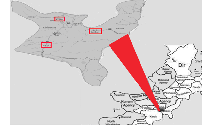
The study was conducted in District Kohat, comprising of three tehsils i.e., Lachi, Seni Gumbat and Kohat, located in Khyber Pakhtunkhwa province of Pakistan (Figure 1) at geographic coordinates of 33° 35’ 13” North latitude and 71° 26’ 32” East longitude with an altitude of 508 meters and total area of 2,545 square kilometres. The district is characterized by mountainous topography with a maximum temperature of 40ºC and average annual rainfall of about 638 mm. Kohat Tehsil exhibits relatively high population and developed agricultural infrastructure as compared to other tehsils.
Collection of Samples
One hundred blood samples were randomly collected in 5cc disposable syringes from each humans and sheep (50 each from both sexes) at three different Tehsils (Lachi, Seni Gumbat and Kohat) of district Kohat, Khyber Pakhtunkhwa province of Pakistan. The collected blood samples were kept in slanting position at room temperature to allow clotting. Sera were separated by centrifugation and kept at -20°C until further processing. Later on, the sera were transported in vacutainers (13x100 capacity) to Veterinary Diagnosis and Research Laboratory, Kohat for the analysis of anti-Brucella antibodies. A total of 50 random milk samples from altogether different unvaccinated animals rearing in various sheep herds in the study areas were also collected and stored at 4°C. The milk samples were examined for the presence of antibodies against Brucella spp. using MRT.
Rose Bengal Plate Test (RBPT)
Rose Bengal plate test (RBPT) was performed according to the procedure described by Zahid et al. 2002. Briefly, the Rose Bengal stained antigen (Veterinary Research Institute (VRI), Lahore) and sera were brought to room temperature. Later on, 40 µl of antigen was added to 40 µl of each serum sample on a clean glass slide and thoroughly mixed by means of a sterilized stirrer. After 4 minutes the slides were checked for presence or absence of agglutination thereby recorded as positive and negative reactions, respectively.
Serum Agglutination Test (SAT)
Phenol saline (0.85% NaCl in 0.5% phenol) was used for the preparation of two fold serial dilutions (1:20 to 1:640) of serum samples in different tubes. Equal amount (0.5 ml) of concentrated antigen (VRI, Lahore) was added to each serum-containing tube and the contents were thoroughly mixed. All tubes were incubated at 37°C for 24 hours. The results were compared with antigen-containing control tube exhibiting 50% agglutination. A titre of 1:40 or above was regarded as positive (Chachra et al., 2009).
Milk Ring Test (MRT)
MRT was performed following the method described by Abbas and Aldeewan, (2009). Milk samples and antigen stored at 4°C were brought to room temperature. About 40 µl Hematoxylin stained Brucella melitenesis antigen (VRI, Lahore) was added to each test tube containing 1 ml of milk sample and the contents were properly mixed. Test tubes were incubated at 37°C for 1 hour. Positive reaction was indicated by the appearance of dark blue ring at the upper portion of milk.
Antibody titration
The antibody titre of positive serum samples was also determined using SAT. All serum samples were subjected to two fold serial dilutions (1:20 to 1:160) using four different test tubes (Din et al., 2013). Later on, Brucella melitensis antigen obtained from VRI, Lahore was added to all dilutions and the antigen-antibody interactions observed at various dilutions were recorded.
Data analysis
Data thus collected for RBPT, SAT and MRT were probed by applying descriptive statistical technique as described previously by Sikandar et al., (2012).
RESULTS AND DISCUSSION
The overall prevalence of ovine brucellosis was recorded as 10.0% in district Kohat. However Iqbal et al. 2013 and Negash et al. 2012 recorded the seroprevalence of ovine brucellosis as 08.07% and 07% in Ethiopia and Southern Punjab (Pakistan), respectively. Lone et al. (2013), reported the prevalence of ovine brucellosis as 06.50% in Kashmir. This slightly higher prevalence in the current study on brucellosis could be attributable to geographical variation and altered systems of vaccination and management. However, Hamidullah et al. (2009) reported the prevalence of ovine brucellosis in Kohat as 34.08%. The likely rationale for this lower prevalence in this study could be the involvement of smaller house hold and nomadic sheep herds with less number of animals than larger animal herds at organized farms analysed by Hamidullah et al. (2009). Extensive animal farming has been documented as a potential risk factor for ovine brucellosis (Al-Majali, 2005; Yesuf et al., 2010). The prevalence of brucellosis was recorded as 07% and 06% in humans (associated with sheep rearing) by SAT and RBPT, respectively. These values of prevalence are in conformity to the findings of Mohmand et al. (2012) and Rahman et al. (2012), who reported a prevalence of 06.09% in adult population of two provinces of Pakistan and persons associated with goat farming in Bangladesh, respectively.
The prevalence of brucellosis in ewes was recorded to be 12.00%, 10.00% and 10.00% by SAT, RBPT and MRT, respectively in the endemic area. Compared with RBPT and MRT, the prevalence of brucellosis was relatively higher in ewes as determined by SAT (Table 1 and Table 2). While, in rams, SAT and RBPT recorded the seroprevalence as 08.00% and 10.00%, respectively. Regardless of the diagnostic techniques applied, a risk of higher prevalence of brucellosis was evident in ewes as compared to rams. Similar results have also been reported by previous studies (Yesuf et al., 2010; Negash et al., 2012; Rahman et al., 2013). The presence of erythritol in allantoic fluid favours the growth and propagation of Brucella organisms thereby enhancing the susceptibility of female sheep to brucellosis (Yesuf et al., 2010; Rahman et al., 2011). Nevertheless, RBPT detected equivalent (10.00%) seroprevalence in rams as well as in ewes. Our results reinforce the findings of earlier studies demonstrating equal seropositivity in male and female sheep using RBPT (Muma et al., 2006; Iqbal et al., 2013). Both SAT and RBPT demonstrated the prevalence of brucellosis to be 06% in men. The prevalence of brucellosis was recorded as 08.00% and 06.00% in women using SAT and RBPT, respectively. Irrespective of the diagnostic techniques applied, a risk of higher prevalence of brucellosis was obvious in women as compared to men. Previous studies have also reported relatively higher prevalence of brucellosis in women than their male counterparts (Khan et al., 2009; Din et al., 2013 and Shahid et al., 2014). The frequent involvement of nomadic and rural females in livestock handling and grazing as compared to their urban counterparts could enhance their susceptibility to brucellosis and other zoonotic problems (Mohmand et al., 2012 and Shahid et al., 2014).
Data regarding the prevalence of ovine brucellosis in three different regions (i.e., Lachi, Seni Gumbat and Kohat) of district Kohat (Pakistan) has been illustrated in table 3. A total of one hundred serum samples from each humans and sheep were examined by SAT and RBPT to determine the prevalence in mentioned areas. The prevalence of ovine brucellosis was recorded as 12.12%, 9% and 8.82% in Lachi, Seni Gumbat and Kohat regions, respectively. The recorded seroprevalence of human brucellosis was 12.12% and 09.09% in Seni Gumbat as determined by SAT and RBPT, respectively. Nevertheless, both SAT and RBPT imparted identical values pertaining to prevalence of human brucellosis in Lachi and Kohat. The prevalence of human brucellosis was documented to be 06.06% and 02.94% in Lachi and Kohat, respectively. Rahman et al. (2012) also found substantial variation in terms of human seropositivity to brucellosis within the three districts of Bangladesh. It was obvious from these results that prevalence of human and ovine brucellosis was significantly higher in Seni Gumbat and Lachi, respectively as compared to Kohat. Unlike Kohat, Seni Gumbat and Lachi are relatively distant regions characterized by hilly terrain, scarce cultivable land and inappropriate health facilities. All these factors could significantly contribute to unproblematic transmission of diseases. Teshale et al. (2006) and Iqbal et al. (2013) also reported similar trend in the prevalence of ovine brucellosis with varying location. Geographical variation has been reported to influence the seroprevalence of brucellosis (Rahman et al., 2011).
Table 1: Prevalence of human and ovine brucellosis in Kohat, Khyber Pukhtunkhawa, Pakistan, determined by different diagnostic techniques
|
Technique |
Sheep |
Humans |
||||
|
Total No. of serum samples examined |
No. of positive samples |
Percentage ofpositive samples |
Total No. of serum samples examined |
No. of positive samples |
Percentage of positive samples |
|
|
SAT |
100 |
10 |
10.00 |
100 |
7 |
7.00 |
|
RBPT |
100 |
10 |
10.00 |
100 |
6 |
6.00 |
|
MRT |
50 |
5 |
10.00 |
- |
- |
- |
Table 2: Sex-wise prevalence of human and ovine brucellosis in Kohat, Khyber Pakhtunkhawa, Pakistan
|
Target Population |
Sex |
Action |
Technique |
||
|
SAT |
RBPT |
MRT |
|||
|
Sheep |
Rams |
Total No. of serum samples examined |
50 |
50 |
- |
|
No. of positive samples |
4 |
5 |
- |
||
|
Percentage of positive samples |
8.00 |
10.00 |
- |
||
|
Ewes |
Total No. of serum samples examined |
50 |
50 |
50 |
|
|
No. of positive samples |
6 |
5 |
5 |
||
|
Percentage of positive samples |
12.00 |
10.00 |
10.00 |
||
|
Humans |
Men |
Total No. of serum samples examined |
50 |
50 |
- |
|
No. of positive samples |
3 |
3 |
- |
||
|
Percentage of positive samples |
6.00 |
6.00 |
- |
||
|
Women |
Total No. of serum samples examined |
50 |
50 |
- |
|
|
No. of positive samples |
4 |
3 |
- |
||
|
Percentage of positive samples |
8.00 |
6.00 |
- |
||
The antibody titer of positive serum samples was determined using SAT and the results were presented in table 4. Of the 50 sera from each male and female sheep, 06 (12%) and 04 (08%) were positive reactors, respectively. Of the 06 positive samples from ewes, 04 and 03 showed antibody titer at dilutions of 1/20 and 1/40, respectively and hence were positive. While out of 04 serum samples from rams, 03 and 01 showed higher antibody titer (declared positive) at dilutions of 1/20 and 1/40, respectively. Similarly, out of 04 positive sera from women, 02 each interacted at dilutions of 1/20 and 1/40, respectively; while of 03 positive sera from men, 02 and 01 interacted at dilutions of 1/20 and 1/40, respectively. While no antigen-antibody interaction was observed at the dilutions of 1/80 and 1/160, hence was declared as negative. Parizadeh et al. (2009) also reported that positive SAT titer base was between 1:20-1:40 among the people of endemic areas in Iran. However, it was concluded that cases of brucellosis was evident in Kohat and relatively higher prevalence with high antibody titer was recorded in women and ewes compared to their male counterparts.
Table 3: Prevalence of human and ovine brucellosis in tested regions of Kohat, Khyber Pukhtunkhawa, Pakistan
|
Target Population |
Area of Study |
Action/ |
Test |
|
|
SAT |
RBPT |
|||
|
Sheep |
Lachi |
Total No. of serum samples examined |
33 |
33 |
|
No. of positive samples |
4 |
4 |
||
|
Percentage of positive samples |
12.12 |
12.12 |
||
|
Seni Gumbat |
Total No. of serum samples examined |
33 |
33 |
|
|
No. of positive samples |
3 |
3 |
||
|
Percentage of positive samples |
9.09 |
9.09 |
||
|
Kohat |
Total No. of serum samples examined |
34 |
34 |
|
|
No. of positive samples |
3 |
3 |
||
|
Percentage of positive samples |
8.82 |
8.82 |
||
|
Humans |
Lachi |
Total No. of serum samples examined |
33 |
33 |
|
No. of positive samples |
2 |
2 |
||
|
Percentage of positive samples |
6.06 |
6.06 |
||
|
Seni Gumbat |
Total No. of serum samples examined |
33 |
33 |
|
|
No. of positive samples |
4 |
3 |
||
|
Percentage of positive samples |
12.12 |
9.09 |
||
|
Kohat |
Total No. of serum samples examined |
34 |
34 |
|
|
No. of positive samples |
1 |
1 |
||
|
Percentage of positive samples |
2.94 |
2.94 |
||
Table 4: Antibody titer of seropositive samples measured using SAT
|
Target Population |
Sex |
Total positive reactors |
Positive reactors (titer/dilutions) |
|||||||
|
1/20 |
1/40 |
1/80 |
1/160 |
|||||||
|
No. |
% |
No. |
% |
No. |
% |
No. |
% |
|||
|
Sheep |
Rams |
4 |
3 |
75.00 |
1 |
25.00 |
0 |
0 |
0 |
0 |
|
Ewes |
6 |
4 |
66.66 |
2 |
33.34 |
0 |
0 |
0 |
0 |
|
|
Humans |
Men |
3 |
2 |
66.66 |
1 |
33.33 |
0 |
0 |
0 |
0 |
|
Women |
4 |
2 |
50.00 |
2 |
50.00 |
0 |
0 |
0 |
0 |
|
Pastoral sheep rearing is commonly practiced for wool and milk production in Kohat and adjoining hilly areas. Organized farming system is typically lacking and animals are customarily subjected to grazing. Combined rearing of multiple species in conjunction with deficient measures of feeding, vaccination, medication and biosecurity enhance the chances of disease transmission. Lack of optimal vaccination program, improper health facilities, poor hygienic measures, close interaction with animals (for farmers and occupationally exposed persons) and consumption of raw animal products (for farmers) were presumed as the potential risk factors. Nomadic life is characterized by seasonal migration, improper health facilities, close interaction with animals, poor hygienic measures and consumption of raw animal products. Owing to extensive livestock raising, the rural population has been reported to be highly susceptible to brucellosis. Earlier studies also found that poverty and low literacy positively correlated with seropositivity to brucellosis in case of humans (Karimi et al, 2003; Kansiime et al., 2014). The control of human brucellosis is contingent upon the prevention of disease in domestic animals as effective vaccines are not available for human immunization (Thakur et al., 2002; Mukhtar and Kokab, 2008; Rahman et al., 2012). Effective control measures in terms of immune-prophylaxis, testing with subsequent culling of infected animals, adequate disposal of aborted fatal material and separation of post-parturient animals could help to circumvent the transmission of ovine brucellosis thereby curtailing the risk of human infection. Appropriate awareness of general public regarding the prevention of brucellosis in humans is also requisite to control this zoonotic problem.
REFERENCES


